Comparative analysis of TNF-α, TLR-2, and TLR-4 in GCF before and after periodontal therapy
Abstract
Periodontitis is a chronic inflammatory disease characterized by progressive destruction of tooth-supporting tissues, primarily driven by a dysregulated host immune response to subgingival biofilms. Toll-like receptors (TLR-2 and TLR-4) and tumor necrosis factor-alpha (TNF-α) are key mediators of immune signaling and tissue breakdown, making them potential biomarkers of periodontal disease activity. This prospective, non-randomized interventional study evaluated TLR-2, TLR-4, and TNF-α levels in gingival crevicular fluid (GCF) for periodontal disease monitoring and assessed the effects of nonsurgical periodontal therapy (NSPT) on clinical parameters. Forty systemically healthy individuals were enrolled and assigned to either periodontitis or healthy groups. Clinical indices—gingival index, plaque index, clinical attachment loss, and probing pocket depth—were recorded at baseline and 8 weeks after NSPT. GCF samples were analyzed using enzyme-linked immunosorbent assay to determine biomarker concentrations. At baseline, TNF-α and TLR-2 levels were significantly higher in periodontitis patients compared with healthy controls (p < 0.001). Following therapy, marked clinical improvements were observed, accompanied by significant reductions in TNF-α and TLR-4 levels (p < 0.05), while TLR-2 levels remained stable. receiver operating characteristic analysis demonstrated that TNF-α and TLR-2 reliably distinguished disease from health. In conclusion, NSPT effectively improved clinical outcomes and reduced TNF-α and TLR-4 levels in GCF, reflecting a decreased local inflammatory burden. The persistence of TLR-2 expression post-treatment suggests a stable role in immune surveillance rather than acute inflammatory response.
Descriptors:
Biomarkers; Gingival Crevicular Fluid; Periodontal Diseases; Periodontitis; Root Planing
Introduction
Periodontitis is a chronic inflammatory disorder characterized by progressive degradation of tooth-supporting structures, primarily resulting from an altered immune response to bacterial biofilms.1 Although biofilms initiate the disease, its severity and progression are largely determined by the host’s immune-inflammatory mechanisms.2 The persistent interaction between bacterial pathogens and the immune system sustains inflammation, ultimately leading to the breakdown of periodontal tissues.3
Toll-like receptors (TLRs), a key subset of pattern recognition receptors, play a central role in the host’s early defense mechanisms against periodontal pathogens. These membrane-bound proteins recognize conserved microbial structures, known as pathogen-associated molecular patterns, and translate these signals into cellular immune responses. Through this recognition, TLRs activate downstream signaling cascades, triggering proinflammatory pathways that influence the onset and severity of periodontal inflammation.4 Among these receptors, TLR-2 and TLR-4 are particularly important in periodontitis due to their ability to detect bacterial lipoproteins and lipopolysaccharides, respectively.5 Upon activation, these receptors stimulate the production of inflammatory mediators such as tumor necrosis factor-alpha (TNF-α), interleukin (IL)-1β, and IL-6, all of which contribute to periodontal tissue destruction.6 Furthermore, genetic variations in TLR-4 have been linked to increased susceptibility to chronic periodontitis, highlighting its role in modulating host–microbial interactions.5
TNF-α is a central proinflammatory cytokine downstream of TLR signaling and plays a critical role in the pathogenesis of periodontal disease.7 It promotes osteoclast differentiation, enhances matrix metalloproteinase activity, and contributes to extracellular matrix degradation, leading to connective tissue destruction and alveolar bone loss.8 Elevated TNF-α concentrations in gingival crevicular fluid (GCF) have been consistently associated with disease severity, underscoring its potential as a biomarker for periodontal inflammation and progression.9 Periodontal therapy primarily aims to reduce microbial challenge, regulate host-mediated inflammation, and restore tissue homeostasis through nonsurgical strategies such as scaling and root planing (SRP). Profiling TNF-α, TLR-2, and TLR-4 levels after therapy may therefore provide valuable insights into immunological modulation and treatment effectiveness.
Correlating these biomarkers with clinical parameters—including gingival index (GI), plaque index (PI), clinical attachment loss (CAL), and probing pocket depth (PPD)—can yield a more comprehensive understanding of the molecular and clinical mechanisms underlying periodontitis. Previous studies have reported increased TLR-2 and TLR-4 expression in gingival tissues11,12 as well as altered TLR-related biomarkers in saliva and GCF.13 Treatment has been shown to influence these signals, with reductions in TLR-4 activity and improvements in GCF markers.14 Similarly, TNF-α has been strongly associated with periodontal status.15 However, only a limited number of studies have concurrently evaluated TLRs and inflammatory mediators in GCF and examined their modulation following periodontal therapy.
Therefore, the present study investigates the impact of periodontal treatment on GCF levels of TNF-α, TLR-2, and TLR-4 and explores their relationships with key clinical periodontal parameters. By analyzing these immune-inflammatory markers at baseline and after therapy, this study aims to provide deeper insights into the biological effects of periodontal treatment and its role in modulating inflammatory pathways involved in disease progression.
Methods
This prospective, non-randomized interventional study included 40 systemically healthy volunteers, comprising 12 males and 28 females aged 20–50 yr. Participants were recruited from patients presenting to the Faculty of Dentistry, Necmettin Erbakan University, Konya, Turkey, between June and December 2021. Ethical approval was obtained from the Non-Interventional Clinical Research Ethics Committee for Pharmaceuticals and Medical Devices of Necmettin Erbakan University (Decision No. 2021/05-63). All methods were performed in accordance with relevant guidelines and regulations, including the 2008 revision of the Declaration of Helsinki. Before enrollment, each participant was informed verbally and in writing about the study objectives, procedures, and potential implications. Written informed consent was obtained from all participants and/or their legal guardians.
Sample size calculation
In the absence of directly comparable data on pre–post changes in GCF TLR-4 levels following nonsurgical periodontal therapy (NSPT), the sample size was determined using pilot study guidance.16,17 A total of 40 systemically healthy individuals were enrolled and assigned to two groups according to periodontal status: Group 1 (periodontally healthy, n = 20) and Group 2 (periodontitis patients, n = 20). All participants completed follow-up. For within-patient comparison in the periodontitis group (paired t test, two-tailed), post hoc analysis indicated an observed standardized mean change (Cohen’s d) of 0.86. With α = 0.05 and n = 20 pairs, the achieved power was 1 − β ≈ 0.96 (G*Power 3.1), confirming adequacy of the sample size.
Study design
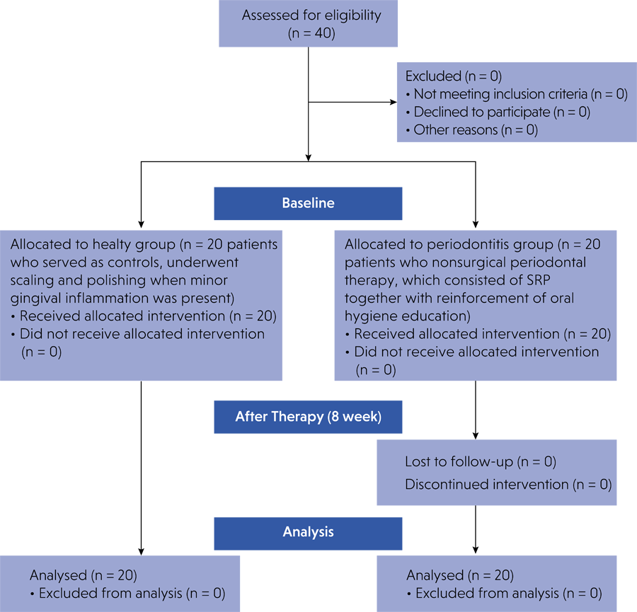
Patients presenting to the Periodontology Clinic were screened for eligibility through detailed medical and dental history reviews, followed by clinical and radiographic examinations. Figure 1 summarizes participant flow throughout the study.

Eligible participants were 20–50 years old with no systemic health problems. Periodontally healthy individuals exhibited no CAL, no gingival inflammation (e.g., redness, swelling, or exudate), probing depths ≤ 3 mm, < 10% bleeding on probing (BOP), and no radiographic evidence of alveolar bone loss. Periodontitis patients were diagnosed according to the 2017 World Workshop on the Classification of Periodontal and Peri-Implant Diseases and Conditions.18 They presented with generalized periodontitis, Stage II or III and Grade B, characterized by PPD > 4 mm, CAL ≥ 3 mm, BOP, and radiographically evident bone loss.
Exclusion criteria included immunodeficiency; systemic conditions requiring prophylactic antibiotics before dental procedures; periodontal therapy within the past 3 months; systemic antibiotic or anti-inflammatory drug (NSAIDs or corticosteroids) use within the past 3 months or during follow-up; any history of tobacco use (cigarettes, e-cigarettes, smokeless tobacco, or waterpipe); and pregnancy, breastfeeding, or menopause.
Clinical periodontal assessments were performed at baseline and 8 weeks after treatment in the periodontitis group. A calibrated examiner (F.U.Y.) using a manual periodontal probe conducted all examinations. Calibration was completed on 15 volunteers not included in the main study, assessed in two sessions 24 h apart. The intraclass correlation coefficient for PPD measurements between sessions was 0.94 ± 0.05, confirming excellent intra-examiner reliability. Healthy participants were examined only at baseline.
Periodontal parameters were assessed across the full dentition. PI was evaluated at six sites per tooth based on supragingival plaque accumulation.19 GI was visually determined using a 0–3 scale, reflecting the severity of inflammation by gingival color, texture, edema, and spontaneous bleeding.20 PPD and CAL were measured at six sites per tooth (mesio-buccal, mid-buccal, disto-buccal, mesio-lingual, mid-lingual, and disto-lingual) and rounded to the nearest millimeter using a UNC-15 periodontal probe (Hu-Friedy, Chicago, Illinois). BOP was recorded after depth measurements and expressed as the percentage of affected sites.
Nonsurgical periodontal therapy
All participants received detailed oral hygiene instructions, including plaque control techniques such as tooth brushing and interdental cleaning, from the calibrated examiner (F.U.Y.). Patients with periodontitis underwent NSPT, consisting of SRP combined with reinforcement of oral hygiene education. All NSPT procedures and post-treatment oral hygiene reinforcement were performed by a single experienced periodontist (F.A.). No antimicrobial agents were administered.
Phase I therapy in the periodontitis group comprised thorough SRP and oral hygiene instruction reinforcement. GCF samples and clinical parameters were collected at baseline and 8 weeks after treatment (± 3 days). Treatment was scheduled within two weeks of the initial examination, with a maximum variation of four days. Healthy participants (controls) underwent scaling and polishing if minor gingival inflammation was observed, along with oral hygiene reinforcement, performed by the same periodontist (F.A.).
All clinical measurements were performed by the calibrated examiner (F.U.Y.). Clinical and GCF assessments were conducted at baseline and 8 weeks after treatmentin the periodontitis group, while the healthy group was assessed only at baseline.
Collection and evaluation of GCF
Gingival crevicular fluid samples were collected from the deepest periodontal pocket of two teeth per participant in the periodontitis group, both at baseline and 8 weeks after treatment. In the periodontally healthy group, samples were obtained from the mesiobuccal sulcus of two maxillary anterior teeth (central incisors) with probing depth ≤ 3 mm and no clinical signs of inflammation.
Before collection, supragingival plaque was gently removed with a manual scaler. The selected sites were isolated with cotton rolls and dried using an air syringe to minimize contamination. Sterile Periopaper strips (ProFlow, Amityville, New York) were inserted 1 mm into the gingival sulcus and left in place for 30 s. Strips contaminated with blood were excluded.
All strips collected from each participant were pooled into labeled Eppendorf tubes and immediately stored at −80°C. On the day of analysis, samples were eluted in 100 μL of phosphate-buffered saline, incubated at room temperature for 30 min without agitation, and centrifuged for 10 min. The resulting supernatant was used for subsequent quantification.
Levels of TNF-α, TLR-2, and TLR-4 in GCF were measured using human-specific sandwich enzyme-linked immunosorbent assay kits (Elabscience, Wuhan, China), following the manufacturer’s instructions. Final concentrations were determined by comparing optical density values against standard curves generated during each assay. Kit specifications were as follows: TLR-2, sensitivity 0.19 ng/mL, detection range 0.31–20 ng/mL; TLR-4, sensitivity 3.75 pg/mL, detection range 6.25–400 pg/mL, total assay time 3 h 30 min; TNF-α, sensitivity 4.69 pg/mL, and detection range 7.81–500 pg/mL.
Statistical analysis
All statistical analyses were performed using SPSS software (version 19.0; IBM Corp., Armonk, USA). Continuous variables were expressed as mean ± standard deviation (SD). The Kolmogorov–Smirnov test was used to assess data normality. Independent-samples t tests were applied to compare the control and periodontitis groups at baseline and at the 8-week follow-up. Paired-samples t tests were used for within-group comparisons across time points. Chi-square tests were applied to analyze categorical variables recorded at baseline and post-treatment.
Pearson correlation analysis was performed to evaluate associations between biomarker concentrations and clinical periodontal parameters. Receiver operating characteristic (ROC) curve analysis was conducted to assess the diagnostic utility of GCF biomarkers in distinguishing periodontitis cases from healthy individuals. Sensitivity and specificity were estimated by calculating the area under the curve (AUC). A p-value < 0.05 was considered statistically significant.
Results
Descriptive data of participants
Table 1 summarizes the demographic characteristics of participants, including gender, education level, oral hygiene frequency, dental visits, and age. No significant differences were observed between groups for gender (p = 0.366) or age (p = 0.057). However, educational attainment differed significantly (p < 0.001): The periodontitis group included a higher proportion of participants with only primary school education, whereas the periodontally healthy group had more participants with university-level education. Brushing frequency also varied significantly; a larger percentage of participants in the periodontitis group reported brushing less than or equal once per day (p < 0.001).
Clinical parameters
Table 2 presents the clinical periodontal measurements. At baseline, individuals with periodontitis had significantly higher PPD, CAL, PI, and GI values compared with healthy controls (p < 0.001). Following nonsurgical therapy, significant reductions were observed in PPD, PI, and GI in the periodontitis group (p < 0.001). Although the reduction in CAL was less marked, it remained statistically significant (p < 0.001). These findings indicate that the intervention produced measurable improvements in clinical periodontal parameters.
Cytokine levels in GCF
As shown in Table 3, baseline concentrations of TNF-α, TLR-2, and TLR-4 were elevated in the periodontitis group compared with controls, with statistically significant differences for TNF-α and TLR-2 (p < 0.001). After nonsurgical therapy, significant reductions were observed in TNF-α and TLR-4 (p < 0.05), whereas TLR-2 levels did not differ significantly within the periodontitis group (p = 0.155).
ROC Curve Analysis
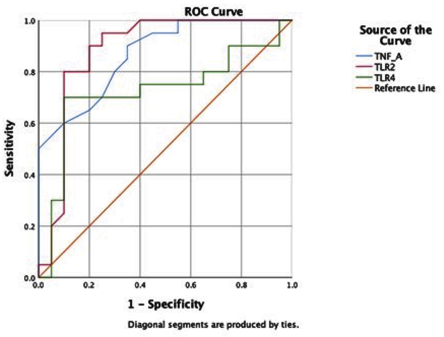
Receiver operating characteristic curve analysis was used to assess the diagnostic performance of TNF-α, TLR-2, and TLR-4 in distinguishing periodontal health from disease (Figure 2). TLR-2 demonstrated the highest diagnostic accuracy (AUC = 0.883), followed by TNF-α (AUC = 0.867), both indicating excellent discriminative ability. TLR-4 showed only moderate diagnostic capacity (AUC = 0.723). These results suggest that TLR-2 and TNF-α are reliable biomarkers for periodontal disease detection, whereas TLR-4 has limited diagnostic utility.
Correlations
Table 4 presents correlations between clinical parameters, biomarker levels, and age. Age correlated strongly with PI (r = 0.832, p < 0.001), GI (r = 0.798, p < 0.001), and PPD (r = 0.688, p < 0.001), and moderately with TLR-2 (r = 0.445, p = 0.004) but not with TNF-α (r = 0.271, p = 0.091) or TLR-4 (r = −0.108, p = 0.507).
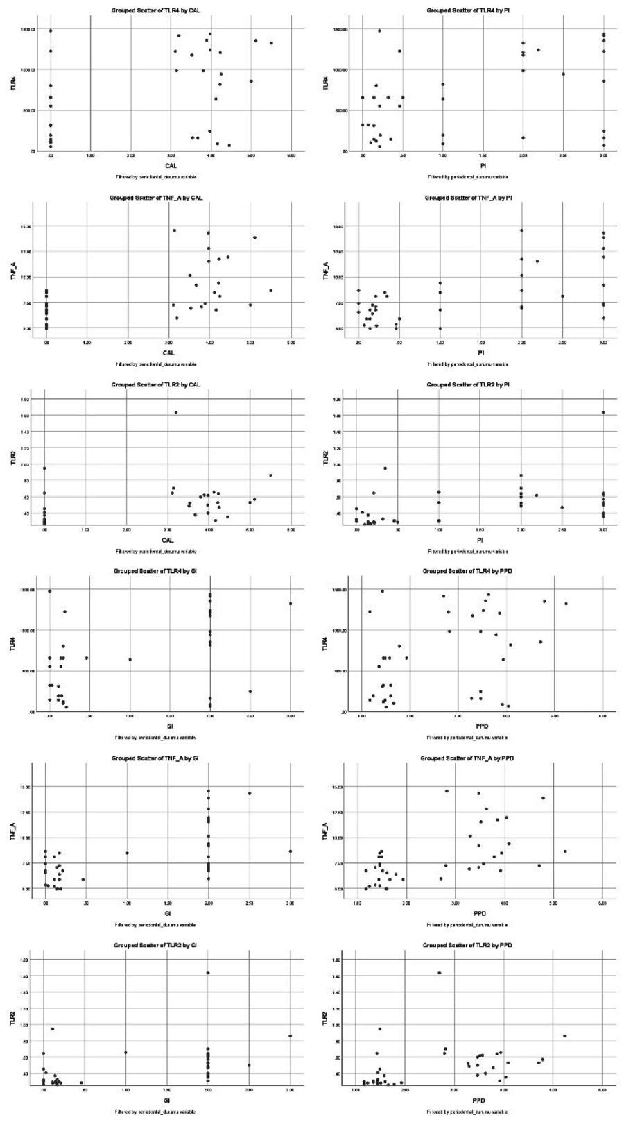
Clinical parameters were positively correlated with biomarker levels: PI with TNF-α (r = 0.427, p = 0.006) and TLR-2 (r = 0.469, p = 0.002); GI with TNF-α (r = 0.417, p = 0.007) and TLR-2 (r = 0.472, p = 0.002); and PPD with TNF-α (r = 0.369, p = 0.019) and TLR-2 (r = 0.371, p = 0.018)
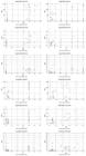
No significant associations were found between clinical parameters and TLR-4 (PI–TLR-4, p = 0.576; GI–TLR-4, p = 0.888; PPD–TLR-4, p = 0.932). Similarly, TNF-α was not significantly associated with TLR-2 (p = 0.941) or TLR-4 (p = 0.922). However, TLR-2 correlated significantly with TLR-4 (r = 0.400, p = 0.011). Figure 3 illustrates scatter plots of the correlations between periodontal parameters and biomarker levels in both groups.
Discussion
The findings of the present study indicate significant differences in clinical periodontal parameters and inflammatory biomarker levels between periodontally healthy individuals and patients with periodontitis. Following treatment, notable improvements in PPD, CAL, PI, and GI were observed, consistent with prior research demonstrating the effectiveness of NSPT in reducing inflammation and supporting tissue repair.21 These results reinforce the established understanding that periodontal therapy reduces clinical disease manifestations by promoting tissue regeneration and modulating inflammatory responses. Furthermore, they highlight the chronic and recurrent nature of periodontal disease, emphasizing that although treatment alleviates symptoms, persistent inflammatory activity requires ongoing management and routine follow-up to sustain periodontal health.22,23
Biomarker analyses confirmed the importance of TNF-α, TLR-2, and TLR-4 in the initiation and progression of periodontal disease.11 TNF-α, a proinflammatory cytokine secreted primarily by macrophages and monocytes, plays a central role in regulating inflammatory cascades within periodontal tissues.24 Elevated TNF-α levels in GCF have been strongly associated with clinical indicators of periodontal disease, including PPD, CAL, and BOP.15,25 In the present study, baseline TNF-α concentrations were significantly higher in the periodontitis group but decreased markedly after NSPT. This decline underscores the capacity of periodontal therapy to modulate host inflammatory responses and stabilize periodontal tissues.26 Our findings are consistent with previous studies.21,27 Ezzat et al.28 reported that NSPT reduced GCF TNF-α levels in parallel with clinical improvements. Similarly, Ceylan et al. observed significantly elevated baseline TNF-α levels that decreased after treatment.25 Beyond local effects, systemic reductions in TNF-α following periodontal therapy have also been reported,15,29 although the present study was restricted to GCF. Collectively, these findings support the value of TNF-α as a biomarker for monitoring periodontal inflammation and assessing therapeutic efficacy.
TLRs, particularly TLR-2 and TLR-4, are key pattern recognition receptors in detecting bacterial pathogens and initiating inflammatory responses in periodontal disease.11,30,31 Immunohistochemical studies have shown elevated expression of these receptors in gingival epithelial cells during periodontitis,11,12 and their activation is closely linked to the dysbiotic biofilm characteristic of chronic disease.12 TLR-2, which primarily recognizes gram-positive bacterial components, is upregulated in periodontitis and promotes cytokine release, including IL-1β and IL-6, contributing to tissue destruction.32,33 Conversely, TLR-4, activated mainly by gram-negative lipopolysaccharides, amplifies inflammation and stimulates osteoclastogenesis, accelerating disease progression.34,35 In the present study, both TLR-2 and TLR-4 levels were elevated in periodontitis patients compared with healthy individuals, suggesting enhanced innate immune activation in periodontal disease. These findings align with earlier reports and indicate that TLRs may serve as potential markers of dysregulated host–microbe interactions in periodontal inflammation.
Evidence further suggests that NSPT modulates these immune pathways by altering biofilm composition and reducing immunogenic components such as lipopolysaccharides, thereby promoting resolution of inflammation and restoring tissue homeostasis.36 In the present study, TLR-4 levels decreased significantly after therapy, whereas TLR-2 remained unchanged, reflecting a differential responsiveness of TLR-mediated pathways at the site-specific level. These results are consistent with an ecological shift from a dysbiotic to a symbiotic microbial community conducive to periodontal health.14 Chew et al.36 likewise reported reduced TLR-4 activation after NSPT, correlating with clinical improvement. Other studies have shown that periodontal therapy can improve periodontal parameters and normalize soluble TLR-2 levels.13 Taken together, these findings suggest that periodontal therapy not only improves clinical outcomes but also regulates host–microbe interactions through TLR-mediated signaling. However, the minimal change in TLR-2 observed in the present study suggests limited utility for early treatment-response monitoring.13
Periodontal diagnosis and monitoring rely on clinical indices, imaging, and biofluid biomarkers. While saliva provides a pooled reflection of overall oral status, GCF offers site-specific information about the local inflammatory milieu and can be sampled minimally invasively, making it preferable for monitoring lesion-level changes.37,38 Previous studies have reported decreases in TLR-4 activation after nonsurgical therapy in parallel with clinical improvement36 and reductions in GCF biomarkers following treatment.25 Although TNF-α and/or TLR-4 have been frequently examined in the context of NSPT, changes in TLR-2 and TLR-4 specifically in GCF remain less well characterized.39,40 In the present study, only TLR-4 decreased significantly after therapy, while TLR-2 levels remained stable. Thus, although GCF biomarkers may contribute to periodontal diagnosis, their utility for evaluating treatment effectiveness appears limited. The present findings contribute to the literature by providing a simultaneous evaluation of TNF-α, TLR-2, and TLR-4 in GCF before and after NSPT, enabling direct comparison of their responsiveness to therapy. This differential pattern requires confirmation in larger longitudinal studies.
Limitations
Several methodological limitations should be acknowledged. First, this was a prospective non-randomized interventional study with a healthy comparison group at baseline, but without a randomized parallel control; therefore, causal inference is limited, and regression to the mean or concurrent influences cannot be excluded. Second, although the sample size was sufficient to detect differences in clinical indices, it may not fully represent inter-individual variability in biomarker expression. Larger and more diverse cohorts are needed to confirm reproducibility and generalizability. Third, the follow-up period was relatively short; longer-term studies are required to assess the persistence and clinical significance of changes in TNF-α, TLR-2, and TLR-4. Finally, we focused only on local inflammatory markers in GCF and did not examine systemic biomarkers or microbiological profiles, which would have provided additional context for the observed local responses.
Conclusion
This study underscores the involvement of pattern recognition receptors and inflammatory mediators in the pathogenesis of periodontitis. Nonsurgical periodontal therapy effectively reduced local inflammation and improved clinical outcomes; however, TLR-4 levels decreased while TLR-2 remained stable, suggesting differential biomarker responsiveness. These findings highlight the importance of monitoring specific inflammatory pathways to better understand host–pathogen interactions in periodontal disease. Future therapeutic strategies should not only target local periodontal inflammation but also address its systemic implications, providing a more comprehensive framework for disease management and prevention.
References
-
1 Hajishengallis G. Immunomicrobial pathogenesis of periodontitis: keystones, pathobionts, and host response. Trends Immunol. 2014 Jan;35(1):3-11. https://doi.org/10.1016/j.it.2013.09.001
» https://doi.org/10.1016/j.it.2013.09.001 -
2 Han N, Liu Y, Du J, Xu J, Guo L, Liu Y. Regulation of the host immune microenvironment in periodontitis and periodontal bone remodeling. Int J Mol Sci. 2023 Feb;24(4):3158. https://doi.org/10.3390/ijms24043158
» https://doi.org/10.3390/ijms24043158 -
3 Al-Ghurabi BH, Mahmood MA, Aldhaher ZA, Al-Hindawi SH. Immunomodulatory role of cytokines in periodontal disease. Adv Life Sci. 2024;11(2):281-5. https://doi.org/10.62940/als.v11i2.2017
» https://doi.org/10.62940/als.v11i2.2017 -
4 Akira S, Takeda K, Kaisho T. Toll-like receptors: critical proteins linking innate and acquired immunity. Nat Immunol. 2001 Aug;2(8):675-80. https://doi.org/10.1038/90609
» https://doi.org/10.1038/90609 -
5 Reddy BH, Jayakumar ND, Akula SR, Sharma R, Kaarthikeyan G, Sankari. Analysis of association between TLR-4 Asp299Gly and Thr399Ile gene polymorphisms and chronic periodontitis in a sample of south Indian population. J Indian Soc Periodontol. 2011 Oct;15(4):366-70. https://doi.org/10.4103/0972-124X.92571
» https://doi.org/10.4103/0972-124X.92571 -
6 Noh MK, Jung M, Kim SH, Lee SR, Park KH, Kim DH, et al. Assessment of IL-6, IL-8 and TNF-a levels in the gingival tissue of patients with periodontitis. Exp Ther Med. 2013 Sep;6(3):847-51. https://doi.org/10.3892/etm.2013.1222
» https://doi.org/10.3892/etm.2013.1222 -
7 Shi LX, Zhang L, Zhang DL, Zhou JP, Jiang XJ, Jin YL, et al. Association between TNF-a G-308A (rs1800629) polymorphism and susceptibility to chronic periodontitis and type 2 diabetes mellitus: a meta-analysis. J Periodontal Res. 2021 Apr;56(2):226-35. https://doi.org/10.1111/jre.12820
» https://doi.org/10.1111/jre.12820 -
8 Kwon T, Lamster IB, Levin L. Current concepts in the management of periodontitis. Int Dent J. 2021 Dec;71(6):462-76. https://doi.org/10.1111/idj.12630
» https://doi.org/10.1111/idj.12630 -
9 Arpag OF, Dag A, Izol BS, Cimitay G, Uysal E. Effects of vector ultrasonic system debridement and conventional instrumentation on the levels of TNF-a in gingival crevicular fluid of patients with chronic periodontitis. Adv Clin Exp Med. 2017 Dec;26(9):1419-24. https://doi.org/10.17219/acem/65410
» https://doi.org/10.17219/acem/65410 -
10 Mori Y, Yoshimura A, Ukai T, Lien E, Espevik T, Hara Y. Immunohistochemical localization of Toll-like receptors 2 and 4 in gingival tissue from patients with periodontitis. Oral Microbiol Immunol. 2003 Feb;18(1):54-8. https://doi.org/10.1034/j.1399-302x.2003.180109.x
» https://doi.org/10.1034/j.1399-302x.2003.180109.x -
11 Hans M, Hans VM. Toll-like receptors and their dual role in periodontitis: a review. J Oral Sci. 2011 Sep;53(3):263-71. https://doi.org/10.2334/josnusd.53.263
» https://doi.org/10.2334/josnusd.53.263 - 12 Song B, Zhang YL, Chen LJ, Zhou T, Huang WK, Zhou X, et al. The role of Toll-like receptors in periodontitis. Oral Dis. 2017 Mar;23(2):168-80.
-
13 Prakasam S, Srinivasan M. Evaluation of salivary biomarker profiles following non-surgical management of chronic periodontitis. Oral Dis. 2014 Mar;20(2):171-7. https://doi.org/10.1111/odi.12085
» https://doi.org/10.1111/odi.12085 -
14 Schulz S, Stein JM, Schumacher A, Kupietz D, Yekta-Michael SS, Schittenhelm F, et al. Nonsurgical periodontal treatment options and their impact on subgingival microbiota. J Clin Med. 2022 Feb;11(5):1378. https://doi.org/10.3390/jcm11051187
» https://doi.org/10.3390/jcm11051187 -
15 Jain P, Ved A, Dubey R, Singh N, Parihar AS, Maytreyee R. Comparative evaluation of serum tumor necrosis factor a in health and chronic periodontitis: a case-control study. Contemp Clin Dent. 2020;11(4):342-9. https://doi.org/10.4103/ccd.ccd_97_18
» https://doi.org/10.4103/ccd.ccd_97_18 -
16 Cocks K, Torgerson DJ. Sample size calculations for pilot randomized trials: a confidence interval approach. J Clin Epidemiol. 2013 Feb;66(2):197-201. https://doi.org/10.1016/j.jclinepi.2012.09.002
» https://doi.org/10.1016/j.jclinepi.2012.09.002 -
17 Moore CG, Carter RE, Nietert PJ, Stewart PW. Recommendations for planning pilot studies in clinical and translational research. Clin Transl Sci. 2011 Oct;4(5):332-7. https://doi.org/10.1111/j.1752-8062.2011.00347.x
» https://doi.org/10.1111/j.1752-8062.2011.00347.x -
18 Chapple IL, Mealey BL, Van Dyke TE, Bartold PM, Dommisch H, Eickholz P, et al. Periodontal health and gingival diseases and conditions on an intact and a reduced periodontium: Consensus report of workgroup 1 of the 2017 World Workshop on the Classification of Periodontal and Peri-Implant Diseases and Conditions. J Periodontol. 2018 Jun;89(S1 Suppl 1):S74-84. https://doi.org/10.1002/JPER.17-0719
» https://doi.org/10.1002/JPER.17-0719 -
19 O'Leary TJ, Drake RB, Naylor JE. The plaque control record. J Periodontol. 1972 Jan;43(1):38. https://doi.org/10.1902/jop.1972.43.1.38
» https://doi.org/10.1902/jop.1972.43.1.38 -
20 Loe H, Silness J. Periodontal disease in pregnancy. I. Prevalence and severity. Acta Odontol Scand. 1963 Dec;21(6):533-51. https://doi.org/10.3109/00016356309011240
» https://doi.org/10.3109/00016356309011240 -
21 Pihlstrom BL, Michalowicz BS, Johnson NW. Periodontal diseases. Lancet. 2005 Nov;366(9499):1809-20. https://doi.org/10.1016/S0140-6736 (05)67728-8
» https://doi.org/10.1016/S0140-6736 (05)67728-8 -
22 Teles FR, Chandrasekaran G, Martin L, Patel M, Kallan MJ, Furquim C, et al. Salivary and serum inflammatory biomarkers during periodontitis progression and after treatment. J Clin Periodontol. 2024 Dec;51(12):1619-31. https://doi.org/10.1111/jcpe.14048
» https://doi.org/10.1111/jcpe.14048 -
23 Loos BG, Van Dyke TE. The role of inflammation and genetics in periodontal disease. Periodontol 2000. 2020 Jun;83(1):26-39. https://doi.org/10.1111/prd.12297
» https://doi.org/10.1111/prd.12297 -
24 Bretz WA, Weyant RJ, Corby PM, Ren D, Weissfeld L, Kritchevsky SB, et al. Systemic inflammatory markers, periodontal diseases, and periodontal infections in an elderly population. J Am Geriatr Soc. 2005 Sep;53(9):1532-7. https://doi.org/10.1111/j.1532-5415.2005.53468.x
» https://doi.org/10.1111/j.1532-5415.2005.53468.x -
25 Ceylan M, Erbak Yilmaz H, Narin F, Tatakis DN, Saglam M. Gingival crevicular fluid lipocalin-2 and semaphorin3A in stage III periodontitis: non-surgical periodontal treatment effects. J Periodontal Res. 2022 Aug;57(4):724-32. https://doi.org/10.1111/jre.12995
» https://doi.org/10.1111/jre.12995 -
26 Dag A, Firat ET, Arikan S, Kadiroglu AK, Kaplan A. The effect of periodontal therapy on serum TNF-a and HbA1c levels in type 2 diabetic patients. Aust Dent J. 2009 Mar;54(1):17-22. https://doi.org/10.1111/j.1834-7819.2008.01083.x
» https://doi.org/10.1111/j.1834-7819.2008.01083.x -
27 Yucel-Lindberg T, Båge T. Inflammatory mediators in the pathogenesis of periodontitis. Expert Rev Mol Med. 2013 Aug;15:e7. https://doi.org/10.1017/erm.2013.8
» https://doi.org/10.1017/erm.2013.8 -
28 Ezzat OH, Ibrahim SS, Gaber HK. The effect of periodontal treatment on circulating tumor necrosis factor-alpha and insulin resistance in patients with type 2 diabetes mellitus. Magalat Game'at al-Malik Abdul Aziz Al-U'lum al-Tibyat. 2012;19(4):3-11. https://doi.org/10.4197/Med.19-4.2
» https://doi.org/10.4197/Med.19-4.2 -
29 Bradley JR. TNF-mediated inflammatory disease. J Pathol. 2008 Jan;214(2):149-60. https://doi.org/10.1002/path.2287
» https://doi.org/10.1002/path.2287 -
30 Jiao Y, Hasegawa M, Inohara N. The role of oral pathobionts in dysbiosis during periodontitis development. J Dent Res. 2014 Jun;93(6):539-46. https://doi.org/10.1177/0022034514528212
» https://doi.org/10.1177/0022034514528212 -
31 Hajishengallis G, Darveau RP, Curtis MA. The keystone-pathogen hypothesis. Nat Rev Microbiol. 2012 Oct;10(10):717-25. https://doi.org/10.1038/nrmicro2873
» https://doi.org/10.1038/nrmicro2873 -
32 Buduneli N, Özçaka Ö, Nalbantsoy A. Salivary and plasma levels of Toll-like receptor 2 and Toll-like receptor 4 in chronic periodontitis. J Periodontol. 2011 Jun;82(6):878-84. https://doi.org/10.1902/jop.2010.100467
» https://doi.org/10.1902/jop.2010.100467 -
33 Rojo-Botello NR, García-Hernández AL, Moreno-Fierros L. Expression of toll-like receptors 2, 4 and 9 is increased in gingival tissue from patients with type 2 diabetes and chronic periodontitis. J Periodontal Res. 2012 Feb;47(1):62-73. https://doi.org/10.1111/j.1600-0765.2011.01405.x
» https://doi.org/10.1111/j.1600-0765.2011.01405.x -
34 Ren L, Leung WK, Darveau RP, Jin L. The expression profile of lipopolysaccharide-binding protein, membrane-bound CD14, and toll-like receptors 2 and 4 in chronic periodontitis. J Periodontol. 2005 Nov;76(11):1950-9. https://doi.org/10.1902/jop.2005.76.11.1950
» https://doi.org/10.1902/jop.2005.76.11.1950 -
35 Yang X, Tao X, Qi W, Liu Z, Wang Y, Han Q, et al. TLR-4 targeting contributes to the recovery of osteoimmunology in periodontitis. J Periodontal Res. 2021 Aug;56(4):782-8. https://doi.org/10.1111/jre.12877
» https://doi.org/10.1111/jre.12877 -
36 Chew RJ, Tang YL, Lin XY, Oh FJ, Sim RP, Anwar EJ, et al. Toll-like receptor-4 activation by subgingival biofilm and periodontal treatment response. Clin Oral Investig. 2023 May;27(5):2139-47. https://doi.org/10.1007/s00784-023-04877-8
» https://doi.org/10.1007/s00784-023-04877-8 -
37 Kinney JS, Ramseier CA, Giannobile WV. Oral fluid-based biomarkers of alveolar bone loss in periodontitis. Ann N Y Acad Sci. 2007 Mar;1098(1):230-51. https://doi.org/10.1196/annals.1384.028
» https://doi.org/10.1196/annals.1384.028 -
38 Miller CS, King CP Jr, Langub MC, Kryscio RJ, Thomas MV. Salivary biomarkers of existing periodontal disease: a cross-sectional study. J Am Dent Assoc. 2006 Mar;137(3):322-9. https://doi.org/10.14219/jada.archive.2006.0181
» https://doi.org/10.14219/jada.archive.2006.0181 -
39 Martínez-Aguilar VM, Carrillo-Ávila BA, Sauri-Esquivel EA, Guzmán-Marín E, Jiménez-Coello M, Escobar-García DM, et al. Quantification of TNF-a in patients with periodontitis and type 2 diabetes. BioMed Res Int. 2019 Jul;2019(1):7984891. https://doi.org/10.1155/2019/7984891
» https://doi.org/10.1155/2019/7984891 -
40 Jiang ZL, Cui YQ, Gao R, Li Y, Fu ZC, Zhang B, et al. Study of TNF-a, IL-1ß and LPS levels in the gingival crevicular fluid of a rat model of diabetes mellitus and periodontitis. Dis Markers. 2013;34(5):295-304. https://doi.org/10.1155/2013/156798
» https://doi.org/10.1155/2013/156798
-
Data Availability Statement:
The datasets generated and/or analyzed during the current study are not publicly available due to participant privacy considerations. However, in line with ethical approval and data confidentiality agreements, the data may be made available to qualified researchers upon reasonable request from the corresponding author.
Edited by
-
Editor-in-Chief:
Lucianne Maia
-
Associate Editor:
Cristina Cunha Villar
The datasets generated and/or analyzed during the current study are not publicly available due to participant privacy considerations. However, in line with ethical approval and data confidentiality agreements, the data may be made available to qualified researchers upon reasonable request from the corresponding author.