 Minimally invasive approach to temporomandibular disorders in a patient with congenital muscular torticollis. Case report
ABSTRACT
BACKGROUND AND OBJECTIVES Temporomandibular disorders (TMD) are often associated with biomechanical and postural factors such as congenital muscular torticollis (CMT). This report describes a minimally invasive therapeutic approach in a patient with TMD and CMT, highlighting the efficacy of arthrocentesis combined with viscosupplementation.
CASE REPORT The 31-year-old female patient with a previous diagnosis of CMT and TMD symptoms (joint pain, crepitation, and limited mouth opening) underwent clinical evaluation according to the Diagnostic Criteria for TMDs (DC/TMD) and imaging tests (computed tomography and magnetic resonance imaging). After unsatisfactory response to conservative treatment (occlusal splint, physical therapy, and dry needling), a minimally invasive approach was adopted, consisting of ultrasound-guided arthrocentesis and viscosupplementation with 1% sodium hyaluronate. Significant improvement was observed after the minimally invasive protocol, with increased mouth opening (from 28 mm to 42 mm), pain reduction, and bone recorticalization in the right temporomandibular joint. The integrated approach demonstrated efficacy in functional restoration and symptom relief.
CONCLUSION The combined use of arthrocentesis and viscosupplementation was associated with clinical and functional improvement in a patient with refractory temporomandibular disorder and congenital muscular torticollis, suggesting a potential role for minimally invasive approaches in complex TMD cases.
Keywords:
Minimally invasive surgical procedures; Temporomandibular joint disorders; Temporomandibular joint; Torticollis
HIGHLIGHTS
Systematic evaluation by the DC/TMD, associated with imaging tests when necessary, is essential for the correct diagnosis and therapeutic planning in complex cases involving muscular, joint, and postural components
Arthrocentesis associated with viscosupplementation with sodium hyaluronate can be effective in cases of refractory joint TMD, promoting significant pain relief and relevant functional gain in mouth opening
The clinical case reinforces the importance of an interdisciplinary and staged approach to the management of TMDs, reserving minimally invasive interventions for situations in which well-conducted conservative treatments are not sufficient
RESUMO
JUSTIFICATIVA E OBJETIVOS As disfunções temporomandibulares (DTM) são frequentemente associadas a fatores biomecânicos e posturais, como o torcicolo muscular congênito (TMC). O objetivo deste estudo foi descrever a abordagem terapêutica minimamente invasiva em uma paciente com DTM e TMC, destacando a eficácia da artrocentese associada à viscossuplementação.
RELATO DO CASO Paciente do sexo feminino, 31 anos, com diagnóstico prévio de TMC e sintomas de DTM (dor articular, crepitação e limitação da abertura bucal), foi submetida a avaliação clínica conforme os Critérios Diagnósticos para DTM (DC/TMD) e exames de imagem (tomografia e ressonância magnética). Após resposta insatisfatória ao tratamento conservador (placa oclusal, fisioterapia e agulhamento seco), optou-se por um tratamento minimamente invasivo com artrocentese guiada por ultrassom e viscossuplementação com hialuronato de sódio 1% na ATM direita. Observou-se melhora significativa após o protocolo minimamente invasivo, com aumento da abertura bucal (de 28 mm para 42 mm), redução da dor e recorticalização óssea na articulação temporomandibular direita. A abordagem integrada demonstrou eficácia na restauração funcional e no alívio sintomático.
CONCLUSÃO O uso combinado da artrocentese e da viscossuplementação foi associado à melhora clínica e funcional em uma paciente com disfunção temporomandibular refratária e torcicolo muscular congênito, sugerindo um papel potencial das abordagens minimamente invasivas em casos complexos de DTM.
Descritores:
Procedimentos cirúrgicos minimamente invasivos; Transtornos da articulação temporomandibular; Articulação temporomandibular; Torcicolo
INTRODUCTION
Temporomandibular disorders (TMD) comprise a group of conditions characterized by pain and/or dysfunction in the masticatory muscles, the temporomandibular joints (TMJ) and associated structures1. These conditions have a multifactorial origin, with both local and systemic factors contributing2-4. The combination and intensity of these factors influence the development and persistence of TMDs, which can be muscular or articular and may lead to functional and psychosocial impacts on patients1.
The clinical diagnosis of TMDs must be systematic and comprehensive. For this purpose, the use of the Diagnostic Criteria for Temporomandibular Disorders (DC/TMD)5 validated in Portuguese is recommended. This instrument provides a structured, dual-axis approach: axis I focus on physical signs and symptoms, while axis II addresses relevant biopsychosocial aspects5-7. In more complex cases, particularly those involving suspected structural abnormalities, clinical evaluation should be supplemented with imaging. Computed tomography (CT) is the gold standard for evaluating bone morphology, whereas magnetic resonance imaging (MRI) is preferred for soft tissue assessment8.
Regarding the treatment of TMDs, although some cases require minimally invasive interventions such as arthrocentesis, arthroscopy, or viscosupplementation, most patients respond well to conservative, non-invasive approaches. Therapeutic counseling plays a pivotal role, as it educates patients about parafunctional habits, promotes healthy postures, and encourages self-management strategies9,10. This approach can be complemented by manual physiotherapeutic exercises and mobile apps10.
In this context, it is important to consider that systemic musculoskeletal conditions may act as predisposing or perpetuating factors for TMDs. One such condition is congenital muscular torticollis (CMT), a disorder typically identified at birth or during early infancy, characterized by unilateral shortening and fibrosis of the sternocleidomastoid muscle11. Without early intervention, CMT may induce compensatory adaptations affecting stomatognathic system structures and TMJs, potentially contributing to TMD development12,13.
Among the different types of TMDs, articular disc displacement is one of the most common conditions, including among individuals with postural alterations such as CMT. Studies suggest that this type of dysfunction may be associated with joint degeneration, characterized by structural changes affecting both the articular disc and the bony components of the mandibular fossa and articular eminence14. These alterations impair TMJ biomechanics, leading to pain, functional limitations, and joint sounds15. When these dysfunctions do not respond adequately to conservative treatments, minimally invasive interventions such as arthrocentesis followed by viscosupplementation may become necessary1,16.
In light of these considerations, this case report describes the therapeutic management of a TMD patient with pre-existing CMT, treated at the Petrópolis Medical School (FMP/FASE) clinic.
The objective was to present the integrated therapeutic approach employed, emphasizing the efficacy of minimally invasive strategies in addressing this clinically complex condition involving both functional and postural components.
CASE REPORT
A 31-year-old female dental health professional with a known history of CMT not corrected during childhood presented to the FMP/FASE outpatient clinic with right TMJ pain and restricted mouth opening. The patient reported long-standing cervical postural alteration associated with CMT, but without significant impairment in daily activities or functional independence. She also reported a 21-year history of right TMJ clicking that had progressed to crepitus, accompanied by persistent right TMJ pain for the past decade. These symptoms exacerbated during mandibular movements and showed significant worsening over the last three years, resulting in functional limitations and reduced mandibular range of motion. While she denied episodes of joint locking, the patient acknowledged both awake and sleep bruxism.
The patient reported no previous treatment for temporomandibular disorders prior to the initial evaluation of the present study’s researchers. She had not used occlusal splints, undergone physical therapy, or received any specific intervention for TMD before enrollment in the proposed treatment protocol. No cervical pain was reported at the time of evaluation.
Diagnostic evaluation
The patient underwent evaluation using the DC/TMD5 protocol. Axis I assessment revealed pain on palpation of the right masseter muscle with radiation to the ipsilateral TMJ, along with familiar pain during right TMJ palpation. Maximum mouth opening measurements showed 28 mm actively and 30 mm with passive assistance. Lateral excursions measured 9 mm to the left and 7 mm to the right, with the right lateral movement eliciting pain in the contralateral TMJ. Crepitation was consistently observed in the right TMJ during both opening and closing movements.
According to DC/TMD Axis II evaluation, the patient demonstrated a characteristic pain intensity score17 of 56, consistent with frequent and clinically significant pain. The pain-related disability index18 was 6.7, indicating mild interference with daily activities, though the patient reported no days of functional disability during the previous month. This discrepancy suggests maintained functional capacity despite subjective pain perception. Psychosocial assessment revealed minimal symptom scores, with both depressive19 and anxiety20 measures at 1 (absent or subclinical). The score for nonspecific physical symptoms19 was 4, which may indicate the presence of some somatic symptoms, but without clinically significant intensity.
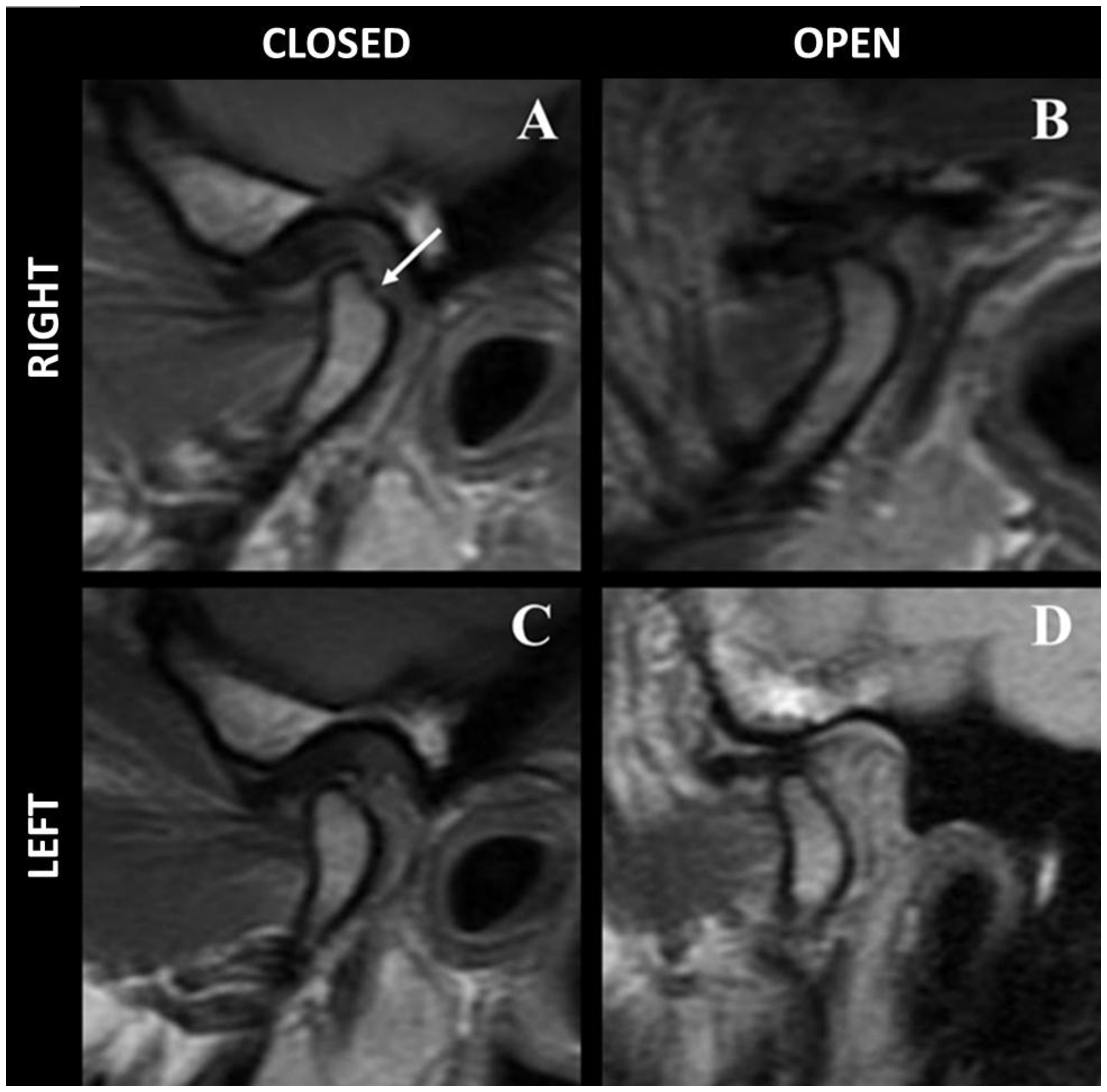
Complementary imaging evaluation included multidetector computed tomography (MDCT) and magnetic resonance imaging (MRI) of the temporomandibular joints. MDCT findings demonstrated cortical discontinuity in the right mandibular condyle, consistent with inflammatory degenerative changes (Figure 1), while the right glenoid fossa appeared preserved and the left TMJ showed no morphological abnormalities. MRI revealed concentric positioning of the right condyle within the mandibular fossa, contrasting with anterior positioning of the left condyle resulting in increased joint space (Figure 2). Degenerative erosive changes were evident in the superoposterior region of the right condyle, with preservation of left-sided bony architecture. Bilateral condylar hypoexcursion was noted during dynamic evaluation. Disc analysis showed proper interposition between the condyle and temporal bone in the left TMJ during both closed- and open-mouth positions. In contrast, in the right TMJ, the disc was anteriorly displaced in the closed-mouth position and did not return to its proper position during mouth opening.
Sagittal multidetector computed tomography images of temporomandibular joints. (A) Right TMJ in closed-mouth position demonstrating cortical discontinuity (white arrow) indicative of degenerative changes. (B) Right TMJ in open-mouth position showing persistent cortical irregularity (white arrow). (C) Left TMJ in closed-mouth position with intact cortical bone. (D) Left TMJ in open-mouth position showing normal morphology. All images are displayed in bone window settings.
Magnetic resonance imaging of the temporomandibular joints. (A) Right TMJ in the closed-mouth position showing anterior displacement of the articular disc, associated with cortical irregularity and erosive changes of the mandibular condyle (white arrow). (B) Right TMJ in the open-mouth position demonstrating absence of disc recapture, with the articular disc remaining anterior to the mandibular condyle, consistent with anterior disc displacement without reduction. (C) Left TMJ in the closed-mouth position showing a normally positioned articular disc, with the posterior band interposed between the mandibular condyle and the articular surface of the temporal bone. (D) Left TMJ in the open-mouth position demonstrating maintenance of a physiological disc–condyle relationship, with appropriate disc translation during condylar movement.
The complementary rheumatological evaluation excluded inflammatory or autoimmune etiologies, confirming the local nature of the degenerative joint pathology. Based on the clinical and imaging findings, the following Axis I DC/TMD diagnoses were established: myalgia, myofascial pain with referral, arthralgia, disc displacement without reduction with limited opening in the right TMJ, and degenerative joint disease of the right TMJ.
Therapeutic intervention
The patient's treatment followed a conservative multidisciplinary protocol combining behavioral counseling for parafunctional habit modification, pharmacotherapy with cyclobenzaprine hydrochloride (10 mg once daily at bedtime for 30 days), and occlusal stabilization splint therapy prescribed for use during sleep. The regimen was complemented by physiotherapy referral for manual therapy and guided exercises, along with dry needling sessions targeting myofascial pain components.
Following four months of conservative therapy with unsatisfactory clinical improvement, the treatment strategy was escalated to minimally invasive interventions. Initial pharmacological management included 30-day chondroprotective therapy with combined glucosamine (1500 mg) and chondroitin (1200 mg) once daily.
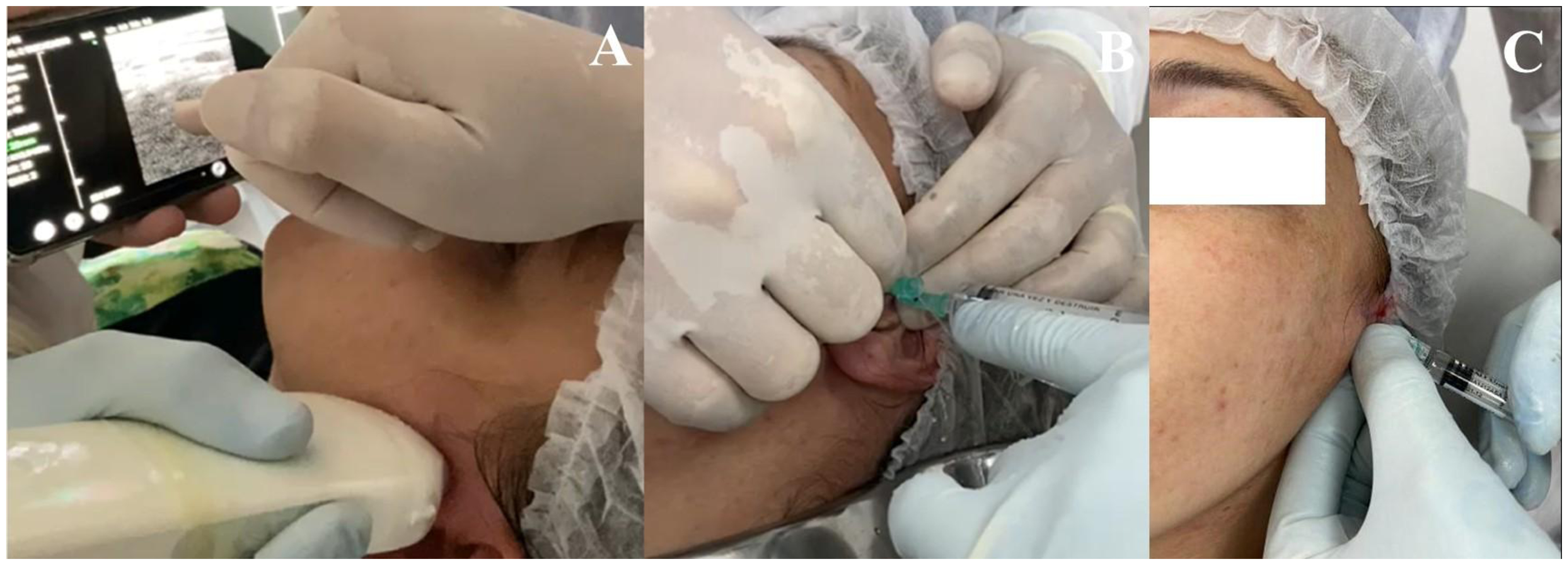
Prior to arthrocentesis, a local anesthetic block of the temporomandibular joint region was performed through an auriculotemporal nerve block using 2% lidocaine with epinephrine 1:100,000 (1.8 mL), administered slowly after negative aspiration and under strict aseptic conditions, without any anesthetic-related complications. Arthrocentesis of the temporomandibular joints was subsequently performed using the classical two-needle technique in the superior joint compartment, as originally described by authors with continuous real-time ultrasound guidance. The use of ultrasound was adopted to ensure accurate needle placement, enhance procedural safety, and standardize the technique in a teaching-clinic setting, where the procedure was simultaneously performed and demonstrated for educational purposes (Figure 3).

Insertion of needles into the superior compartment of the right temporomandibular joint under ultrasound guidance. (A) for arthrocentesis (B), followed by the first application of 1% sodium hyaluronate into the superior compartment of the right TMJ (C).
Two sterile disposable 18-G needles were inserted into the superior joint compartment under ultrasound-guided visualization, followed by joint lavage with approximately 200 mL of sterile 0.9% saline solution under low and continuous pressure to promote hydraulic distension and removal of inflammatory mediators. Immediately after arthrocentesis, intra-articular viscosupplementation with 1% sodium hyaluronate (Osteonil®, 20 mg; TRB Pharma) was performed. Additional viscosupplementation sessions were conducted at monthly intervals, in accordance with the organizational structure of the specialization program in which clinical care was delivered on a monthly basis, allowing for standardized follow-up and maintenance of the therapeutic effect.
After the first month of implementing the minimally invasive protocol, the patient showed significant improvement, with a marked reduction in articular and muscular pain levels and achieving a pain-free maximum mouth opening of 42 mm. The prognosis was considered favorable. This favorable response was sustained at six-month follow-up, with follow-up MDCT revealing bone recorticalization of the right mandibular condyles, indicating positive structural remodeling in response to treatment (Figure 4).

Follow-up multidetector computed tomography of the right temporomandibular joint six months post-intervention. (A) Closed-mouth view demonstrating complete recorticalization of the mandibular condyle (compare with Figure 1A). (B) Open-mouth view showing maintained cortical integrity during function. (A) Closed-mouth view demonstrating complete recorticalization of the mandibular condyle (compared with Figure 1A). (B) Open-mouth view showing maintained cortical integrity during function.
It is noteworthy that conservative measures, including occlusal splint therapy and behavior counseling, were maintained throughout the treatment and follow-up period.
DISCUSSION
Although it is recognized that, in some cases, clinical improvement may occur over time even in the absence of active intervention, reflecting the natural course of the condition and individual adaptive mechanisms, current clinical guidelines emphasize minimally invasive approaches as first-line interventions for TMDs1,6,16. The literature demonstrates that such conservative strategies yield optimal outcomes when implemented early in the disease course and combined with systematic follow-up. Furthermore, contemporary management paradigms highlight the critical importance of interdisciplinary collaboration to address the multifactorial nature of TMDs1,6.
Congenital muscular torticollis (CMT), considered a comorbid postural condition in this case, may influence temporomandibular joint biomechanics by promoting long-standing cervical asymmetry and compensatory postural adaptations. Unilateral shortening of the sternocleidomastoid muscle can result in persistent head tilt and rotation, leading to imbalance of the craniofacial–cervical system and altered mandibular positioning at rest and during function11-13. These alterations may contribute to asymmetric loading of the temporomandibular joints, increasing intra-articular stress and impairing disc–condyle coordination. Such biomechanical conditions have been associated with disc displacement and degenerative joint changes, particularly in individuals exposed to prolonged functional overload and postural alterations14,15. When combined with parafunctional habits such as bruxism, these factors may further increase joint stress and favor the development and persistence of anterior disc displacement1,6, as observed in the present case.
The initial conservative management in this case incorporated patient education regarding parafunctional habit avoidance and sleep hygiene optimization6. This approach is relevant as it promotes autonomy in symptom management and is effective in 30% to 50% of cases9,10.
Pharmacological therapy plays a well-documented role in TMD management6,20. In the present case, cyclobenzaprine hydrochloride was selected as first-line pharmacotherapy during the waiting period for the fabrication of the stabilization splint. As a centrally acting muscle relaxant with demonstrated efficacy in musculoskeletal pain control21, its prescription aligned with current evidence showing significant pain reduction in TMD patients. Although its mechanisms of action are not fully understood, a meta-analysis has shown that this device can reduce pain and contribute to the management of TMD22.
During the sessions, dry needling was employed to deactivate myofascial trigger points. According to author23, these points are hypersensitive nodules in skeletal muscles of patients with referred myofascial pain, typically resulting from muscle overload. The study24 describe the technique as the insertion of a solid needle directly into the trigger points, without the use of drugs, provoking an involuntary spinal reflex with contraction of the affected fibers. This stimulus activates an axonal reflex via Aβ and C fibers, leading to the release of vasoactive substances, vasodilation, and increased local blood flow. As a result, enhanced muscle oxygenation contributes to the reduction of pain intensity24.
Subsequently, the patient was referred to physical therapy, considering that randomized clinical trials have demonstrated that mandibular mobilization combined with stretching can improve mouth opening range and reduce both muscular and joint pain6,21. This parameter, which is essential for assessing mandibular mobility25, was used to monitor the effectiveness of the interventions and to guide therapeutic decisions.
Another reference study1 emphasizes that, in cases of persistent acute pain accompanied by restricted mandibular movement, conservative treatment may be insufficient. Before surgical intervention, chondroprotective therapy was prescribed, based on the role of glucosamine and chondroitin in the synthesis of proteoglycans, key components in the regeneration of articular cartilage26.
Arthrocentesis is indicated for cases of persistent pain and functional limitation, as it promotes joint lavage and removal of inflammatory mediators16,27. When combined with 1% sodium hyaluronate, its anti-inflammatory and analgesic effects are enhanced, improving synovial fluid lubrication and joint biomechanics, and this association is recommended for patients who do not respond adequately to conservative treatment27-29. In the present case, this integrated approach was associated with significant improvement in pain and mouth opening, consistent with previous evidence demonstrating superior outcomes with the combination of arthrocentesis and viscosupplementation30. The reduction of intra-articular inflammation and mechanical stress achieved through this protocol may also favor adaptive bone remodeling, potentially explaining the condylar recorticalization observed during follow-up1,16,27-29. However, this process is multifactorial, and causal inferences should be interpreted with caution.
In addition, imaging examinations were not performed immediately after the conservative treatment phase, which limits causal inference regarding the observed radiographic changes. Therefore, the condylar remodeling identified during follow-up cannot be attributed exclusively to the minimally invasive intervention, as it may also reflect the effects of prior conservative management and subsequent biological adaptation. It is important to emphasize that imaging exams are essential complementary tools in the evaluation of patients with TMDs, as they enable accurate diagnosis and guide therapeutic planning31,32. MRI is considered the gold standard for this evaluation14,15.
CONCLUSION
Thus, it is important to highlight that TMD treatment does not follow a single protocol. It is essential to consider signs, symptoms, duration, and causes in order to plan an individualized approach. Imaging exams assist in this process, supporting the stages of treatment. In more complex cases, conservative therapies, although considered the first line, may prove insufficient.
-
Sponsoring sources:
none.
-
Ethics statement:
This clinical case report was reviewed by the Research Ethics Committee of the Universidade Federal de Juiz de Fora. The study was registered under CAAE number 66808222.4.0000.5147 and approved according to opinion number 5.960.733.
-
Data availability
The data are available in the Institutional Repository of the Federal University of Juiz de Fora (UFJF) at: https://repositorio.ufjf.br/jspui/handle/ufjf/15664.
-
The study was carried out at Departamento de Odontologia, Faculdade de Medicina de Petrópolis, Petrópolis, RJ, Brasil.
References
-
1 Briggs K, Breik O, Ito K, Goss AN. Arthrocentesis in the management of internal derangement of the temporomandibular joint. Aust Dent J. 2019;64(1):90-5. https://doi.org/10.1111/adj.12665 PMid:30422323.
» https://doi.org/10.1111/adj.12665 - 2 Manfredini D, Piccotti F, Ferronato G, Guarda-Nardini L. Etiopathogenesis of temporomandibular disorders: a review of the literature. J Oral Rehabil. 2005;32(6):379-88.
- 3 Vickers ER, Boocock H. Chronic orofacial pain is associated with psychological morbidity and negative personality changes: a comparison to chronic low back pain. J Orofac Pain. 2005;19(3):191-8.
- 4 Manfredini D, Bandettinidi Poggio A, Romagnoli M, Dell’Osso L, Bosco M. Psychosocialsubtypes of temporomandibular disorders. J Orofac Pain. 2003;17(3):234-9.
-
5 Schiffman E, Ohrbach R, Truelove E, Look J, Anderson G, Goulet JP, List T, Svensson P, Gonzalez Y, Lobbezoo F, Michelotti A, Brooks SL, Ceusters W, Drangsholt M, Ettlin D, Gaul C, Goldberg LJ, Haythornthwaite JA, Hollender L, Jensen R, John MT, De Laat A, de Leeuw R, Maixner W, van der Meulen M, Murray GM, Nixdorf DR, Palla S, Petersson A, Pionchon P, Smith B, Visscher CM, Zakrzewska J, Dworkin SF, International RDC/TMD Consortium Network, International association for Dental Research, Orofacial Pain Special Interest Group, International Association for the Study of Pain. Diagnostic criteria for temporomandibular disorders (DC/TMD) for clinical and research applications. J Oral Facial Pain Headache. 2014;28(1):6-27. https://doi.org/10.11607/jop.1151 PMid:24482784.
» https://doi.org/10.11607/jop.1151 -
6 Kapos FP, Exposto FGC, Oyarzo JF, Durham J. Temporomandibular disorders: a review of current concepts in aetiology, diagnosis and management. Oral Surg. 2020;13(4):321-34. https://doi.org/10.1111/ors.12473 PMid:34853604.
» https://doi.org/10.1111/ors.12473 -
7 Ohrbach R, Dworkin SF. The evolution of TMD diagnosis. J Dent Res. 2016;95(10):1093-101. https://doi.org/10.1177/0022034516653922 PMid:27313164.
» https://doi.org/10.1177/0022034516653922 -
8 Costa MRM, Christofolleti LZM, Barbosa KGN, Nóbrega DF, Pinheiro MLN, Cavalcante MK. Disfunção temporomandibular, sintomas otológicos e sua associação com achados de imagem de ressonância magnética. Diversitas J. 2019;4(2):622-30. https://doi.org/10.17648/diversitas-journal-v4i2.779
» https://doi.org/10.17648/diversitas-journal-v4i2.779 -
9 Melo RA, de Resende CMBM, Rêgo CRF, Bispo ASL, Barbosa GAS, de Almeida EO. Conservative therapies to treat pain and anxiety associated with temporomandibular disorders: a randomized clinical trial. Int Dent J. 2020;70(4):245-53. https://doi.org/10.1111/idj.12546 PMid:32153038.
» https://doi.org/10.1111/idj.12546 -
10 Takeuchi-Sato T, Ono Y, Funato M, Sato H, Suganuma T, Baba K. Efficacy of an email-based recording and reminding system for limiting daytime non-functional tooth contact in patients with temporomandibular disorders. J Oral Rehabil. 2020;47(2):158-63. https://doi.org/10.1111/joor.12875 PMid:31418908.
» https://doi.org/10.1111/joor.12875 - 11 Lopes I, Monteiro S, Moreira C. Torcicolo muscular congénito: a propósito de um caso clínico. ArquiMed. 2009;23(3):7-9.
- 12 Ribeiro R. Anomalias congénitas e manifestações orais. Porto: Universidade Fernando Pessoa; 2014.
- 13 Magalhães BO. Tratamento da assimetria dentofacial em adultos: uma revisão da literatura. Belo Horizonte: Universidade Federal de Minas Gerais; 2016.
-
14 Poluha RL, Canales GT, Costa YM, Grossmann E, Bonjardim LR, Conti PCR. Temporomandibular joint disc displacement with reduction: a review of mechanisms and clinical presentation. J Appl Oral Sci. 2018;26:e20170289. https://doi.org/10.1590/1678-7757-2017-0578 PMid:30810641.
» https://doi.org/10.1590/1678-7757-2017-0578 -
15 Bedran LM, Santos AASMD. Changes in temporomandibular joint anatomy, changes in condylar translation, and their relationship with disc displacement: MRI study. Radiol Bras. 2019;52(2):85-91. https://doi.org/10.1590/0100-3984.2018.0020 PMid:31019336.
» https://doi.org/10.1590/0100-3984.2018.0020 -
16 Grossmann E. Técnicas de artrocentese aplicadas às disfunções artrogênicas da articulação temporomandibular. Rev Dor. 2012;13(4):374-81. https://doi.org/10.1590/S1806-00132012000400013
» https://doi.org/10.1590/S1806-00132012000400013 - 17 Von Korff M. Assessment of chronic pain in epidemiological and health services research: empirical bases and new directions. In: Turk DC, Melzack R, editors. Handbook of pain assessment. 3rd ed. New York: Guilford Press; 2011. p. 455-73.
- 18 Ohrbach R, Larsson P, List T. The jaw functional limitation scale: development, reliability, and validity of 8-item and 20-item versions. J Orofac Pain. 2008;22(3):219-30. PMid:18780535.
-
19 Kroenke K, Spitzer RL, Williams JB. The PHQ-9: validity of a brief depression severity measure. J Gen Intern Med. 2001;16(9):606-13. https://doi.org/10.1046/j.1525-1497.2001.016009606.x PMid:11556941.
» https://doi.org/10.1046/j.1525-1497.2001.016009606.x - 20 Dantas LS, Tenório MSA, Lins GPF, Figueiredo Filho AOF, Duarte LLS, Santana BMS, Rocha NS. A evolução do tratamento das desordens temporomandibulares. Braz J Surg Clin Res. 2019;27(3):108-13.
-
21 Fischoff D, Spivakovsky S. Are pharmacological treatments for orofacial pain effective? Evid Based Dent. 2018;19(1):28-9. https://doi.org/10.1038/sj.ebd.6401294 PMid:29568025.
» https://doi.org/10.1038/sj.ebd.6401294 -
22 Ramfjord SP, Ash MM. Reflections on the Michigan occlusal splint. J Oral Rehabil. 1994;21(5):491-500. https://doi.org/10.1111/j.1365-2842.1994.tb01164.x PMid:7996334.
» https://doi.org/10.1111/j.1365-2842.1994.tb01164.x -
23 Brahim CB, Araújo JO, Queiroz DA, Dessaune N No, Tinelli D, Bachour C, Cury SEV. Eficácia da técnica de agulhamento seco no controle da síndrome da dor miofascial. CadUniFOA. 2017;12(34):105-24. https://doi.org/10.47385/cadunifoa.v12.n34.328
» https://doi.org/10.47385/cadunifoa.v12.n34.328 -
24 Castro RC, Almeida CA, Silva TS. Efeitos do agulhamento seco profundo no tratamento da disfunção temporomandibular: estudo de casos. Res Soc Dev. 2021;10(16):e452101623770. https://doi.org/10.33448/rsd-v10i16.23770
» https://doi.org/10.33448/rsd-v10i16.23770 -
25 Benevides SD, Silva HJ, Cruz CF. Determinação da amplitude dos movimentos mandibulares em crianças do estado da Bahia. Rev CEFAC. 2016;18(1):95-103. https://doi.org/10.1590/1982-0216201618110515
» https://doi.org/10.1590/1982-0216201618110515 -
26 Moreira JJ, Coelho JM, Machado TSL, Morais APL, Michelacci YM, Baccarin RYA. Oral glucosamine and chondroitin sulfate on synovial fluid biomarkers from osteoarthritic equine joints. Cienc Rural. 2019;49(9):e20180833. https://doi.org/10.1590/0103-8478cr20180247
» https://doi.org/10.1590/0103-8478cr20180247 -
27 Santos SCAV, Silva MJC, Rocha FA. Viscosupplementation in the treatment of articular temporomandibular disorders: systematic review. Braz J Health Rev. 2020;3(6):18616-27. https://doi.org/10.34119/bjhrv3n6-253
» https://doi.org/10.34119/bjhrv3n6-253 -
28 Urbano ES, Santiago RC, Candeia AJP, Faria LFF, Luz JGC. Avaliação da eficácia da artrocentese associada à viscossuplementação no tratamento das desordens temporomandibulares: relato de caso. HU Rev. 2020;46:1-8. https://doi.org/10.34019/1982-8047.2020.v46.30538
» https://doi.org/10.34019/1982-8047.2020.v46.30538 - 29 Cipriano MS, Lima FB, Freitas AS. Viscosuplementação de ATM nos tratamentos de DTM: revisão de literatura. Braz J Surg Clin Res. 2021;36(3):44-8.
-
30 Alpaslan GH, Alpaslan CE. Efficacy of TMJ arthrocentesis with and without injection of sodium hyaluronate in treatment of internal derangements. J Oral Maxillofac Surg. 2001;59(6):613-8. https://doi.org/10.1053/joms.2001.23368 PMid:11381380.
» https://doi.org/10.1053/joms.2001.23368 -
31 Bertram S, Rudisch A, Innerhofer K, Pümpel E, Grubwieser G, Emshoff R. Diagnosing TMJ internal derangement and osteoarthritis with MRI. J Am Dent Assoc. 2001;132(6):753-61. https://doi.org/10.14219/jada.archive.2001.0272 PMid:11433854.
» https://doi.org/10.14219/jada.archive.2001.0272 -
32 Hunter A, Kalathingal S. Diagnostic imaging for temporomandibular disorders and orofacial pain. Dent Clin North Am. 2013;57(3):405-18. https://doi.org/10.1016/j.cden.2013.04.008 PMid:23809300.
» https://doi.org/10.1016/j.cden.2013.04.008
Edited by
-
Associate editor in charge:
Eduardo Grossmann https://orcid.org/0000-0002-1238-1707
-
Editor in charge:
Juliana Barcellos de Souza https://orcid.org/0000-0003-4657-052X
The data are available in the Institutional Repository of the Federal University of Juiz de Fora (UFJF) at: https://repositorio.ufjf.br/jspui/handle/ufjf/15664.