Relationship between hepatic foamy macrophages and performance of cattle raised extensively on Brachiaria spp. pastures
Abstract
The aim was to evaluate the relationship between the area occupied by foamy macrophages and hot carcass weight as a measure of performance in cattle raised extensively on Brachiaria spp. pastures. The carcasses of 100 Nellore cattle, aged between 20 and 36 months, were weighed and distributed into two groups: Group 1 (247.15 to 275.39 kg) and Group 2 (312.72 to 343.48 kg). The livers of the cattle were weighed, and samples from the right lobe were collected for histopathological analysis. The areas of the hepatic parenchyma containing foamy macrophages were measured using the ImageJ® software. Statistical analysis of the parameters (liver weight, hot carcass weight, and compromised liver tissue area) was performed quantitatively, calculating the mean, standard deviation, and coefficient of variation. These data were analyzed for normality and homogeneity of variances using the Kolmogorov-Smirnov and Bartlett tests. As the variables were homogeneous and had a normal distribution, they were evaluated by a parametric analysis of variance test. The correlation of the variables was performed using Spearman's test. The significance level adopted was 5 %, and all descriptive analyses were performed using Microsoft Excel program, while the statistical tests were done with the R program. Cattle grazing on Brachiaria spp. exhibited hepatic lesions characterized by degeneration (5 %), necrosis (7 %), and the presence (94 %) of a mild inflammatory infiltrate and small areas of hepatic tissue containing foamy macrophages (23 %). The compromised area of the hepatic parenchyma containing foamy macrophages did not influence the hot carcass weight of the cattle.
Keywords:
Bovine carcass; progressive weight loss; stereology
Resumo
O objetivo foi verificar a relação da área ocupada por macrófagos espumosos com o peso de carcaça quente na performance de bovinos criados extensivamente em pasto de Brachiaria spp. As carcaças dos 100 bovinos nelore, com idade entre 20 e 36 meses, foram pesadas e distribuídas em dois grupos: Grupo 1 (247,15 a 275,39 kg) e Grupo 2 (312,72 a 343,48 kg). Os fígados dos bovinos foram pesados e amostras do lobo direito coletadas para análise histopatológica. As áreas do parênquima hepático contendo macrófagos espumosos hepáticos foram mensuradas utilizando-se o programa ImageJ®. A análise estatística dos parâmetros (pesos dos fígados, pesos da carcaça quentes e área comprometida do tecido hepático) foi realizada de forma quantitativa, calculando a média, desvio padrão e coeficiente de variação. Esses dados foram analisados quanto à normalidade e homogeneidade das variâncias, por meio dos testes de Kolmogorov-Smirnov e Bartlett. Como as variáveis foram homogêneas e tiveram uma distribuição normal, foram avaliadas pelo teste paramétrico de análise de variâncias. A correlação das variáveis foi realizada pelo teste de Spearman. O grau de significância adotado foi de 5 % e todas as análises descritivas foram realizadas pelo programa Microsoft EXCEL e os testes estatísticos pelo programa R. Os bovinos sob pastejo de Brachiaria spp. apresentaram lesões hepáticas caracterizadas por degeneração (5 %), necrose (7 %) e presença (94 %) de infiltrado inflamatório de intensidade leve e pequena área do tecido hepático contendo macrófagos espumosos (23 %). A área comprometida do parênquima hepático contendo macrófagos espumosos não influenciou no peso da carcaça quente dos bovinos.
Palavras-chave:
Carcaça bovina; estereologia; perda progressiva de peso
1. Introduction
Improvements in pasture quality and increased production capacity lead to greater livestock efficiency (1, 2). In 2016, approximately 50 % of Brazil’s total planted or natural pastures (100 million hectares) were considered degraded (2). In 2017, the same percentage was found in 173 municipalities in the Brazilian Cerrado (3), which reduces national livestock productivity (2). By 2023, the Cerrado already had 67.18 % of its pasture areas with severe or moderate degradation (4).
Brachiaria spp. grasses have gradually replaced natural pastures; however, they have been reported as a hepatotoxic plants in numerous studies (5-8). Its toxicity has caused liver disease in ruminants in Brazil and Mexico (5, 9-15), including fibrotic lesions in the liver (12, 16-18), which accounted for 1.09 % of the reasons for rejection of this organ at slaughterhouses (16). Studies have reported that the occurrence of this condition was underestimated, leading to economic losses such as delayed development, low weight gain, and reduced milk production (11, 14, 19, 20).
Starting in the 1970s, Brachiaria decumbens (Urochloa decumbens) became the most common grass in Brazilian pastures (21), accompanied by reports of liver changes (5,22,23). Accumulated macrophages with foamy cytoplasm were observed in organs such as the liver, spleen, and hepatic and mesenteric lymph nodes (21, 24, 25). Their presence was also reported in myocardial cells (26) and in the iliac artery (27), and has also been reported in other ruminants (28-30).
There are reports of the presence of these cells in cattle ranging from 30 days (31) to 150 days after the animals began grazing on Brachiaria spp. (21). An increase in the number of this type of macrophage in animals grazing on Brachiaria spp. has been correlated with lower live weight (5,32,33). This is thought to result from the severity of the histopathological liver changes caused by these cells (32). Histopathological lesions often do not result in clinical signs or macroscopic changes, but they affect organ function, leading to poor weight gain (32, 33). Weight losses of approximately 2.52 %, 28.01 %, and 39.28 % have been reported for cattle with hepatogenic photosensitization in the subclinical, moderate chronic, and severe chronic forms, respectively (34).
Although there is a negative correlation between the number of foamy macrophages (FMs) and lower live weight in cattle, this same relationship has not been established when considering the area occupied by these cells, which can vary in size (32, 33). Areas of the hepatic parenchyma may be replaced by clusters of FMs, thereby interfering with normal liver metabolism (32). Since FMs vary in number and size, measuring the area they occupy may represent a more efficient and accurate measure of the extent of the alteration (35), which can be assessed using ImageJ® software, providing a more objective analysis compared to the subjectivity of traditional methods (36). The aim of this study was to investigate the relationship between the area occupied by FMs and hot carcass weight, one of the parameters used to evaluate cattle performance.
2. Material and methods
Liver samples were obtained from cattle aged 20 to 36 months, as determined by dental chronology (37), from a federally inspected slaughterhouse located in the city of Goiânia, Goiás, Brazil. The animals were intact male Nellore cattle, raised extensively on Brachiaria spp. pastures and originating from a farm located in the municipality of Montividiu do Norte, Goiás, Brazil.
To evaluate performance and hepatic changes, the hot carcass and liver weights of 100 animals selected from a batch of 200 animals sent for humane slaughter were recorded. Hot carcass weight was obtained by summing the weights of the half-carcasses. As a classification criterion, the lower (group 1) and upper (group 2) quartiles of the batch’s weight distribution were used as cutoff points. Thus, liver samples were collected from the 50 cattle with the lowest carcass weights (247.15 to 275.39 kg) and from the 50 with the highest carcass weights (312.72 to 343.48 kg).
It was decided to focus on the right lobe and standardize all samples because Fioravanti (32), when studying apparently healthy Nellore cattle aged between 2.5 and 4 years and raised extensively on Brachiaria spp. pastures, demonstrated that there was no statistical difference (p>0.05) in the distribution pattern of FMs and in inflammatory changes (in terms of intensity and extent) between the right and left lobes of the liver. Nevertheless, he observed greater amounts of degeneration and necrosis in the right lobe. The study was approved by the Animal Use Ethics Committee of the Federal University of Goiás (CEUA/UFG) under number 094/2015, in accordance with current legislation and ethical principles.
2.1 Liver samples
A sample was collected from the same region of the right lobe of each liver using a punchlike instrument with the following dimensions: 23 mm inner diameter × 100 mm length (Figure 1). The samples were then placed in a container with 10 % buffered formalin at a ratio of 1 part tissue to 10 parts fixative for subsequent processing (38).
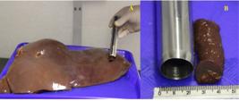
Liver at the sample collection. A - Sample collection from the right lobe. B - Dimensions of the sampling instrument.
2.2 Histopathological analyses
The samples were kept in containers containing 10 % buffered formalin, processed according to the routine method, and then stained with hematoxylin and eosin (HE) (39). The criteria for analysis were described based on the definition of a functional hepatic acinus proposed by Rappaport (1973) (40), in which the lobule is divided into three zones: zone 1 or centroacinar; zone 2 or mediozonal; and zone 3 or periacinar.
The changes were described based on the following criteria, which have already been used for liver tissue: focal lesion (a single, small lesion); multifocal (lesions in at least three fields and randomly distributed); diffuse (lesions throughout the organ) (32). The lesions were also classified according to the Rappaport zone. The intensity of inflammatory infiltrate, degeneration, and necrosis was described as: mild (few cells distributed sparsely or around the bile ducts); moderate (occupying part of the portal space); severe (occupying the entire portal space). Degeneration was further classified as microvacuolar and macrovacuolar.
The number of FMs on the slides was counted, both as isolated cells and in clusters. The final number of FMs was obtained by summing the average number of cells in each cluster with the FMs found in isolation. The average number of cells found was estimated at five cells per cluster:
The area occupied by FMs in the hepatic parenchyma was delineated in each field of the slide and then measured. All slides in which FMs were observed were evaluated, using ten randomly selected fields per slide, which were photographed under a 10x objective (41). Delineation began with the mandatory criterion of FM presence in the first field.
This procedure was performed using the ImageJ® software, which allowed for greater accuracy in image interpretation (42, 43). For this purpose, the slides were photographed using a Leica D750 optical microscope coupled with an ICC50E digital imaging module, and their micrometer scales were added to the images (100 µm).
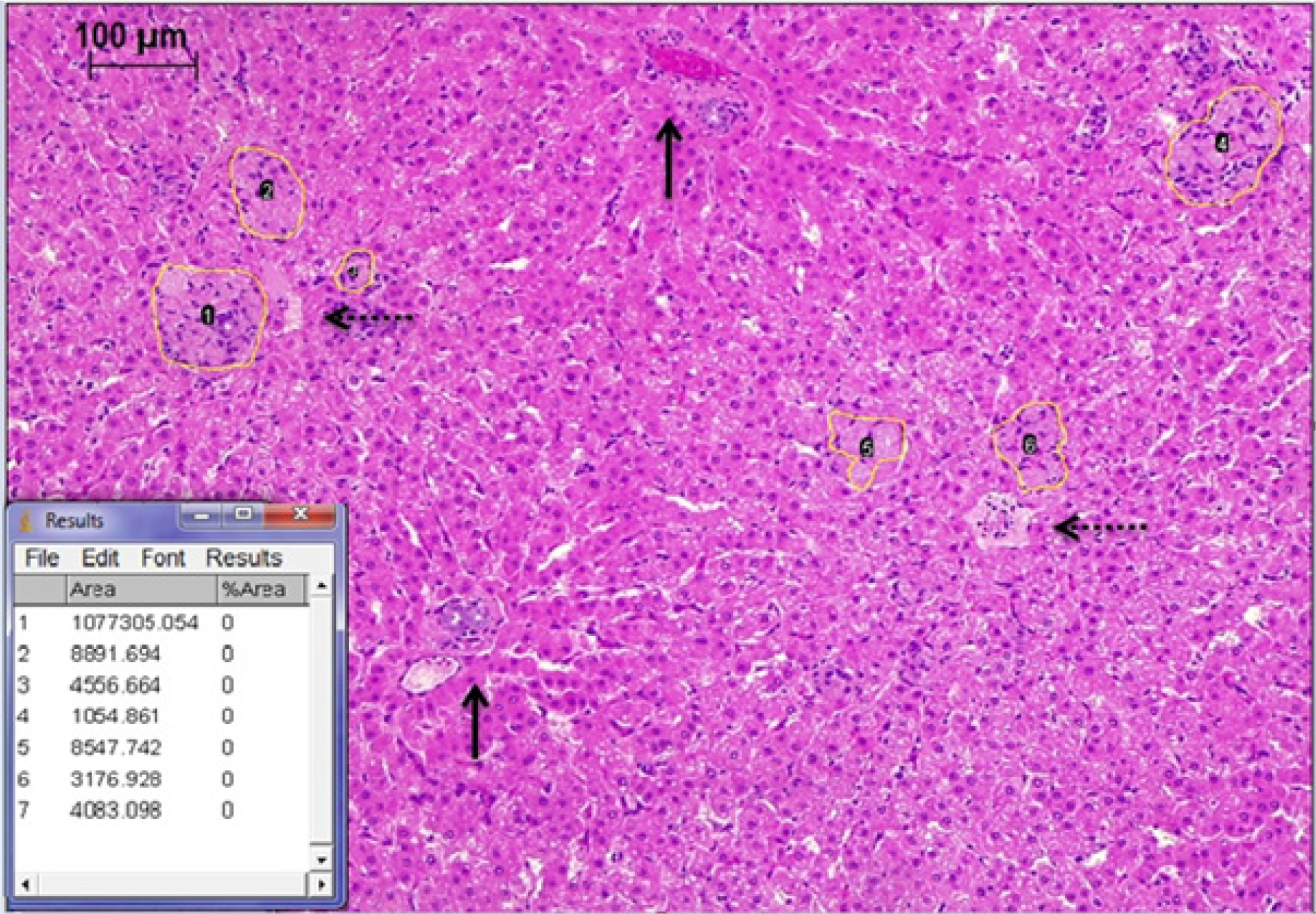
Subsequently, the photomicrographs were calibrated and analyzed using ImageJ® software (version 1.60, Wayne Rasband, National Institutes of Health, USA) across ten fields; the region occupied by FMs was delineated manually, and the percentage was calculated by subtracting the area occupied by FMs from the total image area (Figure 2). The choice to quantify the percentage of area, rather than relying on isolated cell counting, is based on the need to mitigate the bias caused by variations in the size and volume of the damaged cells. Furthermore, the effectiveness of proportional area analysis in liver tissues (44) and its established applicability in the field of veterinary medicine (45) provide the methodological rigor necessary for the precise characterization of tissue damage in cattle.
Manual delineation of the area occupied by FMs in the photomicrograph of a HE-stained liver specimen slide. Total area of the field indicated within the rectangle. Area occupied by FM indicated by the numbered boundaries. Vertical solid arrows indicate portal spaces. Horizontal dotted arrows indicate terminal hepatic veins.
2.3 Statistical analyses
Statistical analysis of the parameters (hot carcass weight, liver weight, and area of compromised hepatic parenchyma) was initially performed quantitatively, calculating the mean, standard deviation, and coefficient of variation. Subsequently, these data were assessed for normality and homogeneity of variances using the Kolmogorov-Smirnov and Bartlett tests. Since the variables were homogeneous and had a normal distribution, they were analyzed using the parametric analysis of variance (ANOVA) test.
The analysis of histopathological changes was performed by studying frequencies and was evaluated using the nonparametric chi-square test. Correlation of the variables was also performed using Spearman’s correlation. The significance level adopted was 5 %, and all descriptive analyses (presence of necrosis, degeneration, inflammatory infiltrate, and FMs) were performed using Microsoft Excel, while the R program was used for statistical tests.
3. Results and discussion
3.1 Weight of the carcass and liver
The mean, standard deviation, coefficient of variation, and correlation of carcass and liver weights in cattle are shown in Table 1.
Mean values (), standard deviation (S), coefficient of variation (CV), and correlation (r) for the carcass and liver weights of Nellore cattle with lower (Group 1) and higher (Group 2) weights slaughtered in the municipality of Goiânia.
The mean carcass weight in Group 2 was 25.58 % higher (p < 0.05) than those for Group 1. This difference contributed to the mean liver weight of Group 2 animals being 14.48 % higher (p<0.05) than that of Group 1. This difference occurred because, according to Vaz et al. (46), there is a strong positive correlation between internal organ weight and empty body weight. Similar results regarding the relationship between empty body weight and liver weight were reported by Jorge and Fontes (47) (r = 0.64), explaining that heavier animals have greater metabolic capacity than lighter ones (42). In the present study, the positive correlation between these variables was significant and strong (r = 0.72). The liver has high metabolic rates because it actively participates in nutrient metabolism, and both the liver and the gastrointestinal tract are responsive to changes in food intake. The weight and metabolic activity of visceral organs are frequently associated with weight gain efficiency (48).
3.2 Histopathological evaluation
The histopathological changes observed (Table 2) in the liver tissue of the cattle in this study were: microvacuolar degeneration (5 %), necrosis (7 %), cholangiohepatitis (94 %), and the presence of FMs (23 %).
Frequency of changes found in the liver tissue of Nellore cattle slaughtered in the municipality of Goiânia.
The changes observed in each experimental group (degeneration, necrosis, inflammatory infiltrate, and the presence of FMs) were mild in intensity and showed no significant differences (p > 0.05) (Table 3).
Frequency, distribution, and location of histopathological changes observed in liver tissue samples from Nellore cattle with lower (G1 - Group 1) and higher (G2 - Group 2) body weights slaughtered in the municipality of Goiânia.
The distribution of degeneration in Group 1 was exclusively diffuse (100 %), while in Group 2 it was diffuse (66.67 %) and multifocal (33.33 %). In both groups, this change was of the microvacuolar type, and the lesions were identified in zones 2 and 3. The distribution of necrosis in Group 1 was predominantly focal (80 %) and multifocal (20 %), whereas in Group 2 it was exclusively focal (100 %). The foci were located primarily in zone 2.
In outbreaks of hepatogenic photosensitization caused by Brachiaria spp., necrosis has been reported by some authors (25, 30, 49-54), while others (22, 56, 57), in addition to describing the lesion, quantified its presence in approximately 50 % of cases. In the present study, the mean frequencies of degeneration and necrosis across both groups were low (5 % and 7 %, respectively), consistent with Alessi et al. (57), Fioravanti (32), and Moreira et al. (33), who also noted the absence or rare occurrence of necrosis. However, they noted the presence of degeneration in most animals, mainly in zone 3, and judged both changes to be nonspecific in cattle with liver changes kept on Brachiaria decumbens pasture.
The presence of inflammatory infiltrates and FMs warrants closer attention, given that these changes have been linked by some authors (10, 11, 13, 17, 18, 32, 33, 53, 58) to hepatic abnormalities, including hepatogenic photosensitization, in animals grazed on Brachiaria spp. pastures. Additionally, elevated serum gamma-glutamyl transferase (GGT) activity and cholangiohepatitis (presence of inflammatory infiltrate in the bile ducts and liver parenchyma (59)) have been used to characterize the subclinical form of hepatogenic photosensitization (14, 21 ,54, 58).
Inflammatory infiltrates were present with a multifocal distribution in both groups (over 70 %), predominantly in the portal space (over 91 %) and in zone 2 (between 29 % and 40 %). Inflammatory infiltrate was described by Dobereiner et al. (60) in 80 % of cattle and in all sheep evaluated during outbreaks of hepatogenic photosensitization in the state of Mato Grosso. Similarly, it was described in 43 % to 66 % of cattle groups with clinical signs of photosensitization studied by Alessi et al. (57). These inflammatory changes were predominantly located in or near the portal space and had a multifocal distribution in cattle (21, 22, 25, 32, 33, 61, 62), buffalo (63, 64), sheep (14, 28, 48, 55, 56, 65-71), goats (50, 52), and horses (51) grazing on Brachiaria spp.
Considered a finding suggestive of sporodesmin toxicosis, the presence of cholangiohepatitis was associated with hepatic FMs in the form of clusters located predominantly in zone 3 (32,33). Cruz et al. (65), however, associated this inflammatory change with plants containing steroidal saponins, and noted that this and other changes, such as lymphocyte and macrophage infiltration, were induced by the experimental administration of saponins obtained from the extract of Brachiaria decumbens. This was later confirmed by Riet-Correa et al. (64), who suggested the presence of FMs as a consequence of ingesting the lithogenic steroidal saponins of Brachiaria spp.
The first reports of hepatic FMs in cattle coincide with the introduction of Brachiaria spp. as the primary forage in Brazil (11, 13, 18, 21, 53). As demonstrated in the present study, these cells were frequently observed in various animal species raised on this type of grass(11, 13, 16-18, 21, 22, 24, 25, 28, 30, 49, 53, 55, 56, 61, 62, 64, 68-70, 72).
Analysis of the FMs revealed a multifocal distribution (53.85 %) in Group 1 and a focal distribution (60 %) in Group 2. In both groups, the presence of FMs in the form of clusters was identified (over 69 %) in zone 3 (over 90 %). The predominant localization in zone 3 and the presence of clusters have also been described by numerous authors (21, 22, 24, 25, 32, 33, 61). Several hypotheses have been proposed to explain the higher prevalence of FMs in zone 3. According to Bogliolo and Brasileiro Filho (73), this is related to the fact that this region is responsible for biotransformation. Assumaidaee and Mustapha (6) suggest that saponins, upon biotransformation, generate free radicals that may trigger the presence of FM. According to Fioravanti (32), superoxide radicals, products of the auto-oxidation of sporidesmin, would cause lipid peroxidation and the emergence of FMs, and the higher presence in zone 3 would result from the involvement of endothelial cells and monocytes, which are necessary for the formation of these cells and may even appear in the form of giant cells. The lesions observed in the liver parenchyma did not affect hot carcass weight (Table 4).
Correlation coefficients between hot carcass weight and histopathological changes in the liver of Nellore cattle slaughtered in the municipality of Goiânia.
Fioravanti (32) demonstrated a significant negative correlation between live weight and the number of hepatic FMs in cattle of both sexes, a finding later confirmed by Moreira et al. (33) when working with two groups of male cattle raised on different forage species (Andropogon gayanus and Brachiaria spp.). On the other hand, Fioravanti (32) did not observe the same correlation between the number of hepatic FMs and either hot carcass weight or carcass yield. The lesions caused by these cells in the liver also did not alter the hot carcass weight of buffalo (64), nor did they affect performance in sheep (74).
A statistical difference was observed between the cattle groups for mean hot carcass weights, unlike the findings for the mean percentage of the area compromised by FMs in the liver parenchyma (Table 5).
Average weight of hot carcasses, frequency, total number, and area affected by FMs in the liver parenchyma of Nellore cattle with lower (Group 1) and higher (Group 2) weights slaughtered in the municipality of Goiânia.
Porto et al. (70) hypothesized that weight loss in animals grazing on Brachiaria spp. was due to some alteration in hepatic metabolism. Tissue injury caused by toxic agents increases cytokine production by endothelial cells, monocytes, and Kupffer cells, triggering the cascade of reactions involved in the liver regeneration process (75). Sporidesmine and saponin were identified as toxic substances to cattle (76). Hepatic functional reserve and regenerative capacity may have contributed to maintaining normal metabolism and the consequent maintenance of hot carcass weight, since, according to Jesus et al. (75), the regenerative process represents a mechanism of organ protection against the loss of functional liver tissue.
In one study, FMs were detected in 42.4 % of liver samples (53), which were collected from animals slaughtered between 1976 and 2008. Fioravanti (32) found FMs in 67.57 % of bovine livers. Souza et al. (22), Caicedo et al. (62), and Faccin et al. (17) found prevalence rates between 70 % and 80 %, while Gomar (25) and Moreira et al. (33) reported rates above 90 %. It was suspected in the present study that the small percentage of animals with FMs (23 %), combined with the low proportion of affected area on the slide (less than 0.5 %), explains the lack of difference between the groups regarding liver involvement, as reflected in the correlation analysis.
Fioravanti (32), upon quantifying the FMs, observed 14,574 cells in the slides evaluated from the right lobe. Moreira et al. (33), in turn, observed 20,099 cells in the slides evaluated from cattle grazing on Brachiaria spp., whereas only half that number was reported for those grazing on Andropogon. In the present study, a higher number of FMs was observed in the group 1, totaling only 2,750 cells. Similarly, the highest number of cells found in this study (2,750 cells) represents only 18.86 % of Fioravanti’s (32) total and 13.68 % of Moreira et al.’s (33) total, which may explain the absence of a significant correlation between the number of foamy macrophages and hot carcass weight.
Both the liver and the small intestine play a role in modulating food intake (48). Fioravanti (32) reported the replacement of areas of the hepatic parenchyma by clusters of FMs, a finding also observed in the present study. Zitnan et al. (77) and Montanholi et al. (78) observed that a lower number of cells in high-metabolism structures affected body weight and/or feed efficiency.
The greatest replacement of hepatocytes by FMs was observed in the group with the lowest hot carcass weight; however, the difference in the areas affected among the groups was not statistically significant.
As the slaughter weight increased in Nellore cattle with two permanent incisors (a category identical to that of the present study), Vaz et al. (79) observed that carcass yield decreased. This decrease in yield could explain the lack of correlation between hot carcass weight and the area occupied by FMs, and conversely, the existence of a correlation between the latter and live weight, since the yield formula is hot carcass weight * 100/live weight. In this case, the animals could have had the same hot carcass weights even while gaining weight, thus failing to demonstrate the correlative relationship expected in the hypothesis of the present study.
It was also shown that the animals’ age ranged from 20 to 36 months, a period considered less susceptible to poisoning; according to Fagliari et al. (80), the weaning phase (between 7 and 12 months, which causes stress) is the most susceptible age. The fact that slaughter and sampling were conducted in May (the beginning of the dry season) may have influenced the frequency of hepatic FMs. According to Brum (19), this was the season with the lowest frequency of Pithomyces chartarum fungal spores. The unfavorable environment for this fungus can be explained by the near absence of senescent material in the pasture and the high stocking rate (80).
Similar average hot carcass weights were found in herds of cattle with healthy livers and in herds of cattle with livers condemned due to fibrosis, demonstrating that this type of lesion did not affect the animals’ weight gain (17). The association of the small area of compromised tissue with the low presence of FMs found in this study, together with the rare observation of necrosis and degeneration (considered nonspecific changes), did not interfere with hot carcass weight in cattle. This may be explained by the fact that the changes were mild, even though inflammatory infiltrate was present in almost all animals.
4. Conclusion
Cattle grazing on Brachiaria spp. exhibit liver lesions characterized by degeneration, necrosis, and the presence of mild inflammatory infiltration, with small areas of liver tissue containing foamy macrophages. The size of the area of liver parenchyma containing foamy macrophages was not associated with reduced hot carcass weight.
Generative AI use statement
The authors did not use generative artificial intelligence tools or technologies in creating or editing any part of this manuscript.
Data availability statement
Data will be provided upon request to the corresponding author.
References
-
1 Barcellos AO, Ramos AKB, Vilela L, Martha Junior GB. Sustentabilidade da produção animal baseada em pastagens consorciadas e no emprego de leguminosas exclusivas, na forma de banco de proteína, nos trópicos brasileiros. R. Bras. Zootec. 2008;37(suplemento especial):51-67. https://doi.org/10.1590/S1516-35982008001300008
» https://doi.org/10.1590/S1516-35982008001300008 -
2 Dias-Filho MB. Uso de pastagens para a produção de bovinos de corte no Brasil: passado, presente e futuro [Internet]. Belém (PA): Embrapa Amazônia Oriental; 2016 [citado em 5 set. 2025]. Documentos 418. Available at: https://www.infoteca.cnptia.embrapa.br/infoteca/bitstream/doc/1042092/1/DOCUMENTOS418.pdf
» https://www.infoteca.cnptia.embrapa.br/infoteca/bitstream/doc/1042092/1/DOCUMENTOS418.pdf -
3 Andrade RG, Bolfe EL, Victoria DC, Nogueira SF. Avaliação das condições de pastagens no cerrado brasileiro por meio de geotecnologias. Rev Bras Agropec Sustentável. 2017;7(1):34-41. https://doi.org/10.21206/rbas.v7i1.376
» https://doi.org/10.21206/rbas.v7i1.376 -
4 Atlas das pastagens, UFG. Atlas das pastagens. [Internet]. 2023 [acesso em 2025 ago 20]. Available at: https://atlasdaspastagens.ufg.br/map
» https://atlasdaspastagens.ufg.br/map -
5 Riet-Correa B, Castro MB, Lemos RAA, Riet-Correa G, Mustafa V, Riet-Correa F. Brachiaria spp. poisoning of ruminants in Brazil. Pesqui. Vet. Bras. 2011;31(3):183-92. https://doi.org/10.1590/S0100-736X2011000300001
» https://doi.org/10.1590/S0100-736X2011000300001 -
6 Assumaidaee AAM, Mustapha NM. Toxicity of signal grass (Brachiaria decumbens) : a review article. J. Adv. Med. Res. 2012;2:18-39. Available at: https://www.researchgate.net/publication/339815783_Toxicity_of_Signal_Grass_Brachiaria_Decumbens_a_Review_Article_Article_Info
» https://www.researchgate.net/publication/339815783_Toxicity_of_Signal_Grass_Brachiaria_Decumbens_a_Review_Article_Article_Info -
7 Melo GKA, Ítavo CCBF, Silva JA, Ferelli KLSM, Silva PCG, Pupin RC, et al. Poisoning by Brachiaria spp. in various lamb breeds at increasing levels of supplementation during growth. Pesq. Vet. Bras. 2019;39(12):978-982. https://doi.org/10.1590/1678-5150-PVB-6478
» https://doi.org/10.1590/1678-5150-PVB-6478 -
8 Muniandy KV, Chung ELT, Jaapar MS, Hamdan MH, Reduan M F H, Salleh A, et al. The influence of feeding low and high level of Brachiaria decumbens diets on the hematology, serum biochemistry, and acute phase proteins of sheep. Trop. Anim. Health Prod. 2021;53(372):1-7. https://doi.org/10.1007/s11250-021-02820-1
» https://doi.org/10.1007/s11250-021-02820-1 -
9 Skalski J, Dittrich JR. Epidemiologia de intoxicações de herbívoros por plantas na região metropolitana de Curitiba, Paraná. Arch. Vet. Sci. [Internet]. 6º de julho de 2017 [citado 5º de setembro de 2025];22(1). Available at: https://revistas.ufpr.br/veterinary/article/view/46047
» https://revistas.ufpr.br/veterinary/article/view/46047 -
10 de Melo GKA, Ítavo CCBF, Silva JA, Monteiro KLS, Faccin TC, Pupin RC, et al. Poisoning by Brachiaria spp. in suckling lambs supplemented and unsupplemented in a creep-feeding system. Small Ruminant Res. 2018;158:30-34. https://doi.org/10.1016/j.smallrumres.2017.11.014
» https://doi.org/10.1016/j.smallrumres.2017.11.014 -
11 Moreira N, Martin CC, Hilgert AR, Tostes RA, Viott ADM. Surto de fotossensibilização hepatógena em bovinos por ingestão de Brachiaria decumbens no município de Cascavel - PR. Arch Vet Sci. 2018;23(1):52-62. https://doi.org/10.5380/avs.v23i1.41659
» https://doi.org/10.5380/avs.v23i1.41659 -
12 Ogliari D, Molossi FA, Savaris T, Wicpolt NS, Zancan I, Gava A. Intoxicação espontânea por Brachiaria híbrida cv mulato I em ovinos e caprinos e experimental por Brachiaria híbrida cv mulato II em ovinos. Pesq. Vet. Bras. 2018;38(2):229-233. https://doi.org/10.1590/1678-5150-PVB-4747
» https://doi.org/10.1590/1678-5150-PVB-4747 -
13 Amado GP, Silva CCB, Barbosa FMS, Nascimento HHL, Malta KC, Azevedo MV, et al. Surtos de fotossensibilização e dermatite alérgica em ruminantes e equídeos no nordeste do Brasil. Pesq. Vet. Bras. 2018;38(5):889-895. https://doi.org/10.1590/1678-5150-PVB-5583
» https://doi.org/10.1590/1678-5150-PVB-5583 -
14 Montoya-Ménez CB, Ruíz-Ramírez JA, Márquez LJG, Méndez-Bernal A, Morales-Salinas E, Ramírez-Romero R, et al. Hepatogenous photosensitization by Brachiaria spp. in sheep: first report in Mexico. Braz. J. Vet. Pathol. 2019;12(3):128-133. https://doi.org/10.24070/bjvp.1983-0246.v12i3p128-133
» https://doi.org/10.24070/bjvp.1983-0246.v12i3p128-133 -
15 Guimarães Carmo J dos S, Nunes Costa J, Almeida Biscarde CE, da Anunciação Pimentel L, de Oliveira Filho JC, Abreu Mendonça AP. Hepatogenous photosensitization in steer by Brachiaria decumbens Acta Scientiae Vet. [Internet]. 1º de janeiro de 2021 [citado 28º de agosto de 2025];49. Available at: https://seer.ufrgs.br/index.php/ActaScientiaeVeterinariae/article/view/114124
» https://seer.ufrgs.br/index.php/ActaScientiaeVeterinariae/article/view/114124 -
16 Faccin TC, Brumatti RC, Lima SC, Madureira RC, Fernandes CES, Kommers GD, et al. Perdas econômicas pela condenação em matadouro frigorífico de fígados de bovino por fibrose causada por ingestão de Braquiaria spp. Pesq. Vet. Bras. 2015;35(6):547-551. https://doi.org/10.1590/S0100-736X2015000600010
» https://doi.org/10.1590/S0100-736X2015000600010 -
17 Faccin TC, Kommers GD, Galiza GJN, Pupin RC, Madureira RC, Lemos RAA. Chronic liver disease in cattle associated with ingestion of Brachiaria spp. Cienc. Rural. 2016;46(11):2036-2042. https://doi.org/10.1590/01038478cr20160297
» https://doi.org/10.1590/01038478cr20160297 -
18 Araújo RR, Pupin RC, Madureira RC, Morais LG de, Quevedo L de S, Lemos RAA de, et al. Characterization and frequency of histological changes in bovine livers from slaughterhouses in Brazil kept on Brachiaria spp. Acta Scientiae Vet. [Internet]. 1º de janeiro de 2017 [citado 28º de agosto de 2025];45(1):9. Available at: https://seer.ufrgs.br/index.php/ActaScientiaeVeterinariae/article/view/79385
» https://seer.ufrgs.br/index.php/ActaScientiaeVeterinariae/article/view/79385 - 19 Brum KB. Papel das saponinas e do Pithomyces chartarum como agentes hepatotóxicos para ruminantes em sistema de pastejo [tese]. Goiânia: Universidade Federal de Goiás; 2006. 93 p.
-
20 Faccin TC, Riet-Correa F, Rodrigues FS, Santos AC, Melo GKA, Silva JA, et al. Poisoning by Brachiaria brizantha in flocks of naïve and experienced sheep. Toxicon. 2014;82:1-8. https://doi.org/10.1016/j.toxicon.2014.02.008
» https://doi.org/10.1016/j.toxicon.2014.02.008 -
21 Driemeier D, Döbereiner J, Peixoto PV, Brito MF. Relação entre macrófagos espumosos ("foam cells") no fígado de bovinos e ingestão de Brachiaria spp no Brasil. Pesq. Vet. Bras. 1999;19(2):79-83. https://doi.org/10.1590/S0100736X1999000200005
» https://doi.org/10.1590/S0100736X1999000200005 -
22 Souza RIC, Riet-Correa F, Brum KB, Fernandes CE, Barbosa-Ferreira M, Lemos RAA. Intoxicação por Brachiaria spp. em bovinos no Mato Grosso do Sul. Pesq. Vet. Bras. 2010;30(12):1036-1042. https://doi.org/10.1590/S0100736X2010001200006
» https://doi.org/10.1590/S0100736X2010001200006 -
23 Furlan FH, Colodel EM, Lemos RAA, Castro MB, Mendonça FS, Riet-Correa F. Poisonous plants affecting cattle in central-western Brazil. IJPPR. [Internet]. 2012 [citado 29 de agosto de 2025];2:1-13. Available at: https://www.ars.usda.gov/ARSUserFiles/oc/np/PoisonousPlants/Fall2012/PoisonousPlantResearch.pdf
» https://www.ars.usda.gov/ARSUserFiles/oc/np/PoisonousPlants/Fall2012/PoisonousPlantResearch.pdf -
24 Driemeier D, Barros SS, Peixoto PV, Tokarnia CH, Döbereiner J, Brito MF. Estudos histológico, histoquímico e ultraestrutural de fígados e linfonodos de bovinos com presença de macrófagos espumosos ("foam cells"). Pesq Vet Bras. 1998;18(1):29-34. https://doi.org/10.1590/S0100-736X1998000100005
» https://doi.org/10.1590/S0100-736X1998000100005 -
25 Gomar MS, Driemeier D, Colodel EM, Gimeno EJ. Lectin histochemistry of foam cells in tissues of cattle grazing Brachiaria spp. J Vet Med A. 2005;52(1):18-21. https://doi.org/10.1111/j.1439-0442.2004.00683.x
» https://doi.org/10.1111/j.1439-0442.2004.00683.x -
26 Santos JW, Wafae N, Beletti ME. Myocardial bridge associated with cardiovascular injuries in bovines adult of canchim race. Arq Bras Cardiol. 2012;98(1):22-29. https://doi.org/10.1590/s0066-782x2012005000003
» https://doi.org/10.1590/s0066-782x2012005000003 -
27 França LHG, Pereira AH, Perini SC, Aveline CC, Argenta R, Mollerkes RO, et al. Aterogênese em artéria ilíaca comum de suínos submetidos à homocisteinemia induzida pela ingestão de metionina. Rev Bras Med Vet. 2006;28(1):20-26. https://doi.org/10.1590/S1677-54492006000100003
» https://doi.org/10.1590/S1677-54492006000100003 -
28 Brum KB, Haraguchi M, Ricardo AAL, Riet-Correa F, Fioravanti MCS. Crystal-associated cholangiopathy in sheep grazing Brachiaria decumbens containing the saponin protodioscin. Pesq Vet Bras. 2007;27(1):39-42. https://doi.org/10.1590/S0100-736X2007000100007
» https://doi.org/10.1590/S0100-736X2007000100007 -
29 Carollo V, Di Giancamillo A, Vitari F, Schneider R, Domeneghini C. Immunohistochemical aspects of Ito and Kupffer cells in the liver of domesticated and wild ruminants. Open J Vet Med. 2012;2:129-136. https://doi.org/10.4236/ojvm.2012.23022
» https://doi.org/10.4236/ojvm.2012.23022 -
30 Oliveira CHS, Barbosa JD, Oliveira CMC, Bastianetto E, Melo MM, Haraguchi M, et al. Hepatic photosensitization in buffaloes intoxicated by Brachiaria decumbens in Minas Gerais state, Brazil. Toxicon. 2013;73:121-129. https://doi.org/10.1016/j.toxicon.2013.07.001
» https://doi.org/10.1016/j.toxicon.2013.07.001 - 31 Torres MB, Coelho KI. Foamy macrophages in the liver of cattle fed Brachiaria brizantha hay. Vet Hum Toxicol. 2003 Jun;45(3):163-164. PMID: 12776798.
- 32 Fioravanti MCS. Incidência, avaliações clínica, laboratorial e anatomopatológica da intoxicação subclínica por esporidesmina em bovinos [tese]. Botucatu (SP): Faculdade de Medicina Veterinária e Zootecnia, Universidade Estadual Paulista; 1999. 256 p.
-
33 Moreira CN, Morais M, Garcia EC, Neto SC, Araújo EG de, Fioravanti MCS. Bovinos alimentados com Brachiaria spp e Andropogon gayanus: alterações histológicas de fígado e linfonodos. Cienc. Anim. Bras. [Internet]. 3º de abril de 2009 [citado 29º de agosto de 2025];10(1):206-218. Available at: https://revistas.ufg.br/vet/article/view/1057
» https://revistas.ufg.br/vet/article/view/1057 - 34 Fagliari JJ, Oliveira JA, Kuchembuck MRG, Curi PR. Intoxicação natural de bovinos pela micotoxina esporidesmina III. Desenvolvimento ponderal de bovinos intoxicados. Arq Bras Med Vet Zootec. 1993;45(3):283-289.
- 35 Banks WJ. Histologia veterinária aplicada. 2. ed. São Paulo: Manole; 1992. 629 p.
-
36 Lertora WJ, Villordo GI, Mussart NB, Catuogno MS, S.Negrette M. Una leche bubalina rica en ácidos grasos trans incrementó la colesterolemia y tendió a agravar la ateroesclerosis en conejos. Rev Vet. 2019;30(1):32-38. https://doi.org/10.30972/vet.3013895
» https://doi.org/10.30972/vet.3013895 -
37 Lawrence TE, Whatley JD, Montgomery TH, Perino LJ. A comparison of the USDA ossification-based maturity system to a system based on dentition. J Anim Sci. 2001;79(7):1683-1690. https://doi.org/10.2527/2001.7971683x
» https://doi.org/10.2527/2001.7971683x -
38 Assis E, editor. Manual de boas práticas em patologia [online]. São Paulo: Sociedade Brasileira de Patologia; 2021 [citado 2025 Set 5]. 92 p. Available at: https://www.sbp.org.br/wp-content/uploads/2020/09/miolo-final-ebook-FINALCOM-CORRECOES-23-09.pdf ISBN 978-65-88327-00-5.
» https://www.sbp.org.br/wp-content/uploads/2020/09/miolo-final-ebook-FINALCOM-CORRECOES-23-09.pdf - 39 Luna LG. Manual of histologic staining methods of the Armed Forces Institute of Pathology. 3. ed. New York: McGraw-Hill; 1968. 258 p.
- 40 Rappaport AM. The microcirculatory hepatic unit. Microvasc Res. 1973;6(2):212-228.
-
41 Grecco FB, Schild AL, Soares MP, Marcolongo-Pereira C, Estima-Silva P, Sallis ESV. Aspectos epidemiológicos e padrões de lesões hepáticas em 35 surtos de intoxicação por Senecio spp. em bovinos no sul do Rio Grande do Sul. Pesq Vet Bras. 2010;30(5):389-397. https://doi.org/10.1590/S0100-736X2010000500003
» https://doi.org/10.1590/S0100-736X2010000500003 -
42 Henriksen SM, Nybing JD, Bouert R, Havsteen I, Christensen AF, Jensen K, et al. A computer-based method for precise detection and calculation of affected skin areas. Clin Physiol Funct Imaging. 2016;36(6):464-467. https://doi.org/10.1111/cpf.12251
» https://doi.org/10.1111/cpf.12251 -
43 Sorrentino G, Rezakhani S, Yildiz E, Nuciforo S, Heim MH, Lutolf MP, Schoonjans K. Mechano-modulatory synthetic niches for liver organoid derivation. Nature Communications. 2020; 11(3416): 1-14. Available at: https://doi.org/10.1038/s41467-020-17161-0
» https://doi.org/10.1038/s41467-020-17161-0 -
44 Tan S, Bai J, Xu M, Zhang L, Wang Y. Thioredoxin-1 activation by pterostilbene protects against doxorubicin-induced hepatotoxicity via inhibiting the NLRP3 inflammasome. Frontiers in Pharmacology. 2022; 13(841330):1-15. Available at: https://doi.org/10.3389/fphar.2022.841330
» https://doi.org/10.3389/fphar.2022.841330 -
45 TOktanella Y, Fahlevi MR, Untari H, Hendrawan VF, Mahalita NR, Jamilaturrosyidah J, Poetranto AL. Alterations in liver enzymes and histopathological changes in an animal model of polycystic ovary syndrome. Open Veterinary Journal. 2025; 15(7): 3277-3284. Available at: https://doi.org/10.5455/OVJ.2025.v15.i7.38
» https://doi.org/10.5455/OVJ.2025.v15.i7.38 -
46 Vaz FN, Restle J, Pádua JT, Fonseca CA, Pascoal LL, Severo MM. Componentes não carcaça de bovinos nelore abatidos com diferentes pesos. Rev Bras Ciênc Anim. 2015;16(3):313-323. https://doi.org/10.1590/10896891v16i318694
» https://doi.org/10.1590/10896891v16i318694 -
47 Jorge AM, Fontes CAA. Crescimento relativo dos componentes do corpo de zebuínos de diferentes raças. Acta Sci Anim Sci. 2000;22(3):801-805. Available at: https://periodicos.uem.br/ojs/index.php/ActaSciAnimSci/article/view/3202/2251
» https://periodicos.uem.br/ojs/index.php/ActaSciAnimSci/article/view/3202/2251 -
48 Ferrell CL, Jenkins TG. Body composition and energy utilization by steers of diverse genotypes fed a high-concentrate diet during the finishing period: ii. angus, boran, brahman, hereford, and tuli sires. J Anim Sci. 1998;76(2):647-657. https://doi.org/10.2527/1998.762647x
» https://doi.org/10.2527/1998.762647x -
49 Lemos RAA, Ferreira LCL, Silva SM, Nakazato L, Salvador SC. Fotossensibilização e colangiopatia associada a cristais em ovinos em pastagem com Brachiaria decumbens Ciênc Rural. 1996;26(1):109-113. https://doi.org/10.1590/S0103-84781996000100020
» https://doi.org/10.1590/S0103-84781996000100020 -
50 Lemos RAA, Nakazato L, Herrero Junior GO, Silveira AC, Porfirio LC. Fotossensibilização e colangiopatia associada a cristais em caprinos mantidos sob pastagens de Brachiaria decumbens no Mato Grosso do Sul. Ciênc Rural. 1998;28(3):507-510. https://doi.org/10.1590/S0103-84781998000300026
» https://doi.org/10.1590/S0103-84781998000300026 -
51 Barbosa JD, Oliveira CMC, Tokarnia CH, Peixoto PV. Fotossensibilização hepatógena em eqüinos pela ingestão de Brachiaria humidicola (Gramineae) no estado do Pará. Pesq Vet Bras. 2006;26(3):147-153. https://doi.org/10.1590/S0100-736X2006000300003
» https://doi.org/10.1590/S0100-736X2006000300003 -
52 Rosa FB, Rubin MIB, Martins TB, Lemos RAA, Gomes DC, Pupin RC, et al. Spontaneous poisoning by Brachiaria decumbens in goats. Pesq Vet Bras. 2016;36(5):389-396. https://doi.org/10.1590/S0100-736X2016000500006
» https://doi.org/10.1590/S0100-736X2016000500006 -
53 Seixas JN, Pinto CA, Rodrigues A, Tokarnia CH, França TN, Graça FAS, et al. Estudo comparativo das intoxicações por Brachiaria spp. e Pithomyces chartarum em bovinos. Rev Bras Med Vet. 2016;38(supl.2):1-10. Available at: https://www.researchgate.net/publication/315679631
» https://www.researchgate.net/publication/315679631 -
54 Lelis DL, Rennó LN, Chizzotti ML, Pereira CER, Silva JCP, Moreira LGT, et al. Photosensitization in naïve sheep grazing signal grass (Brachiaria decumbens) under full sunlight or a silvopastoral system. Small Rumin Res. 2018;169:24-28. https://doi.org/10.1016/j.smallrumres.2018.08.018
» https://doi.org/10.1016/j.smallrumres.2018.08.018 -
55 Mustafa VS, Moscardini ARC, Borges JRJ, Reckziegel GC, Riet-Correa F, Castro MB. Intoxicação natural por Brachiaria spp. em ovinos no Brasil central. Pesq Vet Bras. 2012;32(12):1272-1280. https://doi.org/10.1590/S0100736X2012001200010
» https://doi.org/10.1590/S0100736X2012001200010 -
56 Costa SZR, Nespoli PB, Bezerra Júnior PS, Nogueira VA, Driemeier D, Boabaid FM, et al. Avaliação lectinohistoquímica de fígado e rim de ovinos com fotossensibilização causada por Brachiaria decumbens. Pesq Vet Bras. 2013;33(8):955-962. https://doi.org/10.1590/S0100-736X2013000800001
» https://doi.org/10.1590/S0100-736X2013000800001 - 57 Alessi AC, Fagliari JJ, Okuda HT, Passipieri M. Intoxicação natural de bovinos pela micotoxina esporidesmina. 4 - Lesões hepáticas. Arq Bras Med Vet Zootec. 1994;46(3):319-328.
- 58 Fagliari JJ, Passipieri M, Kuchembuuck MRG, Curi PR. Intoxicação natural de bovinos pela micotoxina esporidesmina II aspectos clinicos. Arq Bras Med Vet Zootec. 1993;45(3):275-282.
- 59 McGavin MD, Zachary JJ. Bases da Patologia em Veterinária. 5th ed. Rio de Janeiro, RJ: Elsevier; 2013. 1344 p.
-
60 Döbereiner J, Tokarnia CH, Monteiro M do C da C, Cruz LCH da, Carvalho EG de, Primo AT. Intoxicação de bovinos e ovinos em pastos de Brachiaria decumbens contaminados por Pithomyces chartarum. Pesq. Agrop. Bras. [Internet]. 15º de abril de 2014 [citado 1º de setembro de 2025];11(9):87-94. Available at: https://apct.sede.embrapa.br/pab/article/view/17045
» https://apct.sede.embrapa.br/pab/article/view/17045 -
61 Costa RFR, Demarque KC, Santos IF, Verardino H, Almeida LAM, Amoril JG, et al. Aspectos histológicos das lesões hepatotóxicas no exame post mortem de bovinos. R Bras Ci Vet. 2006;13(3):147-152. https://doi.org/10.4322/rbcv.2014.288
» https://doi.org/10.4322/rbcv.2014.288 -
62 Caicedo JA, Ospina JC, Chaves CA, Peña J, Lozano MC, Doncel B. Lesiones hepáticas en bovinos mantenidos en pasturas de Brachiaria decumbens en Mesetas, Meta (Colombia). Rev Med Vet Zoot. 2012;59(II):102-108. Available at: http://www.scielo.org.co/scielo.php?script=sci_arttext&pid=S0120-29522012000200004&lng=pt&nrm=iso&tlng=en
» http://www.scielo.org.co/scielo.php?script=sci_arttext&pid=S0120-29522012000200004&lng=pt&nrm=iso&tlng=en -
63 Minervino AH, Barrêto Júnior RA, Rodrigues FA, Ferreira RNF, Reis LF, Headley SA, et al. Hepatogenous photosensitization associated with liver copper accumulation in buffalos. Res Vet Sci. 2010;88:519-522. https://doi.org/10.1016/j.rvsc.2009.12.005
» https://doi.org/10.1016/j.rvsc.2009.12.005 -
64 Riet-Correa B, Riet-Correa F, Oliveira Junior CA, Duarte VC, Riet-Correa G. Alterações histológicas em fígados e linfonodos de búfalos (Bubalus bubalis) mantidos em pastagens de Brachiaria spp. Pesq Vet Bras. 2010;30(9):705-711. https://doi.org/10.1590/S0100-736X2010000900001
» https://doi.org/10.1590/S0100-736X2010000900001 -
65 Cruz C, Driemeier D, Pires VS, Schenkel EP. Experimentally induced cholangiohepatopathy by dosing sheep with fractionated extracts from Brachiaria decumbens J Vet Diagn Invest. 2001;13(2):170-172. https://doi.org/10.1177/104063870101300215
» https://doi.org/10.1177/104063870101300215 -
66 Driemeier D, Colodel EM, Seitz AL, Barros SS, Cruz CEF. Study of experimentally induced lesions in sheep by grazing Brachiaria decumbens Toxicon. 2002;40(7):1027-1031. https://doi.org/10.1016/s0041-0101(01)00276-8
» https://doi.org/10.1016/s0041-0101(01)00276-8 -
67 Mendonça FS, Camargo LM, Freitas SH, Dória RGS, Baratella-Evêncio L, Neto JE. Aspectos clínicos e patológicos de um surto de fotossensibilização hepatógena em ovinos pela ingestão de Brachiaria decumbens (Gramineae) no município de Cuiabá, Mato Grosso. Ciênc anim bras. 2008;9(4):1034-41. Available at: https://revistas.ufg.br/vet/article/view/525
» https://revistas.ufg.br/vet/article/view/525 -
68 Albernaz TT, Silveira JAS, Silva NS, Oliveira CHS, Reis ASB, Oliveira CMC, et al. Fotossensibilização em ovinos associada à ingestão de Brachiaria brizantha no estado do Pará. Pesq Vet Bras. 2010;30(9):741-748. https://doi.org/10.1590/S0100-736X2010000900006
» https://doi.org/10.1590/S0100-736X2010000900006 - 69 Oliveira RS, Silva RMM, Dutra PA, Ferreira EA, Pinheiro EEG, Macêdo JTSA, et al. Intoxicação espontânea por Brachiaria decumbens em ovinos no estado da Bahia. Arq Pesq Anim. 2012;1(2):58-63.
-
70 Porto MR, Saturnino KC, Lima EMM, Lee ST, Lemos RAA, Marcolongo-Pereira C, et al. Avaliação da exposição solar na intoxicação experimental por Brachiaria decumbens em ovinos. Pesq Vet Bras. 2013;33(8):1009-1015. https://doi.org/10.1590/S0100-736X2013000800011
» https://doi.org/10.1590/S0100-736X2013000800011 -
71 Pupin RC, Melo GKA, Heckler RF, Faccin TC, Ítavo CCBF, Fernandes CE, et al. Identification of lamb flocks susceptible and resistant against Brachiaria poisoning. Pesq Vet Bras. 2016;36(5):383-388. https://doi.org/10.1590/S0100-736X2016000500005
» https://doi.org/10.1590/S0100-736X2016000500005 -
72 Miranda IC, Riet-Correa G, Oliveira Júnior CA, Riet-Correa B, Peixoto PV, Costa SZR, et al. Avaliação lectinohistoquímica de fígado e linfonodo mesentérico de búfalos mantidos em pastagens de Brachiaria spp. Pesq Vet Bras. 2016;36(9):798-804. https://doi.org/10.1590/S0100-736X2016000900002
» https://doi.org/10.1590/S0100-736X2016000900002 - 73 Bogliolo L, Brasileiro Filho G. Patologia. 7. ed. Rio de Janeiro: Guanabara Koogan; 2006. 1243 p.
-
74 Pacheco JCG. Fotossensibilização em cordeiros recriados e suplementados em pastagem de Brachiaria brizantha cv. Marandu [tese]. Pirassununga (SP): Universidade de São Paulo; 2014. 102 p. Available at: https://www.teses.usp.br/teses/disponiveis/74/74131/tde-25092014-090007/publico/DO2509859COR.pdf
» https://www.teses.usp.br/teses/disponiveis/74/74131/tde-25092014-090007/publico/DO2509859COR.pdf -
75 Jesus RP, Waitzberg DL, Campos FG. Regeneração hepática: papel dos fatores de crescimento e nutrientes. Rev Assoc Med Bras (1992). 2000;46(3):242-254. https://doi.org/10.1590/s0104-42302000000300010
» https://doi.org/10.1590/s0104-42302000000300010 -
76 Tokarnia CH, Döbereiner J, Peixoto PV. Poisonous plants affecting livestock in Brazil. Toxicon. 2002;40(12):16351660. https://doi.org/10.1016/S0041-0101(02)00239-8
» https://doi.org/10.1016/S0041-0101(02)00239-8 -
77 Zitnan R, Voigt J, Kuhla S, Wegner J, Chudy A, Schoenhusen U, et al. Morphology of small intestinal mucosa and intestinal weight change with metabolic type of cattle. Vet Med. 2008;53(10):525-532. https://doi.org/10.17221/1968-VETMED
» https://doi.org/10.17221/1968-VETMED -
78- Montanholi Y, Fontoura A, Swanson K, Coomber B, Yamashiro S, Miller S. Small intestine histomorphometry of beef cattle with divergent feed efficiency. Acta Vet. Scand. 2013;55(1):9. https://doi.org/10.1186/1751-0147-55-9
» https://doi.org/10.1186/1751-0147-55-9 -
79 Vaz FN, Restle J, Pádua JT, Fonseca CA, Pacheco PS. Características de carcaça e receita industrial com cortes primários da carcaça de machos nelore abatidos com diferentes pesos. Cienc. Anim. Bras. 2013;14(2):200-207. https://doi.org/10.5216/cab.v14i2.21123
» https://doi.org/10.5216/cab.v14i2.21123 - 80 Fagliari JJ, Okuda HT, Kuchemburk MRG, Curi PR. Intoxicação natural de bovinos pela micotoxina esporidesmina. I. Aspectos epidemiológicos. Arq Bras Med Vet Zootec. 1993;45(3):263-274.
Edited by
-
Editor:
Luiz Augusto B. Brito