Computed tomography in the assessment of adiposity in dogs
Abstract
Computed tomography (CT) is considered the gold standard for measuring visceral fat in humans; however, there are few studies about its application in veterinary medicine. This study aimed to investigate the efficacy of CT in the quantitative assessment of subcutaneous and visceral fat in dogs undergoing whole-body examinations for diagnostic purposes. A total of 54 dogs were evaluated and classified according to body condition score (BCS) into control (BCS 4-5; n = 19), overweight (BCS 6; n = 13), and obese (BCS 7-9; n = 22) groups, and further subdivided into smallsized (≤10 kg; n = 35) and large-sized (>10 kg; n = 19) dogs. All animals underwent screening blood tests, anesthesia, and CT scanning. A strong correlation was observed among subcutaneous fat measurements at different lumbar vertebrae, with L4 showing the strongest association. Principal component analysis identified L4 as the vertebra that best explained data variability. Regression analysis revealed that body weight was the only factor significantly associated with visceral and subcutaneous fat in small-sized dogs; in large-sized dogs, no factor was statistically significant. No significant difference was observed between sexes regarding visceral fat (p > 0.05); however, largesized males exhibited greater subcutaneous fat deposition. Age influenced subcutaneous fat only in large-sized dogs. The study demonstrates that CT is an accurate and reliable method for the morphometric assessment of obesity in dogs, enabling objective and segmental quantification of subcutaneous and visceral fat, thereby contributing to tissue compartment analysis and improving veterinary diagnosis and prognosis.
Keywords:
canine obesity; subcutaneous fat; veterinary imaging; visceral.
Resumo
A tomografia computadorizada (TC) é considerada padrão ouro na mensuração de gordura visceral em humanos, porém há poucos estudos sobre a sua aplicação na medicina veterinária. Este estudo teve como objetivo investigar a eficácia da TC na avaliação quantitativa da gordura subcutânea e visceral de cães submetidos a exames de corpo inteiro para fins de diagnóstico. Foram avaliados 54 cães, classificados conforme escore de condição corporal (ECC) em grupo controle (ECC 4-5; n=19), sobrepeso (ECC 6; n=13) e obesos (ECC 7-9; n=22), e subdivididos em pequeno porte (≤10 kg; n=35) e grande porte (>10 kg; n=19). Todos os animais foram submetidos a exames de sangue de triagem, anestesia e TC. Observou-se forte correlação entre as medidas de gordura subcutânea em diferentes vértebras lombares, sendo a L4 a mais forte. A análise de componentes principais destacou a L4 como a vértebra mais explicativa da variabilidade dos dados. A análise de regressão mostrou que o peso corporal foi o único fator significativamente associado a gordura visceral e subcutânea de cães de pequeno porte; em cães de grande porte, nenhum fator foi estatisticamente significativo. Não houve diferença significativa entre os sexos na gordura visceral (p>0,05), porém machos de grande porte apresentaram maior gordura subcutânea. A idade influenciou a gordura subcutânea somente em cães de grande porte. Este estudo demonstra que a TC constitui um método acurado e confiável para a avaliação morfométrica da obesidade em cães, permitindo a mensuração objetiva e segmentar da gordura subcutânea e visceral, o que pode contribuir com a análise de compartimentos teciduais e com a melhoria de diagnóstico e prognóstico veterinário.
Palavras-chave:
obesidade canina; gordura subcutânea; imaginologia veterinária; gordura visceral.
1. Introduction
Obesity is a chronic, inflammatory, and multifactorial disease characterized by an imbalance between energy intake and expenditure, resulting in excessive adipose tissue deposition. This condition is associated with metabolic, cardiovascular, and orthopedic comorbidities (1,2). Considered a global public health problem in dogs and cats (3), obesity has raised increasing concern among owners and veterinarians due to its exponential growth (4-6). Companion animals and their owners often share similar lifestyles as a result of anthropomorphism, which contributes to obesity predisposition (2). Healthy dogs have a longer life expectancy and a lower incidence of chronic diseases, such as diabetes mellitus and renal disease, compared with obese dogs (4). Adiposity refers to the accumulation of adipose tissue in the body and can be measured using direct or indirect methods. Understanding the distribution of visceral and subcutaneous fat in dogs can contribute to elucidating the pathophysiology of obesity and improving diagnostic and preventive strategies against the development of obesity. Total body fat quantification can be performed with high accuracy and precision using specific software coupled with dual-energy X-ray absorptiometry (DXA). However, DXA results may be influenced by the animal’s hydration status (10), which represents a limitation of this method. As an alternative, computed tomography (CT) has proven to be a viable tool, as it is minimally affected by hydration status, has greater equipment availability (11,12), allows tissue differentiation (13), and is considered minimally invasive (5). Moreover, CT is widely recognized as the gold standard for visceral adipose tissue (VAT) assessment in humans, enabling identification, differentiation, and analysis of fat compartmentalization and deposition patterns (13).
Visceral adipose tissue is the fat located around internal organs and is strongly associated with metabolic and hemodynamic impairments (14), largely due to its greater capacity to secrete inflammatory cytokines compared with subcutaneous adipose tissue (SAT) (15). SAT, in turn, is located beneath the dermis and above the deep subcutaneous muscle layer and is crucial in energy storage and mechanical protection.
Understanding adipose tissue compartmentalization may elucidate mechanisms underlying metabolic and cardiovascular diseases and other obesity-related comorbidities. Furthermore, studying the distribution of visceral and subcutaneous fat can assist longitudinal monitoring of body composition and contribute in evaluating the effectiveness of interventions such as dietary management, physical activity, or medical treatments. Therefore, the aim of this study was to analyze the applicability of computed tomography for the quantitative assessment of subcutaneous and visceral fat deposition in dogs.
2. Material and methods
The research project was approved by the Animal Use Ethics Committee (CEUA) of the School of Agricultural and Veterinarian Sciences (FCAV/UNESP), Jaboticabal Campus, under protocol no. 3934/202. Animal owners were informed about the study and signed an informed consent form authorizing their dogs’ participation. Research was conducted with a total of 54 dogs, patients of All Vet’s Hospital Animal (Pouso Alegre, Minas Gerais, Brazil), referred for routine abdominal CT examinations for various clinical indications. Dogs with endocrine or oncological diseases that could interfere with adiposity assessment were excluded. Eligible patients included those with gastrointestinal inflammatory diseases, obstructions, investigation of urinary bladder or prostate lesions, lumbar disc disease, and other conditions not affecting body adiposity.
All dogs underwent general clinical examination and hematological and biochemical laboratory testing. Dogs older than six years also underwent cardiological evaluation (electrocardiography and echocardiography) as part of pre-anesthetic assessment. Dogs were classified according to body condition score (BCS) into control (BCS 4-5, n = 19), overweight (BCS 6, n = 13), and obese (BCS 7-9, n = 22) groups. Subsequently, dogs were subdivided by size: small-sized (≤10 kg, n = 35) and large-sized (>10 kg, n = 19), with body weight adopted as the criterion for statistical analyses. After clinical clearance, dogs were fasted for 12 hours and water was withheld for 2 hours. Physical status classification followed the American Society of Anesthesiologists (ASA) guidelines. Dogs classified as ASA I received intramuscular premedication with dexmedetomidine (2 µg/kg) and methadone (0.2 mg/kg). Then, intravenous catheterization (22G) was implanted in the cephalic vein, in the forelimb, for anesthetic induction with ketamine (1 mg/kg) and propofol (5 mg/kg). For ASA II/III patients, premedication consisted of butorphanol (0.2 mg/kg) and acepromazine (0.03 mg/kg) intramuscularly, followed by intravenous catheterization and induction with lidocaine without vasoconstrictor (1 mg/kg), ketamine (1 mg/kg), and propofol (3 mg/kg).
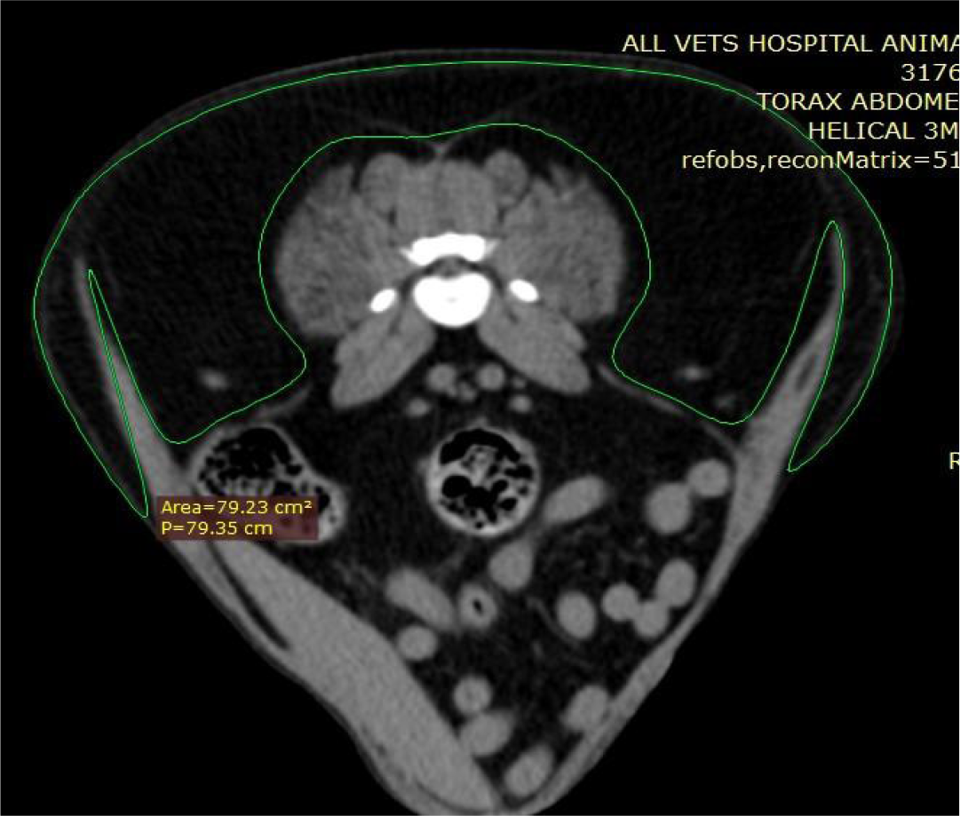
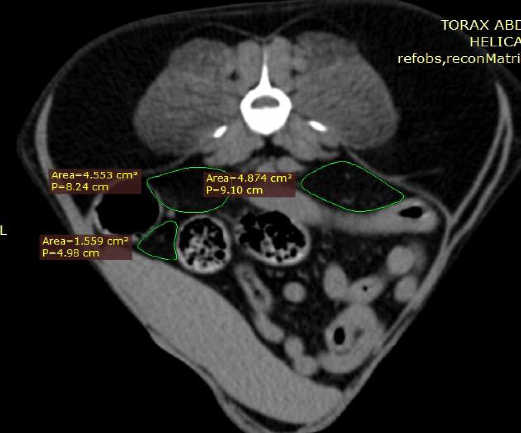
After orotracheal intubation, anesthesia was maintained with isoflurane at 1 minimum alveolar concentration (MAC), with continuous monitoring throughout the procedure and recovery. For CT examination, dogs were positioned in dorsal recumbency with thoracic limbs extended cranially and pelvic limbs extended caudolaterally. Images were acquired using a single-slice helical CT scanner (GE Helical Single Slice). After scout acquisition and ROI definition of the abdominal region, images were obtained using the following parameters: 100 mAs, 80 kV, 3-mm slice thickness, pitch 1, and Standard and Soft acquisition protocols. Transverse images of the lumbar region between vertebrae L3 and L6 were selected for analysis, as previously described by Jang et al (12). Absolute and relative areas (cm2 and cm) of subcutaneous and visceral fat were measured. Relative area (cm2) represents the two-dimensional area of the selected region, while absolute measurement (cm) corresponds to a linear dimension, such as the maximum diameter. Pre-contrast images were used to assess adipose tissue structure and distribution, differentiating SAT and VAT by manually delineating regions of interest (Figures 1 and 2). SAT was defined as the area between the skin and the peritoneal cavity, whereas VAT was measured in three regions with greater adipose deposition.

Measurement of the subcutaneous fat area (green area) on a transverse computed tomography slice at the level of the sixth lumbar vertebra (L6) in a dog. The region of interest (ROI) was highlighted in green using the RadiAnt DICOM Viewer software to obtain relative (cm2) and absolute (cm) fat measurements, with delineation performed between the peritoneal cavity and the skin.
Representation of the assessment of the visceral fat area (green areas) on a transverse computed tomography slice at the level of the fifth lumbar vertebra (L5) in a dog. The region of interest (ROI) was highlighted in green using the RadiAnt DICOM Viewer software to obtain relative visceral fat measurements.
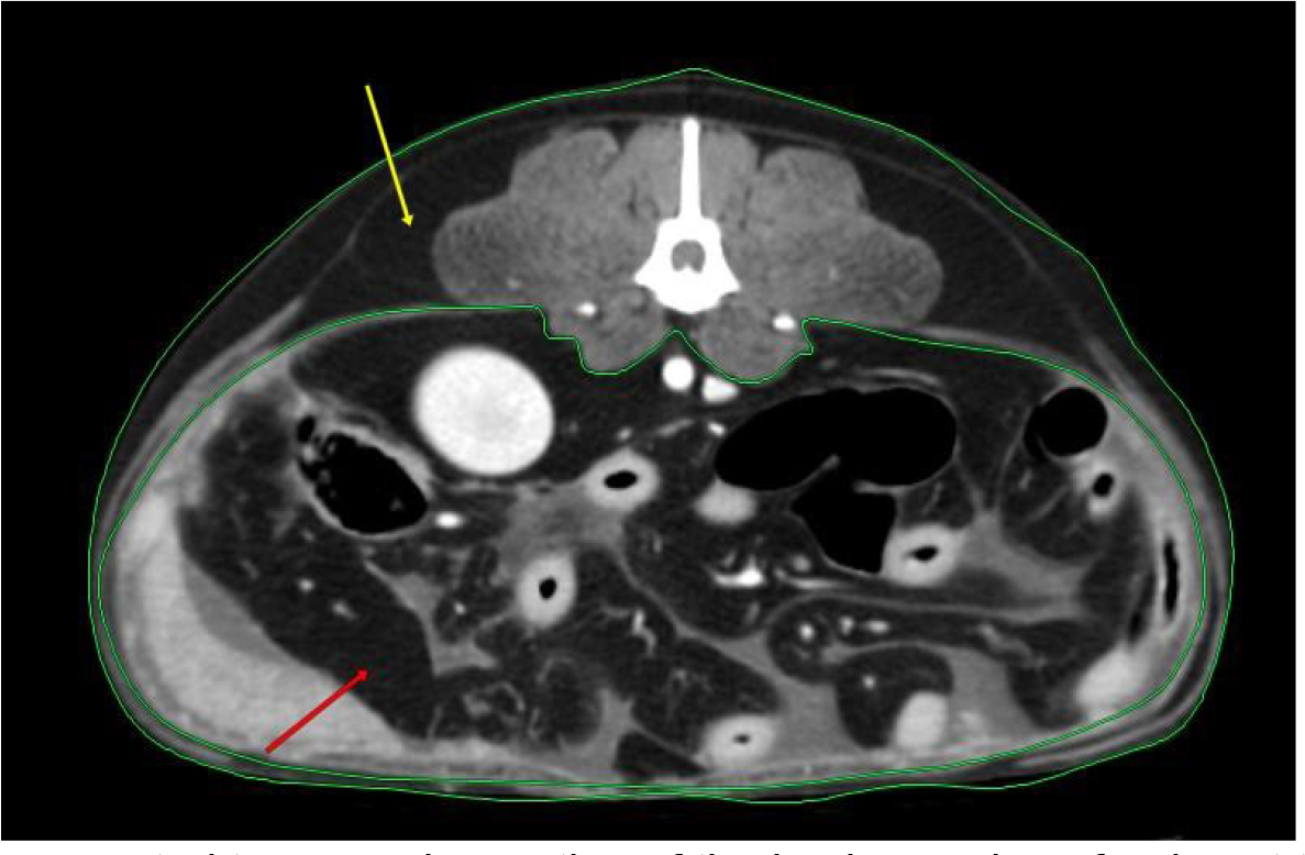
All selected vertebrae were evaluated at a minimum of three points, with results expressed as relative (cm2) and absolute (cm) values (Figure 3).
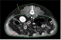
Transverse computed tomography section of the lumbar region of a dog at the level of the fourth lumbar vertebra (L4), showing visceral fat deposition (red arrow) and subcutaneous fat deposition (yellow arrow).
After completion of the examinations, the images were processed and evaluated using the RadiAnt DICOM Viewer software, in which the measurements required to determine visceral and subcutaneous fat (Figure 3) at the lumbar vertebrae L3, L4, L5, and L6 were performed.
For the assessment of subcutaneous and visceral fat deposition, all selected vertebrae were evaluated at a minimum of three points of interest, as shown in Figures 1 and 2, with relative results expressed in cm2 and absolute measurements expressed in cm.
Spearman’s correlation coefficient was used to assess associations among measurements across different vertebrae. Principal component analysis (PCA) was subsequently performed on absolute and relative SAT and VAT measures to identify the vertebra that most contributed to overall data variability. The vertebra with the highest factor loading on the first principal component was selected and used as the dependent variable in multiple regression analyses to evaluate the influence of age, sex, and neutering on fat accumulation.
3. Results
3.1 Descriptive analysis and correlation of fat measurements
Fat measurements were organized by type (visceral and subcutaneous) and anatomical location (L3 to L6). Shapiro-Wilk testing indicated non-normal data distribution (Table 1), justifying the use of Spearman’s correlation coefficient. Strong correlations were observed among measurements at different vertebrae, particularly for relative subcutaneous fat (ρ = 0.95-0.98). Correlations among visceral fat measurements were more moderate (ρ = 0.65-0.89), suggesting regional variability in visceral fat deposition.
Spearman correlation matrix among subcutaneous and visceral fat measurements (relative and absolute) at lumbar vertebrae L3 to L6 in dogs.
3.2 Principal Component Analysis (PCA)
Principal component analysis (PCA) was conducted separately for each type of fat, and was used to identify which vertebra was the most representative for estimating body fat deposition. Table 2 demonstrates the loadings of the first principal component (PC1), which accounts for the largest proportion of variance in the dataset.
Weight measurements of the first principal component (PC1) for each vertebra, considering different types of fat.
PCA identified L3 and L4 as the vertebrae with the highest contributions to total variance, with L4 showing the highest loadings in most models (0.51-0.56). L6 showed the lowest contribution, particularly for absolute visceral fat (0.38). These findings indicate that L4 measurements adequately represent overall body fat deposition in dogs.
3.3 Regression analysis: factors associated with body fat
Multiple linear regression analyses were stratified by size (small and big) and a significance level of 5 % (p < 0.05) was designed. Fat measurements in the L4 vertebra were used as the dependent variable. In small-sized dogs, body weight was significantly associated with both visceral and subcutaneous fat (relative and absolute). Each additional kilogram increased absolute visceral and subcutaneous fat areas by 1.65 cm2 and 6.31 cm2, respectively (p < 0.01). In largesized dogs, males showed significantly greater subcutaneous fat deposition than females, and neutering, age, and body weight also influenced subcutaneous fat accumulation. Table 3 presents the regression effects of factors such as sex, neutering, body weight, and age, with dogs divided into large and small breeds.
Estimates of regression coefficients for the factors sex, neutering, body weight, and age in large and small breed dogs. SAT VAT.
4. Discussion
Nagão et al. (20) conducted a retrospective study with 90 dogs evaluating visceral fat through whole-body analyses and found significant correlations between CT images and visceral fat, corroborating the present study. Other authors have observed the superiority of CT over conventional examinations, such as radiography, in differentiating subcutaneous and visceral adipose tissue (10). In addition, studies by Bae and Oh (17) demonstrated the applicability of tomography for evaluating body fat deposition and weight loss, reaffirming the effectiveness of the method for diagnostic evaluation, monitoring and treatment of obesity.
Grillot et al. (19) highlighted, in their work, the correlation between the assessment of body condition score in humans and the assessment of sarcopenia and obesity through the use of computed tomography and stated that the measurement of only one cross-section in which L3 was used is sufficient to attest to the nutritional status of the patient, corroborating the present research. It is noteworthy that, through the data obtained, even with racial and sex variation, the L3 vertebra, and especially L4, showed a greater positive Spearman correlation with the presence of exacerbated adipose tissue deposition, which corroborates studies carried out previously (6).
Thengchaisri et al. (5) verified a positive correlation of visceral fat with heart disease in dogs. They also observed, similarly to the present work, high precision in the tomographic assessment of visceral fat. Gavin et al. (21) reported that men tend to accumulate more visceral fat, while women deposit more subcutaneous fat. However, in the present study, it is possible to observe only a difference in the deposition of subcutaneous fat in large dogs, in which males showed a tendency to greater deposition when compared to females. Kim et al. and Jang et al. (10, 12) observed that sex was not a determining factor in variation. However, for the body weight factor, in both sizes (small and large), there is a positive correlation between weight and the amount of visceral fat, which was not observed by Muller et al. (6), which suggests that breed variety is a highly relevant factor in the analysis of fat distribution by CT.
The findings of the present research for small dogs demonstrated that the distribution of subcutaneous and visceral fat is uniform, corroborating with Turner (19). However, unlike his study, age presented a positive correlation only in large dogs, both in relation to the age factor and to whether they were intact or neutered, the latter representing the main difference in the deposition of subcutaneous adipose tissue.
5. Conclusion
Computed tomography is an effective and reliable method for quantifying visceral and subcutaneous fat in dogs, allowing precise differentiation of adipose compartments. Vertebra L4 was identified as a reliable marker of overall fat deposition, suitable for clinical and experimental applications. Given the association between visceral fat accumulation and metabolic disorders, CT-based adipose tissue compartment analysis is crucial for improving canine health, supporting individualized treatment strategies, and preventing obesity-related complications.
Supplementary material
(Available only in the online version: https://revistas.ufg.br/vet/article/view/83372) Table S1. Morphometric Analyses of Dogs.
Generative AI use statement
During the preparation of the manuscript, the authors used ChatGPT to improve the quality of the English language writing. The content was verified by the authors, with additional review by a professional, to ensure terminological adequacy and academic consistency. The authors assume full responsibility for the content of the publication. As an expression of the truth, I affirm this declaration..
Data availability statement
The complete dataset supporting the results of this study has been made available in the CAB repository and can be accessed via the link https://doi.org/10.48331/SCIELODATA.Q1KOJ0.
References
- 1 Jericó M, et al. Obesidade Canina e Felina. Associação Brasileira de Endocrinologia Veterinária, 2018;1 p.
-
2 Chandler M, Cunningham S, Lund EM, et al. Obesity and associated comorbidities in people and companion animals: a One Health perspective. J Comp Pathol. 2017;156(4):296-309. https://doi.org/10.1016/j.jcpa.2017.03.006
» https://doi.org/10.1016/j.jcpa.2017.03.006 -
3 Clark M, Hoenig M. Metabolic effects of obesity and its interaction with endocrine diseases. Vet Clin North Am Small Anim Pract. 2016;46(5):797-815. https://doi.org/10.1016/j.cvsm.2016.04.004
» https://doi.org/10.1016/j.cvsm.2016.04.004 -
4 Zoran DL. Obesity in dogs and cats: a metabolic and endocrine disorder. Vet Clin North Am Small Anim Pract. 2010;40(2):221-239. https://doi.org/10.1016/j.cvsm.2009.10.009
» https://doi.org/10.1016/j.cvsm.2009.10.009 -
5 Thengchaisri N, Theerapun W, Kaewmokul S, Sastravaha A. Abdominal obesity is associated with heart disease in dogs. BMC Vet Res. 2014;10:131. https://doi.org/10.1186/1746-6148-10-131
» https://doi.org/10.1186/1746-6148-10-131 -
6 Müller L, Kollár E, Balogh L, et al. Body fat distribution and metabolic consequences - Examination opportunities in dogs. Acta Vet Hung. 2014;62(2):169-179. https://doi.org/10.1556/AVet.2013.057
» https://doi.org/10.1556/AVet.2013.057 -
7 Gomes BF, Accardo CM. Immunoinflammatory mediators in the pathogenesis of diabetes mellitus. Einstein (São Paulo). 2019;17(1):eRB4596. https://doi.org/10.31744/einstein_journal/2019RB4596
» https://doi.org/10.31744/einstein_journal/2019RB4596 -
8 Monteiro AR, Barros LA dos S, Albernaz PCS, Vieira VB de A, Barroso VD de MP, Rodrigues MC da C. Diabetes mellitus, obesidade e maus hábitos de vida: sua relação com a COVID-19. Rev Cient Fac Med Campos. 2020;15(2):56-68. https://doi.org/10.29184/1980-7813.rcfmc.377
» https://doi.org/10.29184/1980-7813.rcfmc.377 -
9 Raposo HF. Tecido adiposo: suas cores e versatilidade. HU Rev. 2020;46:1-12. https://doi.org/10.34019/19828047.2020.v46.31268
» https://doi.org/10.34019/19828047.2020.v46.31268 -
10 Kim D, Noh D, Oh T, Lee K. Body fat assessment by computed tomography and radiography in normal Beagle dogs. J Vet Med Sci. 2018;80(9):1380-1384. https://doi.org/10.1292/jvms.18-0216
» https://doi.org/10.1292/jvms.18-0216 -
11 Sousa JPLBA de, Bekhor D, Saito Filho CF, Bretas EAS, D'Ippolito G. Perfusão por tomografia computadorizada do abdome: aplicações clínicas, princípios e técnica do exame. Radiol Bras. 2012;45(1):39-45. https://doi.org/10.1590/S0100-39842012000100010
» https://doi.org/10.1590/S0100-39842012000100010 -
12 Jang Y, Na H, Choi S, Choi H, Lee Y, Lee K. Assessment of abdominal fat distribution by computed tomography in obese dogs. The Thai Journal of Veterinary Medicine. 2022; 52(2):245-250. https://doi.org/10.56808/2985-1130.3212
» https://doi.org/10.56808/2985-1130.3212 -
13 Nagao I, Ohno K, Nagahara T, et al. Evaluation of visceral fat mass in dogs by computed tomography. J Vet Med Sci. 2019;81(11):1552-1557. https://doi.org/10.1292/jvms.19-0254
» https://doi.org/10.1292/jvms.19-0254 -
14 Ibrahim MM. Subcutaneous and visceral adipose tissue: structural and functional differences. Obes Rev. 2010;11(1):11-18. https://doi.org/10.1111/j.1467-789X.2009.00623.x
» https://doi.org/10.1111/j.1467-789X.2009.00623.x -
15 Leal CC, Rodrigues IG, Leite AL, Arruda IG De, Diniz AS, Pinho C et al. Glomerular filtration rate and its relationship with visceral and subcutaneous adiposity: a study with patients from Northeast Brazil. Rev. Chil. Nutr. 2020; 47(3):449456. http://dx.doi.org/10.4067/S0717-75182020000300449
» http://dx.doi.org/10.4067/S0717-75182020000300449 - 16 Laflamme D. Development and validation of a body condition score system for dogs. Canine practice. 1997;22(4):1015.
-
17 Bae S, Oh T. Anti-obesity effects of Korean red ginseng extract in healthy beagles. Pol J Vet Sci. 2019;22(2):385389. https://doi.org/10.24425/pjvs.2019.129298
» https://doi.org/10.24425/pjvs.2019.129298 -
18 Grillot J, D'Engremont C, Parmentier AL, et al. Sarcopenia and visceral obesity assessed by computed tomography are associated with adverse outcomes in patients with Crohn's disease. Clin Nutr. 2020;39(10):3024-3030. https://doi.org/10.1016/j.clnu.2020.01.001
» https://doi.org/10.1016/j.clnu.2020.01.001 -
19 Turner RBS, Tyrrell D, Hepworth G, Dunshea FR, Mansfield CS. Compartmental fat distribution in the abdomen of dogs relative to overall body fat composition. BMC Vet Res. 2020;16(1):104. https://doi.org/10.1186/s12917-020-023271
» https://doi.org/10.1186/s12917-020-023271 -
20 Nagao, Itsuma et al. Evaluation of visceral fat mass in dogs by computed tomography. Journal of Veterinary Medical Science, v. 81, n. 11, p. 1552-1557, 2019. https://doi.org/10.1292/jvms.19-0254
» https://doi.org/10.1292/jvms.19-0254 -
21 Gavin, K M.; Bessesen, D H. Sex differences in adipose tissue function. Endocrinology and Metabolism Clinics, v. 49, n. 2, p. 215-228, 2020. https://doi.org/10.1016/j.ecl.2020.02.008
» https://doi.org/10.1016/j.ecl.2020.02.008
Edited by
-
Editor:
Luiz Augusto B. Brito