Analysis of risk factors, clinical alterations and laboratory markers in cows with different types of abomasal displacement
Abstract
Depending on the side in which the abomasal displacement occurs, abomasal displacement (AD) can be classified into different types, that directly influence clinical and laboratory alterations. The objective of this study was to analyze the main risk factors, clinical manifestations, and laboratory alterations (energy, hormonal, mineral and enzymatic profile) of cows with different types of AD. We included 104 cows, attended at the Garanhuns Cattle Clinic, Campus of the Federal Rural University of Pernambuco, diagnosed with AD, with 67 cases of left AD (G2), 25 of right AD (G3), and 12 of abomasal volvulus (G4). The control group (G1) consisted of 19 clinically healthy cows. Quantitative data were tested using the ANOVA or Kruskal-Wallis tests, and categorical variables were subjected to Fisher's exact test. A correlation study was carried out between quantitative variables within the groups and all analyses considered p <0.05. Among the risk factors identified, the intensified rearing system (96.15 %), breed (holstein [50.0 %]), and the number of parity (≤ 2 parity [49.04 %]) stand out. Among the clinical findings, dehydration, tachycardia, ruminal tympany, and abdominal distension were more frequent in animals from G3 and G4. Analysis of the energy profile demonstrated an increase in the levels of NEFAs, glucose, and cortisol in animals with AD compared to cows in the control group. Hypocalcemia and hypokalemia were observed in animals with AD, and hyperlactatemia was more pronounced in G4. A clinical-surgical intervention was performed in 86.54 % of animals, with a positive outcome in 76.92 % of cases, with no difference regarding the type of AD. Given the results, it is possible to verify the influence of the type of AD on the intensity of clinical-laboratory alterations, with greater implications for abomasal volvulus, with the predisposing factors for occurrence being identifiable and subject to appropriate control and prevention methods.
Keywords:
abomasal volvulus; displaced abomasum; energetic metabolism; mineral profile.
Resumo
A depender do antímero em que ocorre a paratopia abomasal o deslocamento de abomaso (DA) pode ser classificado em diferentes tipos que influenciam diretamente as alterações clínicolaboratoriais. O objetivo deste estudo foi analisar os principais fatores de risco, manifestações clínicas e alterações laboratoriais (perfil energético, hormonal, mineral e enzimático) de vacas com diferentes tipos de DA. Foram utilizadas 104 vacas atendidas na Clínica de Bovinos de Garanhuns, Campus da Universidade Federal Rural de Pernambuco, diagnosticadas com DA, sendo 67 casos de DA a esquerda (G2), 25 de DA a direita (G3) e 12 de vólvulo abomasal (G4). O grupo controle (G1) foi composto por 19 vacas clinicamente saudáveis. Os dados quantitativos foram testados pelos testes de ANOVA ou Kruskal-Wallis, enquanto as variáveis categóricas foram submetidas ao teste exato de Fisher. Foi realizado um estudo de correlação entre variáveis quantitativas dentro dos grupos e todas as análises consideraram p<0,05. Dentre os fatores de risco identificados destacamse o sistema intensificado de criação (96,15 %), a raça (holandesa [50,0 %]), e número de partos (≤ 2 partos [49,04 %]). Dentre os achados clínicos, a desidratação, taquicardia, timpania ruminal e distensão abdominal foram mais frequentes nos animais do G3 e G4. A análise do perfil energético demonstrou elevação nos níveis de AGNEs, glicose e cortisol dos animais com DA em comparação com as vacas do grupo controle. Hipocalcemia e hipocalemia foram constatados nos animais com DA, e a hiperlactatemia foi mais acentuada no G4. A intervenção clínica-cirúrgica foi realizada em 86,54 % dos animais, com um desfecho positivo em 76,92 % dos casos, sem diferença quanto ao tipo de DA. Diante dos resultados, é possível constatar a influência do tipo de DA na intensidade das alterações clínico-laboratoriais, com maiores implicações no vólvulo abomasal, sendo os fatores predisponentes para sua ocorrência identificáveis e passíveis de métodos de controle e prevenção adequados.
Palavras-chave:
abomaso deslocado; metabolismo energético; vólvulo abomasal; perfil mineral.
1. Introduction
Abomasal displacement (AD) is a digestive disorder that mainly affects high-producing dairy cows in the postpartum period (1-4). Its etiology is multifactorial, but it is believed to primarily involve hypomotility or abomasal atony, with consequent gas accumulation, related, among other predisposing factors, to a diet rich in soluble carbohydrates and the production of excessive amounts of gas and short-chain fatty acids inside the abomasum (2-5).
AD can be classified according to the antimere in which the abomasal displacement occurs, into left displaced abomasum (LDA), right displaced abomasum (RDA), and abomasal volvulus (AV), which is considered a complication of RDA, due to the intense obstruction of the digestive flow, that predisposes the occurrence of the rotation of the organ around its axis” (2,6-9). Depending on the timing of the displacement, different clinical conditions can be identified. In general, affected animals can present varying degrees of apathy, dehydration, inappetence or anorexia, colic, abdominal distension, increased abdominal tension, and a metallic “ping” sound and fluid splash when palpating in the region where the organ is displaced, in addition to changes in the quantity and physical characteristics of feces (2, 10, 11).
The clinical consequences of AD are related to abomasal reflux syndrome (12, 13) and possible concomitant metabolic alterations resulting from the negative energy balance observed in the transition period, as a result of increased energy needs and reduced dry matter consumption (3,14). Thus, the assessment of serum levels of energy, mineral, and enzyme biomarkers can be used for early diagnosis of the condition, institution of treatment, and prediction of prognosis (13, 15-18).
Therefore, the objective of the current work was to carry out an analysis of the main risk factors, clinical manifestations, and laboratory markers (energy, hormonal, mineral, and enzymatic profile) of cows with different types of AD.
2. Material e mtodos
2.1 Animais
In total, 104 cows were used, attended at the Garanhuns Cattle Clinic, Campus of the Federal Rural University of Pernambuco, diagnosed with abomasal displacement (AD), 67 cases of left AD (LDA; G2), 25 of right AD (RDA; G3), and 12 right AD with abomasal volvulus (AV; G4). In addition, 19 clinically healthy cows were included to compose the control group (G1), with productive characteristics similar to those of the groups with AD, of the girolando breed with genetic composition of 3/4 holstein and 5/8 gir, high milk production, early stage of lactation (10 days postpartum), body condition score between 3.0 and 3.5, and rearing in a semi-intensive system. The project was approved by the Animal Use Ethics Committee (CEUA) of the Federal University of Agreste de Pernambuco under license 050942/2024.
2.2. Diagnosis
The diagnosis of AD was established based on physical examination findings associated with increased chloride content in ruminal fluid (> 30 mEq/L) (19). For the diagnosis of abomasal volvulus, in addition to these criteria, it was necessary to identify the torsion of the organ through exploratory laparotomy on the right flank or necroscopic examination.
2.3. Clinical parameters
Information was collected about age, sex, breed, lactation stage, period of occurrence, rearing system, productive phase, therapeutic approach used, period of hospitalization, and clinical outcome, classified as positive (hospital discharge) or negative (natural death or euthanasia).
The animals included in the study were subjected to physical examination according to Dirksen (19). General health aspects, such as body condition score, hydration status, heart and respiratory rates, rectal temperature, and appetite were evaluated. Regarding the specific examination of the digestive system, appetite, degree of filling, stratification and rumen motility (frequency and intensity); location, motility and degree of abomasal filling; abdominal distension and tension; and intestinal motility and fecal production (volume, consistency, color, degree of digestibility, and presence of mixtures) were evaluated.
2.4. Laboratory parameters
At the time of diagnosis, blood samples were collected by venipuncture of the jugular vein, in a sterile vacuum tube (Vacutainer® system) containing EDTA (ethylenediaminetetraacetic acid), for hematological evaluation, and with sodium fluoride/EDTA and without anticoagulant, to obtain plasma and serum, respectively, after centrifugation at 3500 rpm for five minutes. These samples were divided into aliquots and stored at -80°C for subsequent laboratory processing.
The blood count and determination of total plasma protein and plasma fibrinogen were performed according to Harvey (20). Serum levels of non-esterified fatty acids (NEFA assay®), β-hydroxybutyrate (D-3-Hydroxybutyrate/Ranbut assay®), L-lactate (Lactate Enzimático®), and glucose (Glucose Liquiform®) were determined on a semi-automatic biochemical analyzer (BIO 2000®), as well as the serum activity of the enzymes aspartate aminotransferase (AST/GOT Liquiform®) and gamma-glutamyltransferase (GAMA GT Liquiform®).
Rumen fluid samples were analyzed according to Dirksen et al. (19), with chloride content measured using a commercial kit (Clorides Liquiform®). The determination of the concentration of serum electrolytes (Ca2+, Na+, and K+) was carried out on an AVL 9180® Roche® electrolyte analyzer. Serum values of the hormones insulin and cortisol were determined by chemiluminescent immunoassay using commercial kits (Access Immunoassay Systems®).
2.5. Insulin Sensitivity Index
Insulin sensitivity was estimated using the Revised Quantitative Insulin Sensitivity Check Index (RQUICKI) and RQUICKIBHB, which take into account the concentration of glucose, insulin, non-esterified fatty acids (NEFA), and beta-hydroxybutyrate (BHB) according to the equations: RQUICKI=1/ [log10 (glucose mg dL-1) + log10 (insulin μU mL-1) + log10 (NEFA mmol L-1)] and RQUICKIBHB =1/ [log10 (glucose mg dL-1) + log10 (insulin μU mL-1) + log10 (NEFA mmol L-1) + log10 (βHB mmol L-1)]. In both tests, values closer to zero were considered as indicators of disturbance in insulin function (21,22).
2.6. Statistical analysis
The quantitative variables were subjected to normality (Shapiro-Wilk) and homogeneity (Levene) tests to identify their distribution. The means of those that met the assumptions of normality and homogeneity were analyzed using One-Way ANOVA followed by a post-hoc test (Tukey), when significance was observed. In turn, non-parametric or heterogeneous data, even after square root or logarithmic transformation, were subjected to the Kruskal-Wallis test for comparisons of medians, followed by the Dunn test, as a post-hoc test, in the presence of significance. Categorical variables were expressed as frequencies and subjected to Fisher's exact test and, in case of significance, the Bonferroni test for multiple comparisons as post-hoc (23).
To evaluate the relationships between quantitative variables within the groups, missing data were imputed with the MICE (Multiple Imputation by Chained Equations) technique using the PMM method (Predictive Mean Matching) (24,25). After these procedures, the data were subjected to the Spearman correlation test and expressed through heat maps based on the correlation matrix. A high correlation was considered when r > 0.60; a moderate correlation when 0.30 < r < 0.60, and a low correlation when r < 0.30. In all analyses, the statistical software R Core Team (26), version 4.3.3, was used, considering p<0.05.
3. Results
Table 1 presents epidemiological, clinical, and clinical progression for the animals with AD included in the study. The median age of the animals was 48.0 months and the majority of cows were Holstein (50.00 %) or mixed dairy breeds (27.88 %), with up to two parity (49.04 %) and in the first four weeks of lactation (47.12 %).
Epidemiological, clinical, and clinical progression of animals with LDA (G2, n=67), RDA (G3, n=25), and AV (G4, n=12).
A higher incidence of AD was observed during the rainy season in the region (61.54 %), but no difference was observed (p=0.266) between the occurrences of the different types of AD in the rainy and dry seasons (Table 1). The predominant rearing systems were intensive (50.00 %) and semi-intensive (46.15 %), with a median body condition score of 2.5 (on a scale from 0 to 5). The behavior of animals with AD at the time of the initial physical examination (Table 1) was predominantly apathetic (47.12 %) and there were no differences in behavior among the different types of the disease (p=0.462).
Clinical-surgical intervention was the most used (86.54 %) in animals with AD (Table 1), with an average hospitalization period of six days. Regarding the clinical outcomes, 76.92 % of cases were discharged from hospital (positive outcome), while 23.08 % of cases were euthanized or died naturally (negative outcome), with no difference observed in the proportion of positive and negative outcomes between types of AD (p=0.474) (Table 1).
The degree of dehydration was more pronounced in animals from G3 and G4, compared with G2 (40.00 % and 58.33 % vs. 16.42 %; p=0.023) (Table 2). Similarly, tachycardia (Table 2) was also more pronounced in animals from G3 and G4 (p=0.005), but no difference was observed between the proportion of animals with different types of AD in the different heart rate classifications (p=0.279).
Degree of dehydration, heart rate, respiratory rate and rectal temperature of cows with LDA (G2, n=67), RDA (G3, n=25), and AV (G4, n=12).
Tachypnea was identified in the majority of the animals studied (69.23 %) and the mean rectal temperature was 38.7°C (Table 2). There was no difference in the frequencies of these clinical parameters between the groups in the different classifications (p>0.05).
Table 3 presents the gastrointestinal findings of cows with different types of AD. A reduction or absence of appetite was observed in 60.00 % of the animals. The absence of abdominal distension was observed in a greater proportion in animals from G2 (91.04 %), while bilateral distension was observed mainly in animals from G3 (28.00 %) and G4 (18.18 %), as well as increased abdominal tension (G3: 50.00 %, G4: 50.00 %) (p<0.001).
Relative and absolute frequencies of gastrointestinal findings in cows with LDA (G2, n=67), RDA (G3, n=25) and AV (G4, n=12).
Ruminal stratifications were undefined in 67.31 % of cases, while hypomotility or ruminal atony was found in 80.39 % of patients, with no difference in the proportion of these findings among the groups (p>0.05) (Table 3). Ruminal tympany was found in 32.69 % of cows, but was more frequent in animals from G3 (60.00 %) and G4 (41.67 %) (p=0.001). The chloride concentration in rumen fluid (Table 3) was high in all groups, with higher values in animals with AV (p=0.017).
Intestinal hypomotility was found in 65.38 % of cases and fecal consistency was altered in 61.19 %, with sticky consistency being more observed in animals from G3 (52.94 %) and G4 (57.14 %) (p<0.001) (Table 3). The main mixtures observed in the animals under study were mucus (19.73 %) and melena (15.38 %), the latter being most common in G3 (19.05 %) and G4 (27.27 %) (p=0.025).
The hematological findings of animals with AD are shown in Table 4. Hematocrit and band neutrophil count were higher in G4 animals (p<0.05). In all groups, the animals presented mild leukocytosis associated with neutrophilia.
Mean/median and standard deviation (25-75 percentile) of hematological findings of cows with LDA (G2, n=67), RDA (G3, n=25), and AV (G4, n=12).
The energy profile variables indicated metabolic alterations of animals with AD when compared to cows in the control group (Table 5). High values of non-esterified fatty acids (NEFA) indicated negative energy balance in all groups, but more pronounced in animals in G2 and G3 (p=0.001). Similarly, beta-hydroxybutyrate (BHB) levels were higher in these groups (p<0.001), but did not indicate ketosis in any group.
Mean/median and standard deviation (25-75 percentile) of energetic, hormonal, mineral and enzymatic variables, plasma lactate and creatinine of clinically healthy cows (G1, n=19) and cows with LDA (G2, n=67), RDA (G3, n=25), and AV (G4, n=12).
Insulin levels (Table 5) did not differ between the control group and AD groups (p=0.252), and remained within the reference range for the species. On the other hand, cortisol concentrations were high in all groups, but with higher levels in animals from G3 and G4 (p=0.007). The increase in the blood glucose levels was observed in animals with AD, compared to animals in G1, with greater intensity in G4 (p<0.001). Similarly, insulin resistance rates (RQUICKI and RQUICKIBHB), showed greater impairment of the function of this hormone in groups with AD compared to the control group (p<0.05).
Of the serum minerals analyzed (Table 5), only Ca2+ levels showed differences between the groups. Animals with AV had lower concentrations of this mineral compared to the control group (p=0.007), demonstrating hypocalcemia in these animals. Hypokalemia was also identified in all groups of animals with AD. There was no difference in serum Na+ concentration between the AD groups.
The enzymatic profile of animals with AD showed variations when compared to the control group (Table 5). The enzymatic activity of gamma-glutamyltransferase (GGT) was elevated in all groups, but with greater intensity in cases of RDA (G3) and AV (G4) (p<0.001). In turn, serum aspartate aminotransferase (AST) activity remained within normal limits in all groups, although higher in animals with AD compared to the control group (p<0.001).
Plasma lactate values (Table 5) indicated greater hemodynamic impairment in cases of AD, especially with AV (G4) (p<0.001). Similarly, creatinine values were higher in groups with AD, compared with animals in G1 (p=0.017).
In the correlation study of clinical and laboratory variables of cows with different types of AD (Figure 1), the strong correlations between K+ and creatinine (r = -0.73) and insulin and RQUICKIBHB (r = -0.66) in G3 (Figure 1B); as well as between insulin and glucose (r = 0.83), plasma lactate and glucose (r = 0.79), plasma lactate and HR (r = 0.75), creatinine and K+ (r = -0.71), dehydration and HR (r = 0.70), HR and plasma fibrinogen (r = 0.66), glucose and HR (r = 0.64), and dehydration and K+ (r = -0.61). The variables in group G2 (Figure 1A) showed only moderate and weak correlations.
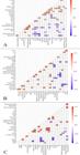
Correlation Heatmap of clinical and laboratory variables of animals with different types of AD. A. LDA (G2, n=67). B. RDA (G3, n=25). C. AV (G4, n=12). The points of intersection between two variables that have a correlation value are significant considering p<0.05.
4. Discussion
Several risk factors have been associated with the occurrence of AD in cattle, including abrupt dietary changes during the postpartum period, combined with hormonal alterations and metabolic stress, which are recognized as predisposing factors for the development of this disease (27). In the present study, 47.12 % of AD cases occurred in the first four weeks postpartum. The proportion of LDA cases in this period (43.29 %) was lower than that reported in other studies (13,18,28) in which occurrence is around 80%. The occurrence of RDA (60.00 %) and AV (45.33 %) in the first four weeks postpartum is in line with the results of Rohn et al. (29) (52.6 % for RDA) and Constable et al. (28) (52.5 % for AV), but lower than the findings by Braun et al. (28) of 82.4 % and 70.5 %, respectively.
The majority of cows with AD (96.15 %) were reared in a semi-intensive or intensive farming system. In these types of farms, the practice of increasing the proportion of fast-fermenting carbohydrates in the diet and low in effective fiber in the postpartum period is common, which is an important risk factor for the development of AD (27,30-32). Providing diets with these energetic and structural characteristics increases the concentrations of short-chain fatty acids in the abomasum, which act directly as motility inhibitors, and reduce ruminal pH, with a consequent increase in osmotic pressure and influx of water, that generates liquid overload (27,33).
In addition to these mechanisms, abrupt feeding of high-energy diets increases the production and release of intra-abomasal gases, particularly methane and carbon dioxide (27). These gases may come from ruminal fermentation or fermentation in the abomasum itself, due to its high bacterial diversity (103-105 aerobic bacteria mL-1 and 103-104 of anaerobic bacteria mL-1) (34). In addition to hypomotility and fluid accumulation, the retention of these gases leads to abomasal distension and contributes to the pathophysiology of its displacement.
Age, parity, breed, climatic conditions, and body condition score, which are predisposing variables for the occurrence of AD (27), did not reveal differences regarding the type of AD (p>0.05). According to Constable et al. (28), AD occurred more frequently in cattle between 4 and 7 years of age, which is a risk factor for the disease. Our results support this hypothesis, since the median age of the affected cows was 4 years. Similarly, Proios and Grünberg (35) identified an average of 4.7 ± 1.7 years for cows with RDA.
Regarding the parity, Wolf et al. (36,37) identified a higher frequency of AD in multiparous cows (>3 calvings), compared to young animals. Constable et al. (28) state that old animals are at greater risk of developing AD due to their greater predisposition to metabolic, reproductive, and musculoskeletal diseases. Despite this, the results found show a higher occurrence of AD in cows with up to two calvings (49.04%). According to Jubb et al. (38), primiparous cows, due to their poor adaptation to new nutritional management, are at a greater risk of developing this disease.
The results of the occurrence of AD according to the number of lactations vary according to the region studied. In the southern region of Brazil (39) and Germany (40) the frequency was higher in multiparous cows, with two or three lactations. While in Ireland (41) and the United States (30) the highest prevalence was in cows with one or two lactations. The variability found in these results can be justified by the different levels of pressure exerted by risk factors on the occurrence of AD.
The holstein breed corresponded to 50.00% of the cows included in the study, which is consistent with data in the literature that show a propensity of animals of this breed to AD (28,38,39). The greater susceptibility observed in high-producing dairy breeds can be attributed to continuous genetic selection for greater milk production associated with increased digestive capacity and body depth, which predisposes to the occurrence of metabolic and digestive diseases (27,42-44).
There was no seasonal occurrence of the AD cases analyzed (p=0.266). This result is in line with that described by Dyck et al. (39) and Freick et al. (40). On the other hand, some studies demonstrate the seasonality of the incidence of AD in countries with a temperate climate due to the greater frequency of calving, lower quality of forage, and increased energy needs of cows in winter (30,28,43).
The average length of hospitalization was six days, with no difference regarding the type of AD (p=0.199). These results demonstrate that although RDA and, mainly, AV are conditions of greater clinical impairment of patients (8,45,46), the treatment instituted was effective in resolving the cases. This fact is supported by the clinical outcome of the animals included in the study, which was positive in 76.92% of cases, with no influence of the type of AD on the outcome of the treatment (p=0.474).
The predominant behavior observed in the animals under study was apathy (47.12%), without differentiation between the types of AD (p=0.462). Despite the greater clinical impairment in cases of RDA and AV (8,46), the lack of difference in the animals' behavior can be associated with Leukocyte alterations characterized by leukocytosis with neutrophilia and regenerative left shift(20) and observed mainly in animals with RDA and AV, were also identified by other studies (1,35,54). These changes may be associated with an inflammatory response, dehydration, high cortisol levels, or a combination of these factors (54). Among the inflammatory causes, abomasitis stands out, an acute disease resulting from the stasis of abomasal contents and ischemic lesions due to hypoperfusion (2,54), more severe in G4, as demonstrated by the chloride content in rumen fluid, serum L-lactate levels, and frequency of animals with melena in this group.
The present results demonstrate high NEFA values in all groups, but more pronounced in G2 and G3, which reveals the energy deficit to which these animals are subjected. During negative energy balance, the mobilization of lipids as an energy source becomes the predominant homeorhetic mechanism, which can be seen by the increase in serum NEFA concentrations, being considered one of the elements involved in the pathogenesis of AD (11,16,55). Despite this, none of the groups analyzed showed ketosis, regardless of the higher BHB levels in G2 and G3.
The higher concentrations of NEFA and BHB in animals with LDA (G2) and RDA (G3), compared to animals in the control group (G1) and with AV (G4), may also be associated with the slower clinical evolution of these diseases, which prolongs the time before diagnosis and appropriate therapeutic intervention and predisposes to greater energy imbalance. Other studies have determined a longer duration of the disease in animals with LDA, followed by RDA and AV with shorter durations. Braun et al. (13) found a shorter average duration in cows with RDA and AV compared to cows with LDA (3.0 and 2.8 days vs. 4.5 days, respectively), while Rohn et al. (29) determined that a duration greater than five days was observed in 43.14 % of cows with LDA and 25.7 % of cows with RDA.
The hyperglycemia in animals with AV, compared to other groups, may reflect the degree of metabolic stress and circulatory impairment of the pancreas to which these animals are subjected. Metabolic stress in these animals can be proven by the increase in serum cortisol concentrations seen in G4, in addition to hyperlactatemia and tachycardia, which showed a strong positive correlation with glycemia. Corroborating these findings, other studies also found elevations in cortisol in animals with RDA and AV (17,18,35).
On the other hand, RDA and, particularly, AV can compromise the release of pancreatic juice and blood circulation in the pancreas, due to changes in the position of the duodenum and omentum (54), predisposing to changes in the secretion of hormones that participate in the regulation of plasma glucose levels. This statement is supported by the results found for GGT levels, an enzyme that indicates cholestasis, which were higher in animals from G3 and G4 (8,56).
Despite the higher concentration of cortisol in G4, all affected animals presented hypercortisolism, possibly associated with postpartum lactational stress (observed in G1) and potentiated by the metabolic stress arising from AD, the intensity of which varies according to the type of AD (p=0.007). This glucocorticoid hormone acts in the postpartum period to increase the availability of oxidizable substrates for milk production, by stimulating glycogenolysis, increasing the release of amino acids from peripheral tissues, inhibiting action on insulin, and stimulating lipolysis (56-58). Cortisol is also released in response to painful stimuli and can be used to determine the intensity of these stimuli in different pathological situations (59). In this sense, it is possible to observe greater clinical impairment in animals with AV (G4), compared to healthy animals and with other types of AD, due to the higher concentration of serum cortisol in these animals.
The results found did not demonstrate elevations in insulin levels in cows with different types of AD in relation to the control group, however, insulin sensitivity indices (RQUICKI and RQUICKIBHB) demonstrated a trend towards lower values i n cows with AD compared to the control group, which may imply reduced insulin sensitivity in these animals. The role of insulin in the pathogenesis of AD is uncertain. Initially, through the observation of hyperglycemia in a state of hyperinsulinemia in cows with AD, it was proposed that insulin resistance would participate in the pathogenesis of this disease (11, 60, 61). Experimentally, intravenous administration of insulin caused a marked decrease in the rate of abomasal emptying in cattle, regardless of blood glucose level(62).
Similar to the results found, other research shows lower values of insulin resistance in cows with AD compared to healthy cows (18) and in animals with AD before and after surgical correction (17). However, the relationship between these indicators of insulin resistance and intravenous glucose tolerance tests in cows with AD should be verified, in order to confirm the participation of this mechanism in the pathogenesis of AD.
Regarding the mineral profile of cows with AD, animals in G4 have lower Ca2+ concentrations compared to the control group, demonstrating a condition of subclinical hypocalcemia in these animals. Hypocalcemia is recognized as a factor inhibiting abomasal motility (63), since the reduction in the plasma concentration of ionized calcium affects smooth muscle contractions (64), and the reduction in abomasal tone can result in the accumulation of ingesta and gas, which are prerequisites for the occurrence of abomasal displacement (27).
We hypothesize that the lower concentration of this ion in cows with AV may be associated with a greater inhibition of abomasal motility, predisposing the organ to torsion. Another hypothesis for the lower serum level in this group is its connection to anions, such as plasma lactate (65), since hyperlactatemia was more significant in these animals. Proios and Grünberg (35) identified more pronounced hypocalcemia in non-surviving RDA animals, which also revealed hyperlactatemia, compared to survivors. As a counterpoint to this argument, the results of the correlation study did not identify a significant association between these two analytes in any of the groups, requiring further evaluations to confirm this hypothesis.
Hypokalemia was found in all groups of animals with AD, but with no statistical difference between them. The reduction in potassium levels in cases of AD is common, being attributed to hyporexia, K+ sequestration in the abomasum, and metabolic alkalosis, which results in increased renal K+ secretion and compartmental displacement of K+ from the extracellular to the intracellular space as compensatory mechanisms (66,67).
In a study carried out with cows with RDA, no association was found between K levels and the patients' clinical outcome. This finding was attributed to more pronounced dehydration in non-surviving animals, where the effects of metabolic alkalosis and depressed food intake on the concentration of this cation, may have been antagonized by an impaired glomerular filtration rate (35). Based on the results of the correlation study, this assumption is not supported, since dehydration (G4) and creatinine concentration (G3 and G4) showed a strong and negative correlation with the K+ concentration in these groups, which demonstrates that the possible reduction in glomerular filtration rate did not affect the intensity of hypokalemia.
In relation to the enzymatic profile of the animals under study, there was an increase in the serum activity of GGT, in animals with RDA and AV, and of AST, in all types of AD. Other studies found similar results (13,29,35,54,68,69), attributing these findings to the negative energy balance, hepatic lipidosis, cholestasis, prolonged recumbency, or inflammatory processes.
The hemodynamic repercussions of clinical conditions of AD, assessed through the determination of plasma L-lactate, were more severe in animals with AD, compared to the control group, but more intense in animals with AV. The increase in this metabolite reflects tissue hypoxia, since this compound is a product of anaerobic glycolysis, acting as a biomarker of hypoperfusion (70,71). In this way, it is possible to verify the greater circulatory impairment in the abomasum in cases of torsion of the organ, reflected in the L-lactate levels in G4.
Similar to the results obtained, other research also verified the occurrence of hyperlactatemia in cases of AD, being more pronounced in cases of RDA (13,17), RDA with a negative outcome (35) and AV (13), where the increase in L-lactate levels was more intense compared to other types of AD.
5. Conclusion
Given the results, it is possible to verify that the type of AD directly influences clinical alterations, mainly gastrointestinal (abdominal shape, ruminal tympany, and alterations in feces) and hemodynamic findings (plasma L-lactate and tachycardia), the energy profile (concentration of NEFA and glucose), and hormonal (cortisol levels and insulin resistance) and mineral findings (hypocalcemia), with AV being the type with the greatest repercussions. Despite there being little difference between the types of AD, the risk factors for its occurrence are identifiable and subject to appropriate control and prevention methods.
Generative AI use statement
The authors did not use generative artificial intelligence tools or technologies in the creation or editing of any part of this manuscript.
Data availability statement
The complete dataset supporting the findings of this study is available within the article.
References
-
1 Câmara ACL, Afonso JAB, Costa NA, Mendonça CL, Souza MI, Borges JRJ. Fatores de risco, achados clínicos, laboratoriais e avaliação terapêutica em 36 bovinos com deslocamento de abomaso. Pesq. Vet. Bras. 2010;30(5):453464. https://doi.org/10.1590/S0100-736X2010000500014
» https://doi.org/10.1590/S0100-736X2010000500014 - 2 Constable PD, Hinchcliff KW, Done SH, Grunberg W. Diseases of the alimentary tract-Ruminant. In: Constable PD, Hinchcliff KW, Done SH, Grunberg W. Veterinary Medicine: A Textbook of the Diseases of Cattle, Horses, Sheep, Pigs, and Goats. 11th ed. St. Louis, Missouri: Elsevier; 2017. p. 436-621.
-
3 Mezzetti M, Cattaneo L, Passamonti MM, Lopreiato V, Minuti A, Trevisi E. The transition period updated: a review of the new insights into the adaptation of dairy cows to the new lactation. Dairy. 2021;2(4):617-636. https://doi.org/10.3390/dairy2040048
» https://doi.org/10.3390/dairy2040048 -
4 Soares GSL, Costa NA, Afonso JAB, Souza MI, Cajueiro JFP, Silva JCR, Ferreira F, Mendonça CL. Digestive diseases of cattle diagnosed at the “Clínica de Bovinos de Garanhuns”-UFRPE: retrospective study and influence of seasonality. Pesq. Vet. Bras. 2021;41:e06800. https://doi.org/10.1590/1678-5150-pvb-6800
» https://doi.org/10.1590/1678-5150-pvb-6800 -
5 Dirksen G. Die Erweiterung, Verlagerung und Drehung des Labmagens beim Rind. Zentralbl. Veterinärmed. 1961;8:977-1015. https://doi.org/10.1111/j.1439-0442.1961.tb00677.x
» https://doi.org/10.1111/j.1439-0442.1961.tb00677.x - 6 Constable PD, St Jean G, Hull BL, Rings DM, Hoffsis GF. Preoperative prognostic indicators in cattle with abomasal volvulus. J. Am. Vet. Med. Assoc. 1991a;198:2077-2085.
- 7 Constable PD, St Jean G, Hull BL, Rings DM, Hoffsis GF. Prognostic value of surgical and postoperative findings in cattle with abomasal volvulus. J. Am. Vet. Med. Assoc. 1991b;199:892-898.
- 8 Meylan M. Prognostic indicators in cattle with right-sided displacement of the abomasum and abomasal volvulus. Schweiz. Arch. Tierheilkd. 1999;141:413-418.
- 9 Smith DF. Right-side torsion of the abomasum in dairy cows: classification of severity and evaluation of outcome. J. Am. Vet. Med. Assoc. 1978;173:108-111.
-
10 Geishauser T. Abomasal displacement in the bovine: a review on character, occurrence, aetiology and pathogenesis. J. Vet. Med. A. 1995;42:229-251. https://doi.org/10.1111/j.1439-0442.1995.tb00375.x
» https://doi.org/10.1111/j.1439-0442.1995.tb00375.x -
11 Kuiper R. Abomasal diseases. Bovine Pract. 1991;26:111-117. https://doi.org/10.21423/bovine-vol1991no26p111-117
» https://doi.org/10.21423/bovine-vol1991no26p111-117 -
12 Breukink HJ, Kuiper R. Digestive disorders following obstruction of flow of ingesta through the abomasum and small intestine. Bovine Pract. 1980;15:139-143. https://doi.org/10.21423/bovine-vol1980no15p139-143
» https://doi.org/10.21423/bovine-vol1980no15p139-143 -
13 Braun U, Nuss K, Reif S, Hilbe M, Gerspach C. Left and right displaced abomasum and abomasal volvulus: comparison of clinical, laboratory and ultrasonographic findings in 1982 dairy cows. Acta Vet. Scand. 2022;64:40. https://doi.org/10.1186/s13028-022-00656-9
» https://doi.org/10.1186/s13028-022-00656-9 -
14 Patelli THC, Fagnani R, da Cunha Filho LFC, Souza FA, Wolf GS, Cardoso MJ, Seiva FRF, Matsuda J. Hypocalcemia in displacement of the abomasum in cattle: study of 39 cases. Pesq. Vet. Bras. 2017;37:17-22. https://doi.org/10.1590/s0100-736x2017000100003
» https://doi.org/10.1590/s0100-736x2017000100003 -
15 Coşkun A, Aydoğdu U, Guzelbektes H. Metabolic profile in dairy cattle with displacement of the abomasum. Turk. Vet. J. 2022;4:18-23. https://doi.org/10.51755/turkvetj.1172715
» https://doi.org/10.51755/turkvetj.1172715 -
16 Leblanc SJ, Leslie KE, Duffield TF. Metabolic predictors of displaced abomasum in dairy cattle. J. Dairy Sci. 2005;88:159-170. https://doi.org/10.3168/jds.S0022-0302(05)72674-6
» https://doi.org/10.3168/jds.S0022-0302(05)72674-6 -
17 Ribeiro ACS, Soares GSL, Coutinho LT, Cajueiro JFP, Souto RJC, Silva BHS, Soares PC, Mendonça CL, Afonso JAB. Cardiac, energy and hormonal blood markers, and lactatemia in cows with displaced abomasum. Acta Sci. Vet. 2020;48:e103310. http://dx.doi.org/10.22456/1679-9216.103310
» http://dx.doi.org/10.22456/1679-9216.103310 -
18 Stengärde L, Holtenius K, Tråven M, Hultgren J, Niskanen R, Emanuelson U. Blood profiles in dairy cows with displaced abomasum. J. Dairy Sci. 2010;93:4691-4699. https://doi.org/10.3168/jds.2010-3295
» https://doi.org/10.3168/jds.2010-3295 - 19 Dirksen G. Sistema Digestivo. In: Dirksen G, Grunder HD, Stober M. Rosenberger. Exame Clínico dos Bovinos. 3ª ed. Guanabara Koogan, Rio de Janeiro; 1993. p. 163-224.
- 20 Harvey JW. Hematology procedures. In: Harvey JW (Ed). Veterinary hematology. A diagnostic guide and color atlas. 1st ed. St. Louis, Missouri: Elsevier, 2012. p. 11-32.
- 21 Djoković R, Dosković V, Cincović M, Belić B, Fratrić N, Jašović B, Lalović M. Estimation of insulin resistance in healthy and ketotic cows during an intravenous glucose tolerance test. Pak. Vet. J. 2017;37:387-392.
-
22 Holtenius P, Holtenius K. A model to estimate insulin sensitivity in dairy cows. Acta Vet. Scand. 2007;49:29. https://doi.org/10.1186/1751-0147-49-29
» https://doi.org/10.1186/1751-0147-49-29 - 23 Petrie A, Watson P, editors. Statistics for veterinary and animal science. 3rd ed. Wiley-Blackwell, Chichester; 2013. p. 396.
-
24 Dohoo IR. Dealing with deficient and missing data. Prev. Vet. Med. 2015;122:221-228. http://dx.doi.org/10.1016/j.prevetmed.2015.04.006
» http://dx.doi.org/10.1016/j.prevetmed.2015.04.006 -
25 Van Buuren S, Groothuis-Oudshoorn K. MICE: Multivariate imputation by chained equations in R. J. Stat. Softw. 2011;45:1-67. https://doi.org/10.18637/jss.v045.i03
» https://doi.org/10.18637/jss.v045.i03 -
26 R Core Team. R: A Language and Environment for Statistical Computing. R Foundation for Statistical Computing, Vienna, Austria, 2024. https://www.R-project.org/
» https://www.R-project.org/ -
27 Doll K, Sickinger M, Seeger T. New aspects in the pathogenesis of abomasal displacement. Vet. J. 2009;181:9096. https://doi.org/10.1016/j.tvjl.2008.01.013
» https://doi.org/10.1016/j.tvjl.2008.01.013 -
28 Constable PD, Miller GY, Hoffsis GF, Hull BL, Rings DM. Risk factors for abomasal volvulus and left abomasal displacement in cattle. Am. J. Vet. Res. 1992;53:1184-1192. https://doi.org/10.2460/ajvr.1992.53.7.1184
» https://doi.org/10.2460/ajvr.1992.53.7.1184 -
29 Rohn M, Tenhagen BA, Hofmann W. Survival of dairy cows after surgery to correct abomasal displacement: 1. Clinical and laboratory parameters and overall survival. J. Vet. Med. A. 2004;51:294-299. https://doi.org/10.1111/j.14390442.2004.00649.x
» https://doi.org/10.1111/j.14390442.2004.00649.x -
30 Cameron REB, Dyk PB, Herdt TH, Kaneene JB, Miller R, Bucholtz HF, Liesman JS, Vandehaar MJ, Emery RS. Dry cow diet, management, and energy balance as risk factors for displaced abomasum in high producing dairy herds. J. Dairy Sci. 1998;81:132-139. https://doi.org/10.3168/jds.S0022-0302(98)75560-2
» https://doi.org/10.3168/jds.S0022-0302(98)75560-2 -
31 Shaver RD. Nutritional risk factors in the etiology of left displaced abomasum in dairy cows: a review. J. Dairy Sci. 1997;80:2449-2453. https://doi.org/10.3168/jds.S0022-0302(97)76197-6
» https://doi.org/10.3168/jds.S0022-0302(97)76197-6 -
32 Stengärde LU, Pehrson BG. Effects of management, feeding, and treatment on clinical and biochemical variables in cattle with displaced abomasum. Am. J. Vet. Res. 2002;63:137-142. https://doi.org/10.2460/ajvr.2002.63.137
» https://doi.org/10.2460/ajvr.2002.63.137 - 33 Svendsen P. Etiology and pathogenesis of abomasal displacement in cattle. Nord. Vet. Med. 1969;21(Suppl. 1):1-60.
-
34 Krey S. Keimgehalt und Gasbildungsvermögen des Labmageninhaltes gesunder Kühe und von solchen mit Labmagenverlagerung. Dissertação, Faculty of Veterinary Medicine, Giessen, 2025. http://dx.doi.org/10.22029/jlupub-12108
» http://dx.doi.org/10.22029/jlupub-12108 -
35 Proios I, Grünberg W. Preoperative and surgical predictors of the treatment outcome of dairy cows with right abomasal displacement: a retrospective study of 234 cases. Animals. 2023;13:2887. https://doi.org/10.3390/ani13182887
» https://doi.org/10.3390/ani13182887 - 36 Wolf V, Hamann H, Scholz H, Distl O. Einflüsse auf das Auftreten von Labmagenverlagerungen bei deutschen Holsteinkühen. Dtsch. Tierärztl. Wochenschr. 2001a;108:403-408.
- 37 Wolf V, Hamann H, Scholz H, Distl O. Systematische Einflüsse auf das Auftreten von Labmagenverlagerungen bei Deutschen Holsteinkühen. Züchtungskunde. 2001b;73:257-265.
-
38 Jubb TF, Malmo J, Davis GM, Vawser AS. Left-side displacement of the abomasum in dairy cows at pasture. Aust. Vet. J. 1991;68:140-142. https://doi.org/10.1111/j.1751-0813.1991.tb03157.x
» https://doi.org/10.1111/j.1751-0813.1991.tb03157.x -
39 Dyck HR, Perotta JH, Rodrigues TC, Galvão JA, Brum JS, Barros Filho IR. Occurrence of abomasal displacement in dairy cows from high-yielding dairy farms of Paraná State, Southern Brazil. Semina: Ciênc. Agrar. 2023;44:9-18. https://doi.org/10.5433/1679-0359.2023v44n1p9
» https://doi.org/10.5433/1679-0359.2023v44n1p9 - 40 Freick M, Sieber I, Endtmann A, Passarge U, Passarge O. Endoskopische Labmagenreposition am stehenden Tier in einem sächsischen Milchviehbetrieb. Tierärztl. Umsch. 2013;68:311-321.
-
41 Sexton MF, Buckley W, Ryan E. A study of 54 cases of left displacement of the abomasum: February to July 2005. Ir. Vet. J. 2007;60:605-609. https://doi.org/10.1186/2046-0481-60-10-605
» https://doi.org/10.1186/2046-0481-60-10-605 -
42 Hansen LB. Consequences of selection for milk yield from a geneticist's viewpoint. J. Dairy Sci. 2000;83:11451150. https://doi.org/10.3168/jds.S0022-0302(00)74980-0
» https://doi.org/10.3168/jds.S0022-0302(00)74980-0 -
43 Van Winden SCL, Jorritsma R, Müller KE, Noordhuizen JPTM. Feed intake, milk yield, and metabolic parameters prior to left displaced abomasum in dairy cows. J. Dairy Sci. 2003;86:1465-1471. https://doi.org/10.3168/jds.s00220302(03)73730-8
» https://doi.org/10.3168/jds.s00220302(03)73730-8 -
44 Van Winden SCL, Jorritsma R, Müller KE, Noordhuizen JPTM. Feed intake, milk yield, and metabolic parameters prior to left displaced abomasum in dairy cows. J. Dairy Sci. 2003;86:1465-1471. https://doi.org/10.3168/jds.s00220302(03)73730-8
» https://doi.org/10.3168/jds.s00220302(03)73730-8 -
45 Buczinski S, Boulay G, Francoz D. Preoperative and postoperative L-lactatemia assessment for the prognosis of right abomasal disorders in dairy cattle. J. Vet. Intern. Med. 2015;29:375-380. https://doi.org/10.1111/jvim.12490
» https://doi.org/10.1111/jvim.12490 - 46 Fubini SL, Gröhn YT, Smith DF. Right displacement of the abomasum and abomasal volvulus in dairy cows: 458 cases (1980-1987). J. Am. Vet. Med. Assoc. 1991;198:460-464
-
47 Soares GSL, Afonso JAB, Coutinho LT, Souto RJC, Silva NAA, Conceição AI, Silva JCR, Mendonça CLD. Clinical and laboratory indicators predictive of the negative outcome of gastrointestinal emergencies in cattle. Cienc. Anim. Bras. 2023;24:e74401. https://doi.org/10.1590/1809-6891v24e-74401P
» https://doi.org/10.1590/1809-6891v24e-74401P -
48 Leek BF. Clinical diseases of the rumen: a physiologist's view. Vet. Rec. 1983;113:10-14. https://doi.org/10.1136/vr.113.1.10
» https://doi.org/10.1136/vr.113.1.10 -
49 Mueller K. Diagnosis, treatment and control of left displaced abomasum in cattle. In Pract. 2011;33:470-481. https://doi.org/10.1136/inp.d6079
» https://doi.org/10.1136/inp.d6079 -
50 Kerby M. Differential diagnosis and management of right-sided abdominal ‘ping’ in dairy cattle. In Pract. 2008;30:98104. https://doi.org/10.1136/inpract.30.2.98
» https://doi.org/10.1136/inpract.30.2.98 -
51 Smith DF. Abomasal volvulus. Bovine Pract. 1987;22:162-164. https://doi.org/10.21423/bovine-vol0no22p162-164
» https://doi.org/10.21423/bovine-vol0no22p162-164 -
52 Munch SL, Nielsen SS, Krogh MA, Capion N. Prevalence of abomasal lesions in Danish Holstein cows at the time of slaughter. J. Dairy Sci. 2019;102:5403-5409. https://doi.org/10.3168/jds.2018-15757
» https://doi.org/10.3168/jds.2018-15757 -
53 Nielsen SS, Krogh MA, Munch SL, Capion N. Effect of non-perforating abomasal lesions on reproductive performance, milk yield and carcass weight at slaughter in Danish Holstein cows. Prev. Vet. Med. 2019;167:101-107. https://doi.org/10.1016/j.prevetmed.2019.04.001
» https://doi.org/10.1016/j.prevetmed.2019.04.001 -
54 Zadnik T. A comparative study of the hemato-biochemical parameters between clinically healthy cows and cows with displacement of the abomasum. Acta Vet. 2003;53:297-310. https://doi.org/10.2298/AVB0306297Z
» https://doi.org/10.2298/AVB0306297Z -
55 Aslan N, Yiğitarslan K, Büyükoğlu T. Investigation of lipid mobilization and oxidative stress parameters in the serum before and after surgery of cows with left displacement abomasum. Int. J. Vet. Anim. Res. 2022;5:80-88. https://doi.org/10.5281/zenodo.7020512
» https://doi.org/10.5281/zenodo.7020512 - 56 Kaneco JJ, Harvey JW, Bruss ML. Clinical biochemistry of domestic animals. 5th ed. Sand Diego: Academic Press, 1997. 932p.
-
57 Kerestes M, Faigl V, Kulcsár M, Balogh O, Földi J, Fébel H, Huszenicza G. Periparturient insulin secretion and whole-body insulin responsiveness in dairy cows showing various forms of ketone pattern with or without puerperal metritis. Domest. Anim. Endocrinol. 2009;37(4):250-261. https://doi.org/10.1016/j.domaniend.2009.07.003
» https://doi.org/10.1016/j.domaniend.2009.07.003 -
58 Hayirli A. The role of exogenous insulin in the complex of hepatic lipidosis and ketosis associated with insulin resistance phenomenon in postpartum dairy cattle. Vet. Res. Commun. 2006;30:749-774. https://doi.org/10.1007/s11259-006-3320-6
» https://doi.org/10.1007/s11259-006-3320-6 -
59 Landa L. Pain in domestic animals and how to assess it: a review. Vet. Med. 2012;57:185-192. http://dx.doi.org/10.17221/5915-VETMED
» http://dx.doi.org/10.17221/5915-VETMED -
60 Muylle E, Van Den Hende C, Sustronck B, Deprez P. Biochemical profiles in cows with abomasal displacement estimated by blood and liver parameters. J. Vet. Med. A. 1990;37:259-263. https://doi.org/10.1111/j.1439-0442.1990.tb00903.x
» https://doi.org/10.1111/j.1439-0442.1990.tb00903.x -
61 Van Meirhaeghe H, Deprez P, Van Den Hende C, Muylle E. Plasma glucose clearance and insulin response in cows with abomasal displacement. J. Vet. Med. A. 1988a;35:221-228. https://doi.org/10.1111/j.1439-0442.1988.tb00026.x
» https://doi.org/10.1111/j.1439-0442.1988.tb00026.x -
62 Van Meirhaeghe H, Deprez P, Van Den Hende C, Muylle E. The influence of insulin on abomasal emptying in cattle. J. Vet. Med. A. 1988b;35:213-220. https://doi.org/10.1111/j.1439-0442.1988.tb00025.x
» https://doi.org/10.1111/j.1439-0442.1988.tb00025.x - 63 Daniel RC. Motility of the rumen and abomasum during hypocalcaemia. Can. J. Comp. Med. 1983;47:276-280.
-
64 Murray RD, Horsfield JE, McCormick WD, Williams HJ, Ward D. Historical and current perspectives on the treatment, control and pathogenesis of milk fever in dairy cattle. Vet. Rec. 2008;163:561-565. https://doi.org/10.1136/vr.163.19.561
» https://doi.org/10.1136/vr.163.19.561 -
65 Ott D, Schrapers KT, Aschenbach JR. Changes in the relationship between ionized and total calcium in clinically healthy dairy cows in the period around calving. Animals. 2021;11:1036. https://doi.org/10.3390/ani11041036
» https://doi.org/10.3390/ani11041036 -
66 Constable PD, Grünberg W, Staufenbiel R, Stämpfli HR. Clinicopathologic variables associated with hypokalemia in lactating dairy cows with abomasal displacement or volvulus. J. Am. Vet. Med. Assoc. 2013;242:826-835. https://doi.org/10.2460/javma.242.6.826
» https://doi.org/10.2460/javma.242.6.826 -
67 Sattler N, Fecteau G. Hypokalemia syndrome in cattle. Vet. Clin. Food Anim. Pract. 2014;30:351-357. https://doi.org/10.1016/j.cvfa.2014.04.004
» https://doi.org/10.1016/j.cvfa.2014.04.004 -
68 Komatsu Y, Itoh N, Taniyama H, Kitazawa T, Yokota H, Koiwa M, Ohtsuka H, Terasaki N, Maeno K, Mizoguchi M, Takeuchi Y, Tanigawa M, Nakamura T, Watanabe H, Matsuguchi Y, Kukino T, Honma A. Classification of abomasal displacement in cows according to histopathology of the liver and clinical chemistry. J. Vet. Med. A. 2002;49:482-486. https://doi.org/10.1046/j.1439-0442.2002.00484
» https://doi.org/10.1046/j.1439-0442.2002.00484 -
69 Itoh N, Koiwa M, Hatsugaya A, Yokota H, Taniyama H, Okada H, Kudo K. Comparative analysis of blood chemical values in primary ketosis and abomasal displacement in cows. J. Vet. Med. A. 1998;45:293-298. https://doi.org/10.1111/j.1439-0442.1998.tb00830.x
» https://doi.org/10.1111/j.1439-0442.1998.tb00830.x -
70 Allen SE, Holm JL. Lactate: physiology and clinical utility. J. Vet. Emerg. Crit. Care. 2008;18:123-132. https://doi.org/10.1111/j.1476-4431.2008.00286.x
» https://doi.org/10.1111/j.1476-4431.2008.00286.x -
71 Radcliffe RM, Buchanan BR, Cook VL, Divers TJ. The clinical value of whole blood point-of-care biomarkers in large animal emergency and critical care medicine. J. Vet. Emerg. Crit. Care. 2015;25:138-151. https://doi.org/10.1111/vec.12276
» https://doi.org/10.1111/vec.12276
Edited by
-
Editor:
Luiz Augusto B. Brito