Comparison between methods of ocular infrared thermography in horses undergoing training for hippotherapy
Abstract
The assessment of stress in horses requires objective, sensitive, and non-invasive methods, with infrared ocular thermography being a promising tool in this context. This study aimed to compare different methods for acquiring thermal images of the ocular region in horses, focusing on areas currently considered to present higher thermal sensitivity to potentially stressful stimuli, in order to contribute to the methodological refinement of this technique. Seven Quarter Horse geldings in training for hippotherapy were evaluated over six months. Thermographic evaluations were performed using a FLIR E40 thermal camera at distances of 0.5m and 1.0m from the animals' eyes, at a 90° angle, on both right and left sides, specifically targeting the lacrimal caruncle, medial canthus, and lateral canthus regions. Data collected over time were analyzed using ANOVA and Tukey’s test for multiple comparison of means. Capture distance significantly influenced ocular temperature (p < 0.001). However, ocular regions and eye side showed no significant differences (p > 0.05). Despite the absence of statistical differences among the three ocular regions evaluated, the lacrimal caruncle exhibited greater thermal stability throughout the experimental period. The 0.5m distance provided greater consistency in image acquisition and lower operational variability, being recommended for future evaluations. Although no differences were observed between eye sides, standardization of the evaluation on the left eye is recommended due to easier handling and evaluator positioning. Adopting the most stable methodological parameters identified in this study significantly contributes to increasing the reproducibility and reliability of sequential thermographic evaluations in horses.
Keywords:
animal welfare; lacrimal caruncle; horses; physiology; quarter horse
Resumo
A avaliação do estresse em equinos demanda métodos objetivos, sensíveis e não invasivos, sendo a termografia infravermelha ocular uma ferramenta promissora nesse contexto. Objetivou-se comparar métodos distintos na aquisição de imagens térmicas da região ocular em equinos consideradas, até o presente momento, de maior sensibilidade térmica a estímulos potencialmente estressores, visando contribuir para o refinamento metodológico dessa técnica. Avaliou-se, por 6 meses, 7 cavalos machos castrados da raça Quarto de Milha em treinamento de doma para equoterapia. As termografias eram realizadas com termógrafo FLIR E40, com distâncias de 0,5m e 1,0m dos olhos dos animais, em ângulo de 90o, dos lados direito e esquerdo, nas regiões da carúncula lacrimal, canto medial e canto lateral dos olhos. Dados obtidos foram comparados ao longo do tempo utilizando ANOVA e múltipla comparação das médias pelo teste de Tukey. A distância de captura influenciou significativamente a temperatura ocular (p < 0,001). Entretanto, as regiões oculares e o lado dos olhos não apresentaram diferenças (p > 0,05). Apesar da ausência de diferenças estatísticas entre as três regiões oculares avaliadas, a carúncula lacrimal apresentou maior consistência térmica ao longo do período experimental. A distância de 0,5m proporcionou maior consistência na aquisição das imagens e menor variabilidade operacional, sendo recomendada para futuras avaliações. Embora não tenham sido observadas diferenças entre os lados oculares, recomenda-se a padronização da avaliação no olho esquerdo, em razão da maior facilidade de manejo e posicionamento do avaliador. A adoção dos parâmetros metodológicos mais estáveis identificados neste estudo contribui significativamente para aumentar a reprodutibilidade e a confiabilidade das avaliações termográficas sequenciais em equinos.
Palavras-chave:
bem-estar animal; carúncula lacrimal; cavalos; fisiologia; quarto de milha
1. Introduction
Animal welfare has gained increasing importance in horse breeding, especially in sporting, therapeutic, and management activities, in which it is essential to guarantee the health, performance, longevity, and quality of life of horses (1). Thus, objective, sensitive, and preferably non-invasive methods are necessary for monitoring animal responses to potentially stressful stimuli (2).
Infrared thermography (IRT) stands out as a promising tool for this purpose, as it allows the assessment of surface thermal distribution without direct physical contact, reducing interference associated with animal restraint and handling (3). This technique acquires real-time images, with wide applicability in training, competition, transport, handling, and veterinary clinical environments (4). Several studies demonstrate its usefulness in identifying musculoskeletal, inflammatory, and physiological changes and investigating thermal responses associated with potentially stressful situations (5-7).
Among the different body regions, the ocular area has been widely used in thermographic studies due to its anatomical and physiological characteristics (4,8). The reduced hair coverage and high superficial vascularization favor the detection of thermal variations, making it particularly sensitive to hemodynamic changes associated with autonomic activation (9). In addition, the anatomical accessibility of the ocular region allows for the proper positioning of the thermographic camera, even in the absence of strict physical restraint, minimizing external interference during image acquisition (10).
The literature describes the use of different ocular points for thermographic evaluation in horses, highlighting the lacrimal caruncle, the medial canthus, and the lateral canthus (1,4,11,12,13). Previous studies indicate that these regions exhibit responsive thermal behavior in potentially stressful contexts, such as transport, physical exercise, and competitions, suggesting their potential as indirect indicators of physiological activation (1,9,12). From an ophthalmological standpoint, the ocular region presents distinct anatomical and functional characteristics that can influence the observed thermal response, especially when evaluated by imaging methods such as IRT that capture superficial variations (14, 15, 16).
The lacrimal caruncle is composed of richly vascularized connective tissue, containing accessory lacrimal glands and intense autonomic innervation, favoring rapid thermal responses to systemic stimuli (15,17,18). In turn, the medial and lateral canthi differ in terms of tissue thickness, vascular density, and proximity to palpebral and lacrimal structures, which can interfere with heat dissipation and local thermal dynamics (15, 16). Recent studies in veterinary ophthalmology emphasize that these anatomical particularities should be considered in the interpretation of imaging examinations of the ocular region, especially in comparative studies between different measurement points (17, 16).
Despite the advances in the application of ocular IRT, important methodological gaps persist regarding the choice of the most appropriate ocular region, the influence of the side evaluated (right or left), and the ideal distance for capturing thermographic images. These operational variables directly impact image quality, the stability of the thermal values, and the reproducibility of the results, making it difficult to compare studies and consolidate consistent experimental protocols.
Therefore, this study aimed to compare different methodologies for acquiring thermographic images of the ocular region in horses, evaluating the effect of the capture distance, the ocular region analyzed, and the side of the eye on surface temperature values. Thus, it seeks to provide support for the refinement and better methodological choice of this technique, favoring its application in studies of horse welfare, management, and training.
2. Material and methods
Before the experimental period, all animals underwent a veterinary inspection, including hematological and general physical status assessments, and were considered clinically healthy. In addition, a prior clinical ophthalmological evaluation was performed, as described in the specialized literature on equine ophthalmology (19,20), ruling out any alterations that would compromise the animals' participation. The study was approved by the Animal Ethics Committee of the Federal University of Goiás (CEUA-UFG), under protocol no. 011/24.
The study used seven quarter horses, all over 3 years old, with an average height ranging from 1.40 m to 1.42 m (1.38±0.04 m), and body weight ranging from 310 to 415 kg (354±34 kg), coat ranging from brown to chestnut, all with black skin and no spots around the eyes. All were castrated males. The animals were undergoing training for equine-therapy services and were kindly provided by the National Equine-Therapy Association (ANDE-BRASIL). The animals were kept working 3 times a week at the ANDE-BRASIL headquarters equestrian center, located in Brasília/DF, Brazil.
At this location, the animals were housed in individual stalls (4 m x 4 m) containing a masonry feeder and an automatic waterer, with ad libitum access to water. Regarding feeding, the dry matter intake corresponded to 2.25 % of the live body weight (LW), divided between commercial concentrate (twice a day) and roughage (three times a day). The animals spent the night and the time of collection in the stalls, subsequently going to work or being released into the paddocks on alternate days. All animals were worked by the same trainer following the same training methodology. On the morning of the collection, they were fed concentrate from 7:00 to 7:30 am and immediately after feeding, the halter was placed on the horse and it was tied inside the stall, free from direct light and wind, where they remained without any type of close contact to the head region until the beginning of the experimental activities/thermographic collections (8:30 am).
The experimental period lasted 6 months, with a monthly thermographic evaluation of each eye of each animal, totaling six collections per individual. The evaluations were always carried out before daily physical exercise to minimize possible interferences of hemodynamic or metabolic changes resulting from physical activity on ocular surface temperature.
The regions of interest (ROIs) were selected according to Soroko; Howell (4); Aragona et al. (12); Kim; Cho (1); and Valera et al. (13), including the lacrimal caruncle, medial canthus, and lateral canthus of the eyes (Figure 1).

Regions of interest evaluated: 1 – Lacrimal caruncle, 2 – Lateral canthus, 3 – Medial canthus. (A) Image of the left eye of a horse; (B) Thermogram of the left eye of a horse; ponto = point; 35,7 °C = 35.7 °C; 35,0 °C = 35.0 °C; 34,6 °C = 34.6 °C.
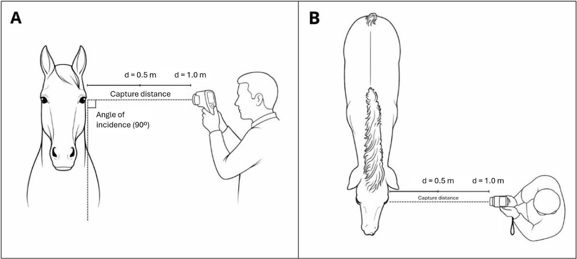
Each ROI was evaluated at two predefined distances of 0.5 m and 1.0 m from the right and left eyes, with the thermographic camera positioned at 90° to the sagittal plane of the animals' eyes for all captures of ocular thermal images (focus on the eyeball) (Figure 2).
Visual diagram of the positioning dynamics in thermographic evaluations. (A) Front view of the horse-thermograph-evaluator positioning; (B) Top view of the horse-thermograph-evaluator positioning. Image generated using Gemini AI.
Distances were measured using a Digital Laser Tape Measure (Mileseey®), directing the laser marker to the lower eyelid, below the eyeball. Thermographic images of the ocular region were captured with a FLIR E40 camera, with a resolution of 160 x 120 pixels, operating in a temperature range of -20 °C to 650 °C, which offered accuracy of ±2 °C or ±2 % in the readings, ensuring reliable results. With a thermal sensitivity of less than 0.07 °C at 30 °C, it detected minimal temperature differences.
Before capturing the images, the FLIR E40 thermograph was calibrated by entering the ambient temperature and relative humidity based on information provided by the weather app (Apple Weather®, iOS) on all days of the experiment, aiming to reduce environmental variables that could influence the ocular temperature result (Table 1). Regarding surface emissivity, it was adjusted to Ɛ = 0.98 in all collections, a suitable value for mammalian skin (4).
Monthly values of ambient temperature (°C) and relative air humidity (%), recorded at the beginning of each collection, carried out between November 2024 and April 2025. Data are expressed as mean ± standard deviation (SD).
Data collection began promptly at 8:30 am. The order of collection was randomly selected. The animals were properly positioned inside the stalls using a halter and a leash to facilitate the capture of thermographic images. Two captures were made for each eye per distance. If there were artifacts, blurs, or partial closure of the eye, new images were taken.
All images were numbered and manually recorded in a spreadsheet containing the date, animal identification, side evaluated, and distance between the camera and the eye. Subsequently, the images were analyzed using IRISoft software (PoliScan®, FLIR Systems Inc., Wilsonville, OR, USA); those with the best technical quality were selected, possessing sharp focus, absence of thermal artifacts, and the eye fully open. All were processed using the same color scale (Rainbow) during the analysis to ensure uniform image presentation. An image of each eye (left and right) per animal was chosen at two distances (0.5 m and 1.0 m), totaling four images per individual. In the software, the ROIs were delimited with a circular selection tool, manually positioned over the regions of the lacrimal caruncle, medial canthus, and lateral canthus of the eye, providing the exact temperature of each area.
The statistical analysis was performed considering three fixed factors in a full factorial model (Table 2) and the temperature variation between months. The months entered the model as a repeated measure over time. The data obtained were compared over time using analysis of variance (ANOVA) in a mixed model, considering the random block (animal) effect. Tukey's test was used for multiple comparisons of means (p < 0.05).
3. Results
All interactions were non-significant; therefore, all factors were analyzed separately. Factor 2 (capture distance) showed a difference in the ocular temperature of the horses, with p < 0.001 (Table 3), where the average observed at 1.0 m (34.5°C) was higher than that recorded at 0.5 m (34.2°C).
There was no difference in Factors 1 and 3 (ocular region and side of the eye) analyzed separately (p = 0.187 and p = 0.112, respectively), as shown in Tables 4 for ocular region and 5 for side of the eye.
The comparison of means of all ocular regions (lateral canthus, medial canthus, and lacrimal caruncle) over 6 months found no significant difference in the overall mean (p = 0.065). However, when evaluated individually per month, the ocular regions showed distinct and significant results (Tables 6, 7, and 8).
Adjusted means of eye temperature (°C), standard deviation (SD), and minimum and maximum temperatures of the lateral canthus of the eyes over the months.
Adjusted means of eye temperature (°C), standard deviation (SD), and minimum and maximum temperatures of the medial canthus of the eyes over the months.
Adjusted means of ocular temperature (°C), standard deviation (SD), and minimum and maximum temperatures of the lacrimal caruncle of the eyes over the months
The month had a significant effect (p = 0.01) on ocular temperature in the lateral canthus. The highest mean was observed in December (35.2 ± 0.98 °C), differing significantly from November (33.7 ± 0.96 °C) and January (33.4 ± 1.69 °C). The means recorded in April (34.2 ± 0.31 °C), February (34.2 ± 0.34 °C), and March (33.9 ± 0.39 °C) were similar, not differing from the other months. The other two ocular regions (medial canthus and lacrimal caruncle) had significant results (Tables 7 and 8).
Regarding the medial canthus (p = 0.005), December maintained the highest mean (35.8 ± 0.78 °C), differing significantly from January (33.4 ± 1.46 °C) and March (32.7 ± 2.09 °C). Intermediate values were found in the means recorded in April (34.4 ± 0.89 °C), February (33.7 ± 1.64 °C), and November (33.7 ± 0.66 °C), without a relevant difference when compared to the other months (Table 7).
As for the lacrimal caruncle, we identified a significant effect of the month (p = 0.002) on ocular temperature, with the highest mean in December (36.1 ± 0.64 °C), differing from November (34.4 ± 0.60 °C), January (33.9 ± 1.34 °C), and March (33.8 ± 1.17 °C), and showing no difference between the means of April (34.7 ± 0.96 °C) and February (34.6 ± 0.68 °C).
4. Discussion
None of the interactions were significant; thus, all three factors were analyzed separately. The capture distance had a significant influence on the collected ocular temperatures. Images obtained at 0.5 m showed lower mean temperatures than those obtained at 1.0 m, a difference that may be associated with how the lens captured and interpreted thermal radiation, being modified according to the framing, focus, and distance of the evaluated area (21,4). Johnson et al. (22) report that distances equal to or greater than 1.0 m can increase the area of interest included in the analysis, influencing the mean temperatures. At shorter distances, such as 0.5 m, there is greater focus on the specific ocular region, reducing the interference of adjacent tissues and possible geometric distortions (1). Thus, rigorous standardization of the capture distance is essential to ensure reproducibility and comparability between studies. Regarding the ocular regions analyzed (lacrimal caruncle, medial canthus, and lateral canthus), when considering the overall means, no significant differences were observed, suggesting similar thermal behavior under controlled handling and environmental conditions (13). This finding indicates that, when the camera positioning and distance are kept constant, the three regions can provide representative readings (4). However, specific differences between months were observed when the regions were analyzed individually, demonstrating that environmental and physiological variations can impact each point differently.
From an anatomical and ophthalmological standpoint, these regions have specific characteristics that can influence their thermal dynamics. The lacrimal caruncle is richly vascularized, has superficial connective tissue with less interference from hair cover, and more evident anatomical delimitation, factors that favor greater stability in thermal capture (4,10). Kim and Cho (1) suggest the medial canthus as a reference due to its correlation with rectal temperature and less variation in response to external changes. The lateral canthus can be influenced by upper eyelid coverage and variations in environmental exposure (1, 11, 14).
As for the side, there was no significant difference between the right and left eyes (23, 24).
Farmer et al. (25) and Krueger et al. (26) report behavioral lateralization in horses, with a possible predominant use of the left eye in assessments with stressful stimuli, associated with activation of the right cerebral hemisphere (responsible for autonomic responses to stress). However, the present study did not observe this effect. The animals were assessed at rest within their respective stalls, under the usual handling and training routine, with the same trainer and handler, and similar housing and feeding, which may have reduced any stimuli capable of triggering differentiated autonomic responses between the sides. Thus, both eyes can be used for methodological purposes, and it is possible to opt for the left side for practical handling reasons (27), provided that sequential assessments maintain consistency.
Data collected over 6 months showed significant monthly variations in eye temperatures in all evaluated regions, with higher values in December. It is recognized that seasonal variations in ambient temperature and relative humidity can influence surface eye temperature (28,24). Studies with a purely methodological objective could concentrate evaluations over shorter periods to reduce environmental interference and isolate the effects of distance, eye region, and side of the eye evaluated in a more controlled manner. However, the present study monitored the horses continuously throughout their entire taming period to reflect practical management conditions. Data were always collected at the beginning of each month, at the same time and before exercise, under standardized environmental and restraint conditions to minimize environmental variability.
The interindividual variation observed throughout the experimental period reinforces the importance of longitudinal designs with repeated measurements, allowing for intraindividual control and greater analytical robustness. Although the number of animals was limited, monthly monitoring for 6 months made it possible to assess consistent thermal patterns within each individual, reducing the influence of biological variability between animals.
In summary, the findings of this study contribute to the better choice of methodology for ocular thermography in equines, demonstrating that the distance from the thermographic camera is the most critical variable for accurate results. Captures at 0.5 m proved to be more stable and less susceptible to anatomical and positional interferences, being recommended for future applications. The lacrimal caruncle stood out among the regions analyzed for its greater temporal consistency and better anatomical definition and can be considered the preferred area for serial evaluations, provided that the acquisition of thermographic images maintains rigorously standardized technical parameters.
Study limitations: This study used the FLIR E40 thermograph, an entry-level piece of equipment for initial thermography studies. Although it is adequate for experimental purposes, devices with higher spatial resolution and thermal sensitivity could provide additional precision in the captures. The assessments were carried out over 6 months with natural environmental variations in temperature and relative humidity. Although equipment calibration recorded and considered these variables, it was not possible to completely eliminate their influence on ocular temperatures. Future studies with assessments concentrated over shorter periods or under more rigorous environmental control could allow for more precise isolation of the effects investigated here.
Additionally, the study did not use independent physiological parameters (plasma cortisol, heart rate, or rectal temperature) for concomitant validation of the thermographic measurements. Thus, the inferences remain restricted to superficial thermal behavior and the methodological analysis of the technique. Finally, the measurements were performed exclusively under basal conditions, prior to training activities, without applying a controlled stress challenge. Therefore, the results should not be extrapolated to contexts of acute stress or intense exercise without further investigation.
5. Conclusion
This study demonstrates that the acquisition distance is the most determining variable in ocular thermographic measurement in horses. The protocol with the most consistent image acquisition for this population involves captures at 0.5 m, with perpendicular positioning in relation to the animal's sagittal plane, for handling convenience, preferably on the left side, and focusing on the lacrimal caruncle as the ROI. This standardization improves physiological sensitivity, reduces environmental interference, and should be adopted in experimental studies and clinical welfare assessments.
Data availability statement
The complete dataset supporting the results of this study is available upon request from the corresponding author.
-
Generative AI Use Statement
During the preparation of this manuscript, the authors used Gemini AI to assist in the creation of Figure 2. After using this tool/service, the authors reviewed and edited the content appropriately, assuming full responsibility for the content of the publication.
Acknowledgments
We would like to thank the National Association for Hippotherapy (ANDE-Brasil) and the Brazilian Association of Quarter Horse Breeders (ABQM) for the support necessary for the development of this project. We also acknowledge Poliscan Brasil for providing the IRISoft software and technical assistance in its use. We are grateful to the Research Support Foundation of the Federal District (FAPDF) for the financial support for this research, and to the Research Support Foundation of the State of Goiás (FAPEG) for granting the scholarship to the first author. We further thank the Graduate Program in Animal Science of the Federal University of Goiás (PPGZ-UFG), the School of Veterinary Medicine and Animal Science of the Federal University of Goiás (EVZ-UFG), and the Center for Studies, Extension and Research in Equine Science of the Federal University of Goiás (NEEPEq-UFG).
References
-
1. Kim SM, Cho GJ. Validation of eye temperature assessed using infrared thermography as an indicator of welfare in horses. Appl Sci. 2021;11(16):7186. Available from: https://doi.org/10.3390/app11167186
» https://doi.org/10.3390/app11167186 -
2. Travain T, Valsecchi P. Infrared thermography in the study of animals' emotional responses: a critical review. Animals (Basel). 2021;11(9):2510. Available from: https://doi.org/10.3390/ani11092510
» https://doi.org/10.3390/ani11092510 -
3. Giannetto C, Cerutti R, Scaglione M, Fazio F, Aragona F, Arfuso F, et al. Simultaneous recording of subcutaneous temperature and total locomotor activity in Bos taurus and Bos indicus raised in a subtropical region of Argentina. Trop Anim Health Prod. 2022;54:371. Available from: https://doi.org/10.1007/s11250-022-03365-7
» https://doi.org/10.1007/s11250-022-03365-7 -
4. Soroko M, Howell K. Infrared thermography: current applications in equine medicine. J Equine Vet Sci. 2018;60:90-96. Available from: https://doi.org/10.1016/j.jevs.2016.11.002
» https://doi.org/10.1016/j.jevs.2016.11.002 -
5. Smith WM. Applications of thermography in veterinary medicine. Ann N Y Acad Sci. 1964;121(1):248-254. Available from: https://doi.org/10.1111/j.1749-6632.1964.tb13700.x
» https://doi.org/10.1111/j.1749-6632.1964.tb13700.x -
6. Lampang KN, Isawirodom A, Rungsri P. Correlation and agreement between infrared thermography and a thermometer for equine body temperature measurements. Vet World. 2023;16(12):2464-2470. Available from: https://doi.org/10.14202/vetworld.2023.2464-2470
» https://doi.org/10.14202/vetworld.2023.2464-2470 -
7. Rankins EM, Reega SJ, Rudd C, Peters BC. Horse behavior and training in adaptive/therapeutic riding: a survey of instructors’ perceptions and knowledge in the United States. Appl Anim Behav Sci. 2025;282:106490. Available from: https://doi.org/10.1016/j.applanim.2024.106490
» https://doi.org/10.1016/j.applanim.2024.106490 -
8. Gulias-Cañizo R, Rodríguez-Malagón ME, Botello-González L, Belden-Reyes V, Amparo F, Garza-Leon M. Applications of infrared thermography in ophthalmology. Life (Basel). 2023;13(3):723. Available from: https://doi.org/10.3390/life13030723
» https://doi.org/10.3390/life13030723 -
9. Redaelli V, Luzi F, Mazzola S, et al. The use of infrared thermography (IRT) as stress indicator in horses trained for endurance: a pilot study. Animals (Basel). 2019;9(3):84. Available from: https://doi.org/10.3390/ani9030084
» https://doi.org/10.3390/ani9030084 -
10. Ijichi C, Evans L, Woods H, Yarnell K. The right angle: validating a standardised protocol for the use of infra-red thermography of eye temperature as a welfare indicator. Anim Welf. 2020;29(2):123-131. Available from: https://doi.org/10.7120/09627286.29.2.123
» https://doi.org/10.7120/09627286.29.2.123 -
11. Stewart M, Schaefer AL, Haley D, et al. Infrared thermography as a non-invasive method for detecting fear-related responses of cattle to handling procedures. Anim Welf. 2008;17:387-393. Available from: https://doi.org/10.1017/S0962728600027895
» https://doi.org/10.1017/S0962728600027895 -
12. Aragona F, Di Pietro S, Arfuso F, Fazio F, Piccione G, Giudice E, et al. Correlation between ocular and rectal temperature with intraocular pressure in horse during exercise. Animals (Basel). 2022;12:1850. Available from: https://doi.org/10.3390/ani12141850
» https://doi.org/10.3390/ani12141850 -
13. Valera M, Bartolomé E, Sánchez-Guerrero M, Molina-Alcalá A, Cook N, Schaefer A. Changes in eye temperature and stress assessment in horses during show jumping competitions. J Equine Vet Sci. 2012;32:827-830. Available from: https://doi.org/10.1016/j.jevs.2012.03.005
» https://doi.org/10.1016/j.jevs.2012.03.005 -
14. Aragona F, Rizzo M, Arfuso F, Acri G, Fazio F, Piccione G, et al. Eye temperature measured with infrared thermography to assess stress responses to road transport in horses. Animals (Basel). 2024;14(13):1877. Available from: https://doi.org/10.3390/ani14131877
» https://doi.org/10.3390/ani14131877 -
15. Carastro SM. Equine ocular anatomy and ophthalmic examination. Vet Clin North Am Equine Pract. 2004;20(2):285-299. Available from: https://doi.org/10.1016/j.cveq.2004.04.013
» https://doi.org/10.1016/j.cveq.2004.04.013 -
16. McMullen RJ. Advancements in equine ophthalmic imaging enhance understanding of ocular and orbital anatomy and disease in standing sedated horses. J Am Vet Med Assoc. 2024;262(S2):S47-S56. Available from: https://doi.org/10.2460/javma.24.06.0376
» https://doi.org/10.2460/javma.24.06.0376 - 17. Gelatt KN, Ben-Shlomo G, Gilger BC, Hendrix DVH, Kern TJ, Plummer CE. Veterinary ophthalmology. 6th ed. Hoboken: Wiley-Blackwell; 2021. English.
-
18. Ollivier FJ. The precorneal tear film in horses: its importance and disorders. Vet Clin North Am Equine Pract. 2004;20(2):301-318. Available from: https://doi.org/10.1016/j.cveq.2004.04.008
» https://doi.org/10.1016/j.cveq.2004.04.008 - 19. Brooks DE. Oftalmologia para veterinários de equinos. São Paulo: Roca; 2005. Português.
-
20. Dwyer AE, de Linde Henriksen M. Examination techniques and treatment of the equine eye. In: Gilger BC. Equine ophthalmology. 4th ed. Ames: Wiley-Blackwell; 2022. p. 1-89. English. Available from: https://doi.org/10.1002/9781119782285.ch1
» https://doi.org/10.1002/9781119782285.ch1 -
21. McManus C, Tanure CB, Peripolli V, Seixas L, Fischer V, Gabbi AM, et al. Infrared thermography in animal production: an overview. Comput Electron Agric. 2016;123:10-16. Available from: https://doi.org/10.1016/j.compag.2016.01.027
» https://doi.org/10.1016/j.compag.2016.01.027 -
22. Johnson SR, Rao S, Hussey SB, Morley PS, Traub-Dargatz JL. Thermographic eye temperature as an index to body temperature in ponies. J Equine Vet Sci. 2011;31(1):63-66. Available from: https://doi.org/10.1016/j.jevs.2010.12.004
» https://doi.org/10.1016/j.jevs.2010.12.004 -
23. Hall C, Kay R, Yarnell K. Assessing ridden horse behavior: professional judgment and physiological measures. J Vet Behav. 2014;9(1):22-29. Available from: https://doi.org/10.1016/j.jveb.2013.09.005
» https://doi.org/10.1016/j.jveb.2013.09.005 -
24. Jansson A, Lindgren G, Velie BD, Solé M. An investigation into factors influencing basal eye temperature in the domestic horse (Equus caballus) when measured using infrared thermography in field conditions. Physiol Behav. 2021;228:113218. Available from: https://doi.org/10.1016/j.physbeh.2020.113218
» https://doi.org/10.1016/j.physbeh.2020.113218 -
25. Farmer K, Krueger K, Byrne RW. Visual laterality in the domestic horse (Equus caballus) interacting with humans. Anim Cogn. 2010;13(2):229-238. Available from: https://doi.org/10.1007/s10071-009-0260-x
» https://doi.org/10.1007/s10071-009-0260-x -
26. Krueger K, Schwarz S, Marr I, Farmer K. Laterality in horse training: psychological and physical balance and coordination and strength rather than straightness. Animals (Basel). 2022;12(8):1042. Available from: https://doi.org/10.3390/ani12081042
» https://doi.org/10.3390/ani12081042 -
27. Baragli P, Scopa C, Felici M, Reddon AR. Horses show individual level lateralisation when inspecting an unfamiliar and unexpected stimulus. PLoS One. 2021;16(8):e0255688. Available from: https://doi.org/10.1371/journal.pone.0255688
» https://doi.org/10.1371/journal.pone.0255688 -
28. Church JS, Hegadoren PR, Paetkau MJ, Miller CC, Regev-Shoshani G, Schaefer AL, et al. Influence of environmental factors on infrared eye temperature measurements in cattle. Res Vet Sci. 2014;96(1):220-226. Available from: https://doi.org/10.1016/j.rvsc.2013.11.006
» https://doi.org/10.1016/j.rvsc.2013.11.006
Edited by
-
Editor:
Luiz Augusto B. Brito