 An exploratory Mendelian randomization study on the genetically predicted effects of circulating blood cells on osteoarthritis risk
Abstract
Background The causal relationship between circulating blood cells and Osteoarthritis (OA) remains unclear. This study aimed to explore this potential relationship using a two-sample Mendelian Randomization (MR) approach.
Methods Data from large-scale GWAS (Blood Cell Consortium and GWAS Catalog) were analyzed using two-sample MR methods, with Inverse Variance Weighting as the primary approach. A Bonferroni correction was applied for multiple testing for the two primary traits. Robustness was assessed through comprehensive sensitivity analyses for heterogeneity and pleiotropy.
Results After Bonferroni correction, Genetically Predicted Hemoglobin (HGB) showed a weak genetic association consistent with a potential causal pathway for OA risk (OR = 1.004, 95% CI 1.001-1.007, p = 0.018). The association for Neutrophil (NEU) count was not statistically significant. While sensitivity analyses did not detect directional pleiotropy, significant heterogeneity was observed for the genetic instruments.
Conclusion This exploratory study provides preliminary genetic evidence for a statistically significant but clinically negligible MR estimate of hemoglobin in OA pathogenesis. The clinical and biological significance of this minuscule effect remains uncertain. The findings should be interpreted as hypothesis-generating, highlighting the need for further research rather than establishing a definitive causal link.
Keywords
Mendelian randomization; Circulating blood cells; Osteoarthritis; GWAS
Introduction
Osteoarthritis (OA) is a prevalent musculoskeletal disorder and a leading cause of disability worldwide.1 It is characterized by the degeneration of articular cartilage and underlying bone, resulting in pain, stiffness, and functional impairment.2 OA imposes a substantial burden globally, impacting both the physiological and psychological health of patients and incurring significant healthcare costs.3 Risk factors for OA include genetic predisposition, age, obesity, and trauma.4-7 However, its etiology remains incompletely understood. Therefore, identifying potential and modifiable risk factors is crucial for elucidating the pathogenesis of OA and is essential for reducing its incidence rates.
In OA research, circulating blood cells and systemic inflammatory mediators play a critical role in the disease's pathogenesis. Numerous studies have demonstrated that various immune cells, including leukocytes (particularly monocytes), macrophages, T-cells, and platelets, are integral to the chronic inflammatory response, bone resorption, and cartilage degradation associated with OA.8,9 Monocytes exhibit altered inflammatory responses in OA patients,8 while macrophages mediate the production of cytokines and Matrix Metalloproteinases (MMPs) that drive joint degeneration.10 Furthermore, specific T-cell subsets, such as pro-inflammatory Th17 and Th9 cells, are elevated in OA patients and contribute to cartilage destruction through T-cell-secreted-cytokine-mediated pathways.11-13 These studies underscore the complex involvement of immune cells in the chronic inflammation seen in OA.
While current studies provide important insights, the present study specifically focuses on widely measured and standardized erythrocyte and leukocyte parameters (hemoglobin and neutrophils). This focus is rationalized because these parameters have strong genetic instruments available in large-scale GWAS, represent fundamental physiological systems (oxygen transport and innate immunity), and have been implicated in systemic inflammation, a known contributor to OA. It is acknowledged that previous Mendelian randomization studies have investigated the role of iron status (which influences hemoglobin).14,15 and broader categories of immune cells in OA.9 This study aims to build upon this by examining these specific blood cell traits. Most existing research relies on observational designs, which are susceptible to confounding factors and reverse causality biases, leading to inconsistent conclusions.13 For example, the role of platelets in OA is complex; they are implicated in OA inflammation but are also used as a basis for therapeutic strategies.16 These inconsistencies highlight the need for new research approaches to clarify the complex relationship between circulating blood cells and OA.
To overcome these limitations, Mendelian Randomization (MR) analysis, a causal inference method, offers a powerful tool. MR uses genetic variations as Instrumental Variables (IVs) to mitigate bias from confounding factors and reverse causality.17,18 It has been successfully applied to investigate various health outcomes.19,20 By using MR, the authors can obtain more reliable evidence on the potential causal role of exposures like circulating blood cells in OA. However, MR relies on key assumptions that must be carefully evaluated and interpreted within its inherent limitations. This study was conducted and reported following the principles outlined in the STROBE-MR guidelines.21
In this study, the authors employ MR to evaluate the potential causal relationship between specific circulating blood cell traits and OA. The authors aim to provide additional insights into the role of these blood cells in the pathogenesis of OA, thereby contributing to the ongoing effort to identify novel prevention and treatment strategies.
Materials and methods
Research design
This study utilizes a two-sample MR approach to assess the causal relationship between circulating blood cells and OA. MR studies must satisfy three essential assumptions: 1) Genetic variants should be significantly associated with the exposure (relevance assumption); 2) The genetic variants used as instruments should be independent of any potential confounders (independence assumption); 3) The genetic variants should influence the outcome solely through their effect on the exposure (exclusion restriction assumption). Fig. 1 depicts the overall MR study process.

Data sources
Data for this study were sourced from publicly available GWAS databases. Genetic variants related to circulating blood cells were obtained from the Blood Cell Consortium (BCX), encompassing data on red blood cells, white blood cells, and platelets.22 The authors analyzed data from European populations, with further details available at Chen et al., Cell 2020.22 Genetic data related to osteoarthritis were retrieved from the GWAS Catalog (Dataset: ebi-a-GCST90038686), which includes 484,598 participants (39,515 cases and 445,083 controls). Specific research data can be found on the website. It is important to note that all GWAS data used are based on European populations, which limits the generalizability of these findings.
Instrumental variables (IVs) selection
To ensure the validity of the MR analysis, the authors implemented a rigorous SNP selection process. First, the authors extracted SNPs related to blood cell types from the GWAS data at a genome-wide significance threshold (p < 5 × 10⁻⁸). Next, to address Linkage Disequilibrium (LD), the authors employed the “clump_data” function with parameters set to κb = 10,000 and r2 = 0.001. Furthermore, the authors validated the selected SNPs against potential confounding factors using the Phenoscanner database (www.phenoscanner.medschl.cam.ac.uk).23 SNPs found to be strongly associated with known OA risk factors independent of the blood cell trait were considered for exclusion. During data coordination, the authors excluded SNPs with inconsistent allele frequencies or those exhibiting palindromic structures. Finally, the authors calculated the F-statistic for each SNP to assess instrument strength, retaining only those with an F-statistic greater than 10 to minimize weak instrument bias.
MR analysis
The authors employed multiple MR analysis methods, including Inverse Variance Weighting (IVW), MR-Egger regression, Weighted Median, Simple Model, and Weighted Model. IVW was selected as the primary analysis method due to its high statistical power, assuming no unbalanced horizontal pleiotropy. To complement the IVW analysis, the authors utilized MR-Egger regression to assess directional horizontal pleiotropy, and the Weighted Median and other models as sensitivity analyses to test the consistency of the results under different assumptions. In this study, the authors focused on two primary blood cell traits (HGB and NEU) to represent the erythroid and myeloid lineages, respectively. Consequently, the authors applied a Bonferroni correction for these two primary tests, setting the significance threshold at p < 0.025 (0.05/2).
Sensitivity analysis
To validate the robustness of the MR analysis, several sensitivity analyses were conducted. Cochran's Q-test evaluated heterogeneity among SNPs. The MR-Egger regression intercept was used to examine directional horizontal pleiotropy. The MR-PRESSO method was used to identify and correct for horizontal pleiotropic outliers. Additionally, the authors applied the Leave-One-Out (LOO) analysis to check whether any individual SNP disproportionately influenced the causal estimates. Finally, the authors utilized Steiger filtering to confirm the causal direction between the exposure and the outcome. All analyses were performed using R software (version 4.1.0) with the “TwoSampleMR” package.
Results
Selection of IVs
The authors implemented a rigorous selection process for genetic IVs (p < 5 × 10⁻⁸). For the HGB analysis, 454 SNPs were used, and for the NEU analysis, 415 SNPs were used. Overall, F-values for the selected instruments ranged from 29.76 to 6218.95, and the variance explained (R2) ranged from 0.01% to 1.27%. The mean F-statistic was 105.4 for HGB and 98.7 for NEU, suggesting a robust instrument strength overall. Summary information for the IVs is available in Supplementary Table 1.
MR analysis
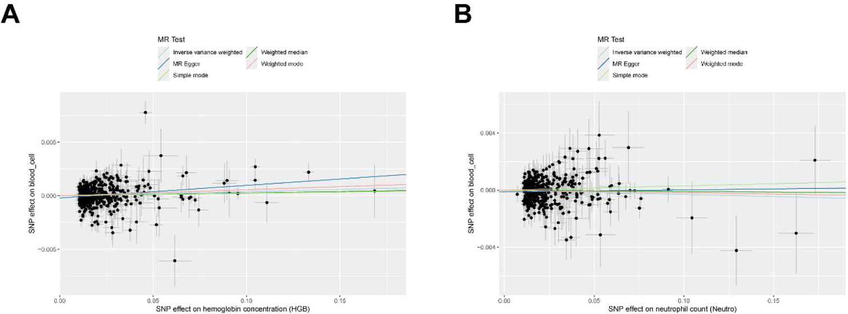
The two-sample MR analysis results are presented in Supplementary Table 2. After applying the Bonferroni correction (significance threshold p < 0.025), a genetically predicted increase in Hemoglobin (HGB) levels was causally linked to a slightly increased risk of OA (OR = 1.004, 95% CI 1.001-1.007, p = 0.018). The observed inverse association for Neutrophil (NEU) levels was not statistically significant after correction (OR = 0.997, 95% CI 0.994-1.000, p = 0.049, Bonferroni-corrected p > 0.025) (Fig. 2). The directional consistency across the other four MR methods (MR Egger, Weighted median, Simple mode, Weighted mode) provided some support for the findings, though statistical significance varied across methods (Fig. 3, Supplementary Fig. 1).
Global heterogeneity test using Mendelian Randomization (MR) of Circulating Blood Cells and Osteoarthritis (MR-PRESSO).

A forest plot showing the causal relationships between HGB, NEU, and osteoarthritis risk from the primary IVW analysis. The association for HGB is statistically significant after Bonferroni correction (p < 0.025), while the association for NEU is not.
Sensitivity analysis
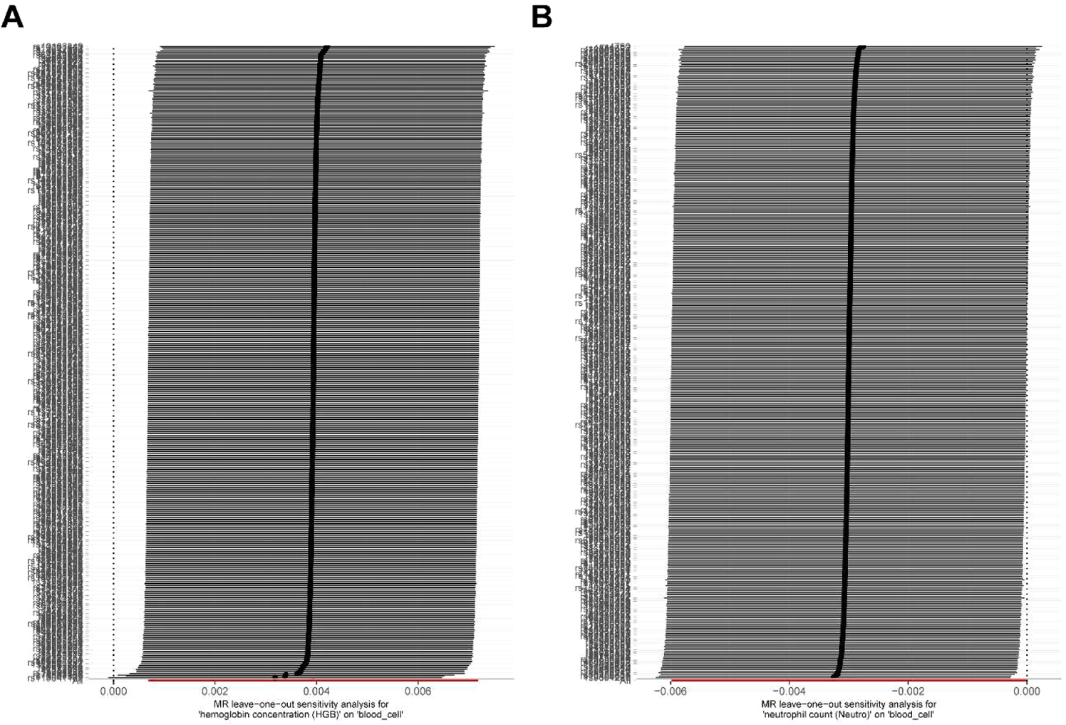
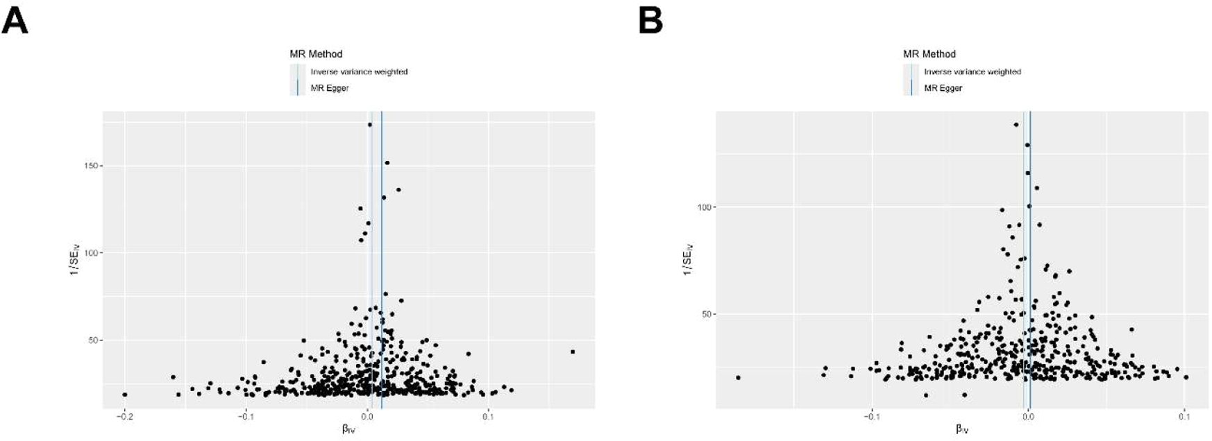
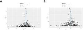
To validate the reliability of the MR analyses, the authors assessed both pleiotropy and heterogeneity. The MR-Egger intercept did not indicate significant directional horizontal pleiotropic effects for HGB (p-intercept = 0.052) or NEU (p-intercept = 0.107) (Table 1). Similarly, the MR-PRESSO global test found no evidence of significant outlier-driven pleiotropy (Table 2). However, the Cochran's Q-statistic from the IVW method showed significant heterogeneity for both HGB (Q p-value < 0.001) and NEU (Q p-value < 0.001) (Table 3). This indicates variability in the SNP-specific estimates. The Leave-One-Out (LOO) analysis revealed that the exclusion of any individual SNP did not substantially alter the causal estimates, confirming the stability of the results (Fig. 4). The Steiger filtering test confirmed the assumed causal direction from blood cell traits to OA (Table 4). Funnel plots for HGB and NEU showed some asymmetry, consistent with the heterogeneity detected by the Cochran's Q-test (Fig. 5).

Discussion
This study employed Mendelian randomization to explore the causal effects of circulating blood cell traits on OA risk in European populations. This analysis identified a statistically significant but clinically minuscule causal association between genetically predicted HGB levels and an increased risk of OA. In contrast, the association for NEU levels did not withstand correction for multiple testing. The odds ratio for HGB (OR=1.004) indicates a mere 0.4% increase in OA risk per unit increase in genetically predicted HGB, an effect size dwarfed by established risk factors like obesity.24 and unlikely to be meaningful for individual risk prediction. It is important to interpret these findings in the context of existing literature; the link between iron status (related to HGB) and OA has been explored in previous MR studies.14,15 and this finding for HGB is consistent with reports of higher iron levels associated with a small increased OA risk. The present study thus adds to this body of evidence but does not represent a transformative discovery.
The finding that higher HGB is a risk factor for OA is biologically plausible. Hemoglobin, the key oxygen-transporting protein in red blood cells.25 is intrinsically linked to iron metabolism. Elevated systemic iron levels, potentially reflected by higher HGB, have been causally associated with increased OA risk in other MR studies.14,15 The proposed mechanism involves iron-induced oxidative stress, which can promote chondrocyte senescence and cartilage degradation.26,27 Excess iron can also lead to ferroptosis in chondrocytes, contributing to joint damage.28 Previous research also found that elevated glycated Hemoglobin (HbA1c) levels are causally associated with an increased risk of knee OA.29 Therefore, these finding aligns with established biological pathways linking iron overload and metabolic dysfunction to OA pathogenesis. Given the tiny effect size, it is also possible that HGB acts as a proxy for other correlated factors such as altitude exposure, cardiorespiratory fitness, or smoking status, rather than exerting a direct biological effect.30,31
The initial, uncorrected finding of a protective association between higher neutrophil levels and OA risk (p = 0.049) was not robust to multiple testing correction. This result is also biologically counterintuitive and contradicts a substantial body of literature. Neutrophils are key mediators of acute inflammation and are implicated in the chronic inflammatory processes of OA. They contribute to cartilage degradation through the release of enzymes like neutrophil elastase and the formation of Neutrophil Extracellular Traps (NETs) within the joint.32,33 The speculative hypothesis that systemic neutrophil levels might be protective via anti-inflammatory Extracellular Vesicles (EVs).34 is not sufficient to overturn this well-established pro-inflammatory paradigm in OA. A more parsimonious explanation for the initial borderline association is that it represents a false positive (Type I error), residual confounding, or complex horizontal pleiotropy, where the genetic variants for neutrophil count influence OA through pathways unrelated to neutrophil function itself.
Limitations and methodological considerations
This study has several important limitations. First, these findings of statistically significant but minuscule genetic effects must be interpreted with extreme caution. The clinical utility of these associations for risk prediction or therapeutic development is likely negligible in their current form. Their primary value is in generating hypotheses about underlying biological pathways.
Second, the significant heterogeneity detected by the Cochran's Q-test (p < 0.001 for both HGB and NEU) is a major concern. Such profound heterogeneity suggests that the genetic instruments are not acting with a consistent effect on the outcome, which challenges the validity of the primary IVW estimate. While the sensitivity analyses did not detect directional pleiotropy, the presence of widespread, balanced pleiotropy cannot be excluded and may be driving the observed heterogeneity. However, it is crucial to note that while the IVW method yielded a significant result, the MR-PRESSO outlier-corrected estimate (Table 2) did not reach statistical significance (p = 0.075), suggesting that the IVW result may be driven by horizontal pleiotropy or outliers. This discrepancy, combined with the extreme heterogeneity, indicates that the IVW estimate may not be a valid causal estimate and should be interpreted with extreme caution.
Third, the present study is based exclusively on data from individuals of European ancestry. This significantly limits the generalizability of these findings to other populations. Genetic architecture and gene-environment interactions can vary across different ancestries, and these results may not be representative of the global OA burden.35 Future replication in large, diverse cohorts is essential to validate these findings.
Fourth, while the authors attempted to address multiple comparisons by applying a Bonferroni correction, this exploratory study scanned multiple blood cell traits initially. The borderline significance of the main findings highlights the risk of false positives inherent in such an approach. Finally, MR studies assess the lifelong effect of genetically predicted exposure levels, which may not correspond to the effects of interventions that modify blood cell counts later in life. Although the mean F-statistics were high, the wide range indicates some instruments were weaker, which, combined with the tiny effect size, could introduce weak instrument bias. Finally, due to the widespread heterogeneity and likely pleiotropy, a formal colocalization analysis was not performed as the assumption of a single causal variant was likely violated.
In conclusion, this exploratory MR study provides weak evidence for a potential causal link between genetically elevated HGB and a very small increase in OA risk. The finding for neutrophils was not statistically significant after appropriate correction. Given the tiny effect size, unresolved heterogeneity, and other limitations, these findings should be considered preliminary and hypothesis-generating.
Conclusion
This Mendelian Randomization study explored the genetically predicted effects of selected blood cell traits on osteoarthritis risk in European-ancestry individuals. This analysis found a statistically significant but minuscule genetic association between higher Hemoglobin (HGB) levels and increased OA risk, while the association for Neutrophil (NEU) counts was not significant after correction for multiple testing. The biological and clinical relevance of the HGB finding remains uncertain due to its negligible effect size. The study’s conclusions are constrained by significant heterogeneity and a lack of generalizability beyond European populations. Therefore, these results should be interpreted as exploratory and are insufficient for making any clinical inferences. Future research with more diverse datasets and functional studies is required to clarify the potential role, if any, of hematological parameters in OA pathogenesis.
Data availability
This study analyzed publicly available datasets. Genetic association data for circulating blood cells were obtained from the Blood Cell Consortium (BCX) as described by Chen et al.22 Genetic association data for osteoarthritis were obtained from the GWAS Catalog (Dataset: ebi-a-GCST90038686; https://www.ebi.ac.uk/gwas/studies/GCST90038686). Original data sources are cited in the manuscript.
Clinical trial number
Not applicable.
Ethics approval and consent to participate
Ethical approval was not required for this study, as all data were sourced from publicly accessible summary-level GWAS databases. The original studies that generated these data obtained appropriate ethical approval and participant consent.
Consent for publication
Not applicable.
-
Funding
No funding was received for this study.
Acknowledgements
The authors thank the investigators and participants of the Blood Cell Consortium (BCX) and the UK Biobank and Arthritis Research UK Osteoarthritis Genetics (arcOGEN) consortia for making their summary statistics publicly available.
Supplementary materials
Supplementary material associated with this article can be found, in the online version, at doi:10.1016/j.clinsp.2026.100959.
References
- 1 Litwic A., Edwards M.H., Dennison E.M., Cooper C. Epidemiology and burden of osteoarthritis. Br Med Bull. 2013:105:185-99.
- 2 Sacitharan P.K. Ageing and osteoarthritis. Subcell Biochem. 2019:91:123-159.
- 3 Motta F., Barone E., Sica A., Selmi C. Inflammaging and osteoarthritis. Clin Rev Allergy Immunol. 2023;64(2):222-38.
- 4 Abramoff B., Caldera F.E. Osteoarthritis: pathology, diagnosis, and treatment options. Med Clin North Am. 2020;104(2):293-311.
- 5 Neogi T., Zhang Y. Epidemiology of osteoarthritis. Rheum Dis Clin North Am. 2013;39(1):1-19.
- 6 Lawrence R.C., Felson D.T., Helmick C.G., Arnold L.M., Choi H., Deyo R.A., et al. Estimates of the prevalence of arthritis and other rheumatic conditions in the United States. Part II. Arthritis Rheum. 2008;58(1):26-35.
- 7 Reyes C., Leyland K.M., Peat G., Cooper C., Arden N.K., Prieto-Alhambra D. Association between overweight and obesity and risk of clinically diagnosed knee, hip, and hand osteoarthritis: a population-based cohort study. Arthritis Rheum (Hob NJ). 2016;68(8):1869-75.
- 8 Guillem-Llobat P., Marín M., Rouleau M., Silvestre A., Blin-Wakkach C., Ferrándiz M.L., et al. New insights into the pro-inflammatory and osteoclastogenic profile of circulating monocytes in osteoarthritis patients. Int J Mol Sci. 2024;25(3):1710.
- 9 Xu C., Wang S., Chen X., Zhang T., Ni Z., Ji G., et al. Causal associations between circulating immune cells and osteoarthritis: a bidirectional mendelian randomization study. Int Immunopharmacol. 2024;142(Pt A):113156.
- 10 Griffin T.M., Scanzello C.R. Innate inflammation and synovial macrophages in osteoarthritis pathophysiology. Clin Exp Rheumatol. 2019;37 Suppl 120(5):57-63.
- 11 Li Y.S., Luo W., Zhu S.A., Lei G.H. T cells in osteoarthritis: alterations and beyond. Front Immunol 2017;8:356.
- 12 Akhter S., Tasnim F.M., Islam M.N., Rauf A., Mitra S., Emran T.B., et al. Role of Th17 and IL-17 cytokines on inflammatory and auto-immune diseases. Curr Pharmac Des. 2023;29(26):2078-90.
- 13 Qi C., Shan Y., Wang J., Ding F., Zhao D., Yang T., et al. Circulating T helper 9 cells and increased serum interleukin-9 levels in patients with knee osteoarthritis. Clin Exp Pharmacol Physiol. 2016;43(5):528-34.
- 14 Xu J., Zhang S., Tian Y., Si H., Zeng Y., Wu Y., et al. Genetic Causal Association between iron status and osteoarthritis: a two-sample mendelian randomization. Nutrients. 2022;14(18):3683.
- 15 Ruan G., Ying Y., Lu S., Zhu Z., Chen S., Zeng M., et al. The effect of systemic iron status on osteoarthritis: a mendelian randomization study. Front Genet. 2023;14:1122955.
- 16 Tramś E., Malesa K., Pomianowski S., Kamiński R. Role of platelets in osteoarthritis-updated systematic review and meta-analysis on the Role of platelet-rich plasma in osteoarthritis. Cells. 2022;11(7):1080.
- 17 Davey Smith G., Hemani G. Mendelian randomization: genetic anchors for causal inference in epidemiological studies. Hum Mol Genet. 2014;23(R1):R89-98.
- 18 Burgess S., Butterworth A., Thompson S.G. Mendelian randomization analysis with multiple genetic variants using summarized data. Genet Epidemiol. 2013;37(7):658-65.
- 19 Larsson S.C., Butterworth A.S., Burgess S. Mendelian randomization for cardiovascular diseases: principles and applications. Eur Heart J. 2023;44(47):4913-24.
- 20 Zhu J., Zhou D., Wang J., Yang Y., Chen D., He F., et al. Frailty and cardiometabolic diseases: a bidirectional mendelian randomisation study. Age Ageing. 2022;51(11):afac256.
- 21 Skrivankova V.W., Richmond R.C., Woolf B.A.R., Yarmolinsky J., Davies N.M., Swanson S.A., et al. Strengthening the reporting of observational studies in epidemiology using mendelian randomization: the STROBE-MR Statement. JAMA. 2021;326(16):1614-21.
- 22 Chen M.H., Raffield L.M., Mousas A., Sakaue S., Huffman J.E., Moscati A., et al. Trans-ethnic and ancestry-specific blood-cell genetics in 746,667 individuals from 5 global populations. Cell. 2020;182(5):1198-1213.e1114.
- 23 Kamat M.A., Blackshaw J.A., Young R., Surendran P., Burgess S., Danesh J., et al. PhenoScanner V2: an expanded tool for searching human genotype-phenotype associations. Bioinformatics. 2019;35(22):4851-3.
- 24 Zheng H., Chen C. Body mass index and risk of knee osteoarthritis: systematic review and meta-analysis of prospective studies. BMJ Open. 2015;5(12):e007568.
- 25 Mansouri A. Methemoglobinemia. Am J Med Sci. 1985;289(5):200-9.
- 26 Yudoh K., Nguyen V.T., Nakamura H., Hongo-Masuko K., Kato T., Nishioka K. Potential involvement of oxidative stress in cartilage senescence and development of osteoarthritis: oxidative stress induces chondrocyte telomere instability and downregulation of chondrocyte function. Arthritis Res Ther. 2005;7(2):R380-391.
- 27 Henrotin Y., Kurz B., Aigner T. Oxygen and reactive oxygen species in cartilage degradation: friends or foes? Osteoarthr Cartil. 2005;13(8):643-54.
- 28 He Q., Yang J., Pan Z., Zhang G., Chen B., Li S., et al. Biochanin a protects against iron overload associated knee osteoarthritis via regulating iron levels and NRF2/system xc-/GPX4 axis. Biomed Pharmacother. 2023;157:113915.
- 29 Chen L., Jia C., Yang H. Causal effect of higher glycated hemoglobin (HbA1c) levels on knee osteoarthritis risk: a mendelian randomization study. Rheumatol Ther. 2023;10(1):239-247.
- 30 Liu Q., You J., Zhong M., Wu Z., Geng Y., Huang C. Hemoglobin level is negatively associated with sarcopenia and its components in Chinese aged 60 and above. Front Public Health. 2023;11:1081843.
- 31 Gassmann N.N., Matthes K.L., Eppenberger P., Brabec M., Panczak R., Zwahlen M., et al. Residential altitude associates with endurance but not muscle power in young Swiss men. Front Physiol. 2020;11:860.
- 32 Chaney S., Vergara R., Qiryaqoz Z., Suggs K., Akkouch A. The involvement of neutrophils in the pathophysiology and treatment of osteoarthritis. Biomedicines. 2022;10(7):1604.
- 33 Carmona-Rivera C., Carlucci P.M., Goel R.R., James E., Brooks S.R., Rims C., et al. Neutrophil extracellular traps mediate articular cartilage damage and enhance cartilage component immunogenicity in rheumatoid arthritis. JCI Insight. 2020;5(13):e139388.
- 34 Thomas B.L., Montero-Melendez T., Oggero S., Kaneva M.K., Chambers D., Pinto A.L., et al. Molecular determinants of neutrophil extracellular vesicles that drive cartilage regeneration in inflammatory arthritis. Arthritis Rheumatol (Hob NJ). 2024;76(12):1705-18.
- 35 Martin A.R., Kanai M. Clinical use of current polygenic risk scores may exacerbate health disparities. Nat Genet. 2019;51(4):584-91.
Edited by
-
Edited by Editor:
José Maria Soares Junior