 Speech profile in patients with Huntington's Disease: cognitive, clinical, and sociodemographic correlations
ABSTRACT
Purpose To describe speech profiles in individuals with Huntington's Disease (HD), correlate them with cognitive and clinical aspects, and compare them with healthy controls.
Methods Symptomatic individuals with a clinical and molecular diagnosis of HD were included. Seven healthy controls, matched by age and sex, were also included. Clinical and sociodemographic data were obtained from medical records. The Unified Huntington's Disease Rating Scale was used to measure severity. Cognitive data were collected using verbal fluency, symbol digit modalities, and Stroop tests. Auditory perceptual assessments were used to evaluate speech, and acoustic analysis extracted information about the following tasks: sustained vowel /a/, utterances with different intonations, oral diadochokinesis, spontaneous speech, and the repeated diphthong /ju:/.
Results Of the seven individuals with HD, four women with a mean age of 48.86 (±16.03), presented severe (57.15%), moderate (28.57%), and mild (14.28%) dysarthria. Speech impairment in HD case subjects was related to overall motor decline; the worse the motor symptoms, the worse the speech impairment. There was no correlation with the other clinical data or cognition. The case subjects were significantly worse than the control group, specifically regarding the subsystems of phonation (fundamental frequency, phonation time, local jitter, local shimmer), respiration (maximum phonation time) and articulation (speech rate, phonation time in spontaneous speech, number of syllables in spontaneous speech, average duration of syllables and duration of spontaneous speech).
Conclusion In HD subjects, the most affected speech subsystems were articulation, phonation, and respiration. Poor motor speech patterns were associated with overall motor decline. Speech assessments may provide biomarkers that predict HD progression.
Keywords:
Huntington's Disease; Speech; Dysarthria; Cognition; Acoustic Analysis
RESUMO
Objetivo Descrever o perfil de fala na Doença de Huntington (DH), correlacionar com aspectos cognitivos e clínicos, e comparar com controles.
Método Foram incluídos indivíduos sintomáticos, com diagnóstico clínico e molecular de DH e controles. Foram obtidos dados clínicos e sociodemográficos. A gravidade foi coletada pela Unified Huntington's Disease Rating Scale (UHDRS). A cognição foi avaliada pelos testes: fluência verbal, dígitos e stroop. A avaliação de fala foi feita por julgamento perceptivo auditivo e análise acústica.
Resultados Foram incluídos 7 indivíduos com DH, sendo 4 mulheres, com idade média de 48,86 (±16,03). Destes, 57,15% apresentaram disartria grave, 28,57% moderada e 14,28% leve. Sete controles saudáveis, pareados por sexo e idade, participaram do estudo. As alterações de fala dos indivíduos com DH estão relacionadas com a evolução dos sintomas motores, quanto piores os sintomas motores, pior o desempenho na fala. Com os demais dados clínicos, não houve correlação. Os indivíduos com DH foram significativamente piores comparados ao grupo controle nos subsistemas da fonação (frequência fundamental, tempo de fonação, jitter local, shimmer local), respiração (tempo máximo de fonação) e articulação (speech rate, tempo de fonação na fala espontânea, número de sílabas na fala espontânea, média de duração das sílabas e na duração da fala espontânea).
Conclusão Os subsistemas da fala mais afetados foram articulação, fonação e respiração. O perfil de fala está relacionado à progressão dos sintomas motores. A avaliação da fala tem a possibilidade de se configurar como um preditor da progressão da DH.
Descritores:
Doença de Huntingon; Fala; Disartria; Cognição; Análise Acústica da Fala
INTRODUCTION
Huntington's disease (HD) is an autosomal dominant neurodegenerative condition characterized by neuropsychiatric and behavioral symptoms(1). In Brazil, the national prevalence of HD is unknown. However, in the state of Rio Grande do Sul, research has revealed a minimum prevalence of 1.85/100,000(2), which is lower than in European countries but similar to other Latin American countries(3,4).
This disorder is caused by CAG (cytosine-adenine-guanine) repeat expansions in exon 1 of the HTT gene, located on the short arm of chromosome 4 (4p16.3)(5). In normal individuals, CAG repeats range from 10 to 35; however, HD patients may have an allele with 36 to 60 CAG repeats. Since HD is inherited in an autosomal dominant pattern, one allele with CAG repeat expansions is sufficient to cause the disease(6). The age of onset varies and depends largely on the number of CAG repeats(7). Affected individuals become symptomatic, on average, at 35 - 44 years of age(8).
As the disease progresses, cognitive impairment, such as reduced planning ability, becomes more pronounced. Initially, memory is one of the least affected functions, but eventually, subcortical dementia syndrome sets in. More recently, imaging tests have shown that subcortical involvement can cause cortical deficits(9). Depression and anxiety are common, and the suicide rate is high among individuals with HD(10). One study estimated that more than 25% of individuals living with HD attempt suicide at some point during the course of the condition(11).
HD-related speech disorders are common, with dysarthria estimated at a 93% to 100% prevalence. Changes in the cortico-basal ganglia-thalamo-cortical loop cause involuntary movements and speech symptoms that are broadly classified as hyperkinetic dysarthria(12-17). This subtype is characterized by prolonged intervals, varying or reduced articulation speed, imprecise consonants, and frequent changes in intensity(18-21). It occurs in 20% of adults with a diagnosis of dysarthria(22). Hertrich and Ackermann reported increased acoustic variability and voice-onset time, in addition to the excessive prolongation of short vowels(23). Skodda et al. identified a pattern of reduced articulation rate, increased pauses, and difficulty in producing single syllables(24). Rusz et al. detected irregular fluctuations in tone, sudden interruptions in phonation, and poor articulation. The authors noted a moderate correlation (r = -0.48) between sudden interruptions in phonation and voluntary domains of the Unified Huntington's Disease Rating Scale (UHDRS)(25).
In clinical settings, auditory-perceptual assessment (APA) is considered the gold standard for evaluating speech and speech disorders(26). While there may not be a specific protocol for every clinical assessment for dysarthria, patients are typically asked to repeat words and phrases and perform other speech tasks(27,28). As a complement to APA, acoustic assessments have proven to be non-expensive, non-invasive, and easy to perform, with the aid of software(22). Data from these tests can serve as diagnostic support, and qualify interpatient and intrapatient comparisons(29,30). Furthermore, computerized acoustic assessments can provide objective information that the human ear cannot detect, increasing the contribution to studies regarding speech biomarkers in neurodegenerative diseases(31,32).
The current literature on speech in symptomatic HD patients has been limited, particularly regarding the acoustic variables of these patients' speech subsystems or speech profiles, within the Brazilian population. By using controls matched by sex and age, this study aims to describe the speech characteristics of patients with HD and correlate them with clinical, cognitive, and sociodemographic aspects.
METHODS
This was a cross-sectional study.
Participants
The participant group was a convenience sample of symptomatic patients from the neurogenetics outpatient clinic at Hospital de Clínicas de Porto Alegre (HCPA), Rio Grande do Sul. Subjects with a clinical and molecular diagnosis of HD were included. Healthy controls were matched by age and sex to case subjects. Subjects were excluded from either group if they were younger than 18, had a history of other neurological events, sensory disorders, or other systemic diseases or structural changes that affect speech or voice. The ethics committee approved and identified the project under number 2019-0648. All subjects gave their informed consent by signing a form.
Clinical and sociodemographic data
Clinical and sociodemographic data were collected from electronic medical records on the same day as the speech-language assessment. The variables were age, sex, disease history, age of onset, time since diagnosis, current neurological status, education level, and the number of CAG repeat expansions.
Clinical assessment
The Unified Huntington's Disease Rating Scale (UHDRS)(33) is the most widely used instrument to monitor the progress of patients with HD(34,35). It consists of 83 items divided into four domains:
-
Motor assessment: 31 items address various aspects of motor function. Each item has five options, from 0 to 4. A score of 4 indicates greater motor impairment.
-
Cognitive assessment: three tests evaluate cognitive capacity - verbal fluency, the Symbol Digit Modalities Test, and Stroop tests. The higher the sum of the correct answers, the better the performance.
-
Behavioral assessment: neuropsychiatric HD symptoms are given severity and frequency scores (from 0 to 4, with 4 being the most severe), to calculate the sum.
d) Functional assessment: three scales are completed (1) List of daily life tasks – 1 point is given for each activity the patient is still able to do. The higher the sum, the higher the capacity; (2) Independence - scores range from 10 (bedbound) to 100 (no need for special care); (3) Total functional capacity - five functional aspects (employment, finances, domestic chores, activities of daily living and care level) are examined. Scores range from 0 to 13, with 13 being normal.
Cognitive assessment
The Frontal Assessment Battery (FAB) evaluates cognitive functions such as phonemic fluency, cognitive flexibility, inhibitory control, and sensitivity to interference. The maximum score for each subtest is three points. Higher scores indicate better performance, and the total test score is calculated by adding the scores of the six subtests (maximum score = 18). It is validated in Brazilian Portuguese(36).
The Stroop Color and Word Test (SCWT) assesses inhibitory control and attention. There is a reading task, a color naming task, and an interference task during which the individual must read the color in which the word is written, even though there is a mismatch between the ink and the words. The scores of the items completed in 120 seconds are calculated. The higher the score, the better the performance(37).
The Montreal Cognitive Assessment (MoCA) is a cognitive screening test that examines visuospatial apraxia, naming, memory, attention, language, abstraction, and orientation. A score of 26 or higher suggests preserved cognition(38).
The Symbol Digit Modalities Test (SDMT) is commonly used to assess psychomotor speed (processing and motor speed). Attention, visual scanning and tracking, and working memory affect scores(39).
Speech assessment
Data collection
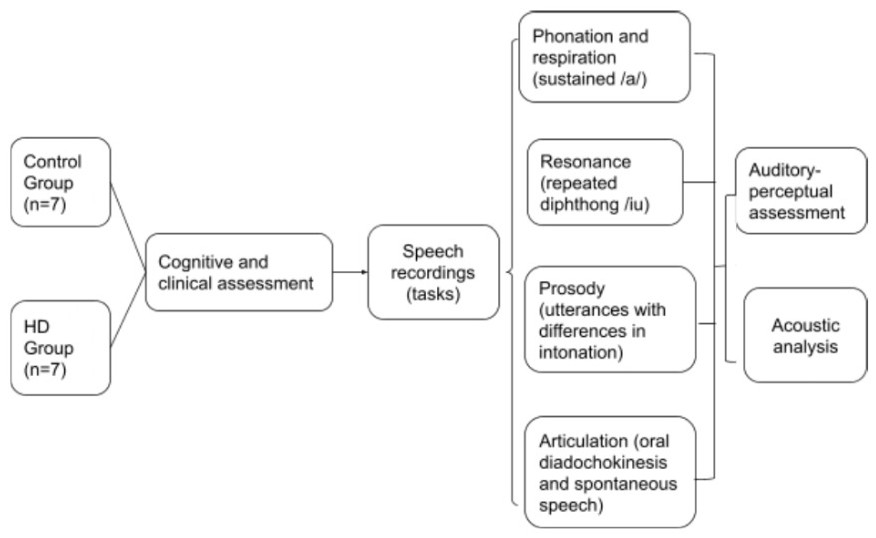
The speech tasks were recorded in a single session, using Audacity software, an Andrea Pure audio USB adapter, and a KARSECT HT-9 microphone positioned approximately 5cm from the patient's mouth. Case subjects and controls recorded 16-bit speech samples at a 44.1 kHz rate in a silent environment with no soundproofing. Both groups were asked to perform five tasks: (a) sustain the vowel /a/ in a single breath, for as long as possible, (b) repeat the diphthong /ju:/ in a single breath, (c) say /pataka/ as quickly as possible in a single breath (DDK) (d) use the correct intonation to say the sentence “It rained a lot this weekend” as a statement, a question and an exclamation, (e) spontaneously answer the question “What have you done today since waking up?” for 60 seconds.
Auditory perceptual assessment (APA)
This is currently the gold standard for assessing dysarthria. Three trained speech-language pathologists with at least five years of experience rated the blinded voice samples, using a kappa value of ≥ 0.90 for interrater agreement. A simulation activity preceded the assessment for the purpose of training. The examiners listened to the blinded speech samples in random order and rated the speech subsystems (phonation, articulation, respiration, resonance, and prosody). Using Duffy’s classification, each sample was rated as (0) normal, (1) mildly impaired, (2) moderately impaired, or (3) severely impaired. Subsequently, the final diagnosis was expressed as (0) normal, (1) mild dysarthria, (2) moderate dysarthria, or (3) severe dysarthria.
Acoustic analysis
Acoustic analysis was performed in Praat(40) (version 6.1.55) software, with a script(41) to automatically detect intensity peaks. Syllable structure in Brazilian Portuguese only allows vowels at the nucleus; thus, counting intensity peaks is the same as determining the number of syllables. De Jong and Wempe(41) were used to check reliability by comparing the results of manual analyses with those of the Praat script. The acoustic parameters recommended by Rusz et al.(42) and Vogel and Maruff(43) were also used. For phonation, we extracted information regarding jitter (rap), shimmer (local), fundamental frequency (F0 in Hz), standard deviation of the fundamental frequency (F0 SD) and the harmonics-to-noise ratio (HNR), measured through the sustained vowel /a/. For articulation, DDK and spontaneous speech recordings were used to analyze the number of syllables, number of pauses, total duration (in seconds), phonation time (total duration minus pauses), phonation rate (phonation time divided by total duration), speech rate (number of syllables divided by total duration), articulation rate (number of syllables divided by phonation time), average syllable duration (ASD) and number of pauses weighted by total time. From the repeated [ju:] task, the ratio between the second formant for the vowel [i] and the 2nd formant for the vowel [u] was used as a measure of vowel centralization. This measure can indicate reduced articulatory amplitude. Although it is not an exclusive respiratory measure, the maximum phonation time (MPT) was used to assess the respiratory subsystem, due to the association with the myoelastic-aerodynamic model of phonation. Regarding prosody, variations in F0 and intensity were evaluated during statement, question, and exclamation utterances. Variations in the F0 - the difference between the maximum and minimum values of the F0 - indicate melodic changes and, therefore, the speaker's ability to vary intonation (Figure 1).
Statistical analysis
A qualitative analysis of the data was performed using interquartile ranges and medians. Quantitative data were analyzed using Spearman's rank correlation coefficient. To compare groups, means and standard deviations were calculated, and the Student’s t-test was applied.
RESULTS
The seven case subjects included in the study - four females and three males - were matched by age and sex with healthy controls. Table 1 describes the clinical and sociodemographic characteristics of the sample. No statistically significant differences were found between the groups. Table 2 presents the results of the clinical severity scales for functional, behavioral, independence (Unified Huntington's Disease Rating Scale), and cognitive capacities. Table 3 describes the auditory-perceptual speech assessment. All case subjects presented impaired prosodic modulation and imprecise articulation; six presented speech and breathing discoordination, and slow speech rates. Table 4 shows the acoustic analysis of speech subsystems in the cases and controls (phonation, respiration, and prosody). The case subjects performed significantly worse than the control group, specifically regarding the variables of fundamental frequency, maximum phonation time, jitter (local), shimmer (local), variations in fundamental frequency in statements, and variations in intensity in questions and exclamatory utterances. Table 5 shows the acoustic analysis of speech tasks in the cases and controls. The HD subjects presented significantly worse results than the control group, specifically regarding the variables of speech rate, phonation time in spontaneous speech, number of syllables, average duration of syllables, and duration of spontaneous speech. In Table 6, we show correlations of significance between speech and clinical aspects.
Acoustic analysis of phonation, respiration and prosody in HD case subjects and healthy controls
DISCUSSION
In this study, all HD case subjects were diagnosed with some degree of dysarthria, with 4 (57.15%) having severe speech impairment, 2 (28.57%) moderate impairment and 1 (14.28%) mild impairment. The most frequent changes detected in the auditory perceptual assessment were impaired prosodic modulation, imprecise articulation, speech and breathing discoordination, and slow speech rate.
The case group presented significantly worse acoustic parameters than the control group regarding phonation (fundamental frequency, phonation time, jitter (local), shimmer (local), respiration (maximum phonation time) and articulation (speech rate, phonation time in spontaneous speech, number of syllables in spontaneous speech, average duration of syllables and duration of spontaneous speech).
The literature(21,23,24,28) that addresses affected speech in HD patients has described language, used discourse analysis, and evaluated speech subsystems. All the articles analyzed the speech of individuals in early stages of the disease and used different software for auditory-perceptual or acoustic assessment. They describe speech profiles with increased speech onset time, vowel prolongation, slower articulation and speech rates, sudden interruptions in phonation, and imprecise articulation.
Hertrich and Ackermann(23) performed an acoustic evaluation of the speech of 13 HD patients (most in more advanced stages of the disease) and 12 controls. The former group showed increased acoustic variability, speech onset time (voice onset time), and prolonged short vowels. The authors interpreted these findings as symptoms of advanced disease, which were consistent with the literature on degenerative cerebellar disorders. In our study, even though the sample size was smaller, the case subjects also presented more pauses during speech, shorter phonation times, and fewer syllables per second, corroborating Hertrich and Ackermann’s findings.
Another study evaluated 21 HD subjects (with an average disease duration of 5 years) and 21 controls. In the former group, the authors noted slower speech (slower articulation rate), increased pauses, and significant difficulty in generating single syllables(24). Our research also revealed slower speech in the HD group (slower articulation rate). However, the number of pauses in articulation was significantly higher when compared to other studies. Our hypotheses for these findings include the group’s low level of education, associated with cognitive and linguistic changes. Furthermore, age, age at onset, and duration of the disease may similarly influence these variables.
There were correlations between the clinical aspects of HD and the speech profiles, such as the correspondence between the motor scale scores and the speech subsystems. In our sample, the worse the subject's overall motor assessment, the greater the impairment in all speech subsystems. There was no correlation between the speech profiles and cognitive or other clinical variables (duration of disease, age at onset, behavior, functional status, or independence).
Rusz et al.(25) evaluated 34 individuals with HD (with a mean disease duration of 5.9 years) and 34 controls. They observed irregular intonation fluctuations, sudden interruptions in phonation, and imprecise articulation. They found a moderate correlation (r = -0.48) between sudden interruptions in phonation and voluntary components of the UHDRS scale. We found a significant decline in the rate of syllables per second, resulting in imprecise articulation, variations in jitter and shimmer, and compromised phonation.
Illes(44) analyzed spontaneous speech in three groups: 10 subjects with Huntington's disease, 10 with Alzheimer's disease (AD), and 10 with Parkinson's disease (PD). He reported that patients with HD present verbal paraphasias in spontaneous speech and simplify complex sentences. These results corroborate our finding of significantly shorter spontaneous speech in HD patients.
As well as HD-associated language deficits, the literature has widely described reduced lexical fluency and important communication difficulties. Similarly, cognitive impairment has been studied at different stages of the disease, and changes in memory, executive function, and attention have been documented, even prior to motor symptoms(45,46). The results from our study reflect these findings. Our case subjects achieved a median score of 12.5 points in the MoCA screening test(47), which is below the cutoff point that distinguishes healthy adults from patients with dementia (15 points) (sensitivity 90%, specificity 77%). The FAB battery(48) scores were similarly low in the HD group, with case subjects achieving a median score of 8.5 (shown in Table 2), while the cutoff point is 13.0 (±2.3). Changes in verbal fluency (both semantic and phonological) may also have impacted speech rate.
The DDK task has been cited as important in identifying neurological cases(49), since affected individuals produce fewer syllables per breath(30). Phonation, oral-motor function - including DDK (articulation) - and prosody are commonly impaired in HD subjects(50). Since speech disorders in individuals with HD are directly related to the overall progression of motor symptoms, health professionals with a view to early intervention should refer patients for speech assessments. The worse the patient’s general motor symptoms, the greater the impairment in speech subsystems.
For all speech variables, the HD group performed worse than the controls, who were included in our research to provide a reference for comparison. This was especially necessary because not all speech variables have a cutoff point or limits of normality for the Brazilian population. In addition, since the sample included a series of cases, comparison with healthy controls aimed to ensure that differences were due to the neurodegenerative condition, and not other external variables like education.
The small sample size was a limitation; however, HD is a rare disorder, and only participants from one specialized center were recruited. Further studies with larger samples from more healthcare centers, and longitudinal monitoring of HD patients, are needed to establish the speech profiles of these individuals and contribute to new diagnostic and therapeutic approaches.
Our present research shows that individuals with HD can have significantly impaired articulation, respiration, and phonation, although articulation was the most affected speech subsystem in our results. Considering these changes, clinicians should be attentive to these three subsystems during assessments, since their scores may define plans of care in speech therapy.
CONCLUSION
The most affected speech subsystems in the HD case subjects were articulation, phonation and respiration. Clinical assessments should include tasks that test these aspects, as they will also become a priority in therapy. Furthermore, HD speech profiles were directly related to the overall progression of motor symptoms. Evaluating the speech profiles of HD patients can support diagnosis, early rehabilitation, and a better quality of life. While the sample size in this study limits generalizations based on the findings, motor speech patterns present potential as biomarkers for predicting disease progression.
ACKNOWLEDGEMENTS
We would like to thank Hospital de Clínicas de Porto Alegre for the support and funding provided through the FIPE program (GPPG HCPA 2018-0648).
-
Study conducted at Universidade Federal do Rio Grande do Sul – UFRGS and Hospital de Clínicas de Porto Alegre – HCPA - Porto Alegre (RS), Brasil.
-
Financial support:
Hospital de Clínicas de Porto Alegre - FIPE (GPPG HCPA 2018-0648).
-
Data Availability:
Research data is available in the body of the article.
References
- 1Hayden MR. Huntington’s Chorea. Berlin: Springer-Verlag; 1981. http://doi.org/10.1007/978-1-4471-1308-9.
- 2Castilhos RM, Santos JA, Augustin MC, Pedroso JL, Barsottini O, Saba R, et al. Minimal prevalence of Huntington’s disease in the South of Brazil and instability of the expanded CAG tract during intergenerational transmissions. Genet Mol Biol. 2019;42(2):329-36. http://doi.org/10.1590/1678-4685-gmb-2018-0032. PMid:31259362.
- 3Alonso ME, Ochoa A, Boll M-C, Sosa AL, Yescas P, López M, et al. Clinical and genetic characteristics of Mexican Huntington’s disease patients. Mov Disord. 2009;24(13):2012-5. http://doi.org/10.1002/mds.22737. PMid:19672992.
- 4Paradisi I, Hernández A, Arias S. Huntington disease mutation in Venezuela: age of onset, haplotype analyses and geographic aggregation. J Hum Genet. 2008;53(2):127-35. http://doi.org/10.1007/s10038-007-0227-1. PMid:18157708.
- 5MacDonald ME, Ambrose CM, Duyao MP, Myers RH, Lin C, Srinidhi L, et al. A novel gene containing a trinucleotide repeat that is expanded and unstable on Huntington’s disease chromosomes. Cell. 1993;72(6):971-83. http://doi.org/10.1016/0092-8674(93)90585-E. PMid:8458085.
- 6Andrew SE, Goldberg YP, Kremer B, Squitieri F, Theilmann J, Zeisler J, et al. Huntington disease without CAG expansion: phenocopies or errors in assignment? Am J Hum Genet. 1994;54(5):852-63. PMid:8178825.
- 7Wexler NS, Lorimer J, Porter J, Gomez F, Moskowitz C, Shackell E, et al. Venezuelan kindreds reveal that genetic and environmental factors modulate Huntington’s disease age of onset. Proc Natl Acad Sci USA. 2004;101(10):3498-503. http://doi.org/10.1073/pnas.0308679101. PMid:14993615.
-
8 <edb[REMOVED IF= FIELD]><edb[REMOVED IF= FIELD]8Caron NS, Wright GEB, Hayden MR. Huntington Disease. In: Adam MP, Feldman J, Mirzaa GM, Pagon RA, Wallace SE, Amemiya A, editors. GeneReviews. Seattle (WA): University of Washington; 2018 [citado em 2019 Set 15]. Disponível em: https://www.ncbi.nlm.nih.gov/books/NBK1305/
» https://www.ncbi.nlm.nih.gov/books/NBK1305/ - 9Teixeira AL, Souza LC, Rocha NP, Furr-Stimming E, Lauterbach EC. Revisiting the neuropsychiatry of Huntington’s disease. Dement Neuropsychol. 2016;10(4):261-6. http://doi.org/10.1590/s1980-5764-2016dn1004002. PMid:29213467.
- 10Sokol LL, Troost JP, Bega D, Paulsen JS, Kluger BM, Applebaum AJ, et al. Death anxiety in Huntington Disease: longitudinal heath-related quality-of-life outcomes. J Palliat Med. 2023;26(7):907-14. http://doi.org/10.1089/jpm.2022.0160. PMid:36607769.
- 11Roman OC, Stovall J, Claassen DO. Perseveration and suicide in Huntington’s Disease. J Huntingtons Dis. 2018;7(2):185-7. http://doi.org/10.3233/JHD-170249. PMid:29614688.
- 12 Duffy JR. Motor speech disorders-e-book: substrates, differential diagnosis, and management. St. Louis: Elsevier Health Sciences; 2013.
- 13Darley FL, Aronson AE, Brown JR. Differential diagnostic patterns of dysarthria. J Speech Hear Res. 1969;12(2):246-69. http://doi.org/10.1044/jshr.1202.246. PMid:5808852.
- 14DeLong M, Wichmann T. Changing views of basal ganglia circuits and circuit disorders. Clin EEG Neurosci. 2010;41(2):61-7. http://doi.org/10.1177/155005941004100204. PMid:20521487.
- 15DeLong MR, Wichmann T. Circuits and circuit disorders of the basal ganglia. Arch Neurol. 2007;64(1):20-4. http://doi.org/10.1001/archneur.64.1.20. PMid:17210805.
- 16Kent RD. Research on speech motor control and its disorders: a review and prospective. J Commun Disord. 2000;33(5):391-427. http://doi.org/10.1016/S0021-9924(00)00023-X. PMid:11081787.
- 17Kent RD, Duffy JR, Slama A, Kent JF, Clift A. Clinicoanatomic studies in dysarthria: review, critique, and directions for research. J Speech Lang Hear Res. 2001;44(3):535-51. http://doi.org/10.1044/1092-4388(2001/042). PMid:11407559.
- 18Darley FL, Aronson AE, Brown JR. Differential diagnostic patterns of dysarthria. J Speech Hear Res. 1969;12(2):246-69. http://doi.org/10.1044/jshr.1202.246. PMid:5808852.
- 19Darley FL, Aronson AE, Brown JR. Clusters of deviant speech dimensions in the dysarthrias. J Speech Hear Res. 1969;12(3):462-96. http://doi.org/10.1044/jshr.1203.462. PMid:5811846.
- 20Darley FL, Aronson AE, Brown JR. Motor speech disorders. Philadelphia: Saunders; 1975.
- 21Rusz J, Klempíř J, Tykalová T, Baborová E, Čmejla R, Růžička E, et al. Characteristics and occurrence of speech impairment in Huntington’s disease: possible influence of antipsychotic medication. J Neural Transm. 2014;121(12):1529-39. http://doi.org/10.1007/s00702-014-1229-8. PMid:24809686.
- 22Miller N, Mshana G, Msuya O, Dotchin C, Walker R, Aris E. Assessment of speech in neurological disorders: development of a Swahili screening test. S Afr J Commun Disord. 2012;59(1):27-33. PMid:23409616.
- 23Hertrich I, Ackermann H. Acoustic analysis of speech timing in Huntington’s disease. Brain Lang. 1994;47(2):182-96. http://doi.org/10.1006/brln.1994.1048. PMid:7953613.
- 24Skodda S, Schlegel U, Hoffmann R, Saft C. Impaired motor speech performance in Huntington’s disease. J Neural Transm. 2014;121(4):399-407. http://doi.org/10.1007/s00702-013-1115-9. PMid:24221215.
- 25Rusz J, Klempíř J, Baborová E, Tykalová T, Majerová V, Cmejla R, et al. Objective acoustic quantification of phonatory dysfunction in Huntington’s Disease. PLoS One. 2013;8(6):e65881. http://doi.org/10.1371/journal.pone.0065881. PMid:23762447.
- 26Duffy JR. Motor speech disorders: substrates, differential diagnosis, and management. 4th ed. Amsterdam: Elsevier; 2020.
- 27Hinzen W, Rosselló J, Morey C, Camara E, Garcia-Gorro C, Salvador R, et al. A systematic linguistic profile of spontaneous narrative speech in pre-symptomatic and early stage Huntington’s disease. Cortex. 2018;100:71-83. http://doi.org/10.1016/j.cortex.2017.07.022. PMid:28859906.
- 28Ergun A, Oder W. Oral diadochokinesis and velocity of narrative speech: A prognostic parameter for the outcome of diffuse axonal injury in severe head trauma. Brain Inj. 2008;22(10):773-9. http://doi.org/10.1080/02699050802372182. PMid:18787987.
- 29Kent RD, Kim Y-J. Toward an acoustic typology of motor speech disorders. Clin Linguist Phon. 2003;17(6):427-45. http://doi.org/10.1080/0269920031000086248. PMid:14564830.
- 30Carrillo L, Ortiz KZ. Análise vocal (auditiva e acústica) nas disartrias. Pró-Fono Rev Atualização Científica. 2007;19(4):381-6. http://doi.org/10.1590/S0104-56872007000400010.
- 31Titze IR, Wong D, Milder MA, Hensley SR, Ramig LO. Comparison between clinician-assisted and fully automated procedures for obtaining a voice range profile. J Speech Hear Res. 1995;38(3):526-35. http://doi.org/10.1044/jshr.3803.526. PMid:7674644.
- 32Kouba T, Frank W, Tykalova T, Mühlbäck A, Klempíř J, Lindenberg KS, et al. Speech biomarkers in Huntington’s disease: A cross-sectional study in pre-symptomatic, prodromal and early manifest stages. Eur J Neurol. 2023;30(5):1262-71. http://doi.org/10.1111/ene.15726. PMid:36732902.
- 33Kieburtz K, Penney JB, Como P, Ranen N, Shoulson I, Feigin A, et al. Unified Huntington’s disease rating scale: reliability and consistency. Mov Disord. 1996;11(2):136-42. http://doi.org/10.1002/mds.870110204. PMid:8684382.
- 34Winder JY, Achterberg WP, Gardiner SL, Roos RAC. Longitudinal assessment of the Unified Huntington’s Disease Rating Scale (UHDRS) and UHDRS-For Advanced Patients (UHDRS-FAP) in patients with late stage Huntington’s disease. Eur J Neurol. 2019;26(5):780-5. http://doi.org/10.1111/ene.13889. PMid:30576046.
- 35Winder JY, Achterberg WP, Marinus J, Gardiner SL, Roos RAC. Assessment scales for patients with advanced Huntington’s Disease: comparison of the UHDRS and UHDRS-FAP. Mov Disord Clin Pract. 2018;5(5):527-33. http://doi.org/10.1002/mdc3.12646. PMid:30515443.
- 36Beato R, Amaral-Carvalho V, Guimarães HC, Tumas V, Souza CP, Oliveira GN, et al. Frontal assessment battery in a Brazilian sample of healthy controls: normative data. Arq Neuropsiquiatr. 2012;70(4):278-80. http://doi.org/10.1590/S0004-282X2012005000009. PMid:22358310.
- 37Periáñez JA, Lubrini G, García-Gutiérrez A, Ríos-Lago M. Construct validity of the stroop color-word test: influence of speed of visual search, verbal fluency, working memory, cognitive flexibility, and conflict monitoring. Arch Clin Neuropsychol. 2021;36(1):99-111. http://doi.org/10.1093/arclin/acaa034. PMid:32514527.
- 38Freitas S, Simões MR, Martins C, Vilar M, Santana I. Estudos de adaptação do Montreal Cognitive Assessment (MoCA) para a população portuguesa. Aval Psicol. 2010;9(3):345-57.
- 39Brody DJ, Kramarow EA, Taylor CA, McGuire LC. Cognitive performance in adults aged 60 and over: national health and nutrition examination survey, 2011-2014. Natl Health Stat Report. 2019;(126):1-23. PMid:31751207.
-
40Boersma P, Weenink D. Praat: doing phonetics by computer [software]. Amsterdam: University of Amsterdam; 2024 [citado em 2019 Set 15]. Disponível em: http://www.praat.org/
» http://www.praat.org/ - 41De Jong NH, Wempe T. Praat script to detect syllable nuclei and measure speech rate automatically. Behav Res Methods. 2009;41(2):385-90. http://doi.org/10.3758/BRM.41.2.385. PMid:19363178.
- 42Rusz J, Cmejla R, Ruzickova H, Ruzicka E. Quantitative acoustic measurements for characterization of speech and voice disorders in early untreated Parkinson’s disease. J Acoust Soc Am. 2011;129(1):350-67. http://doi.org/10.1121/1.3514381. PMid:21303016.
- 43Vogel AP, Maruff P. Comparison of voice acquisition methodologies in speech research. Behav Res Methods. 2008;40(4):982-7. http://doi.org/10.3758/BRM.40.4.982. PMid:19001389.
- 44Illes J. Neurolinguistic features of spontaneous language production dissociate three forms of neurodegenerative disease: alzheimer’s, huntington’s, and parkinson’s. Brain Lang. 1989;37(4):628-42. http://doi.org/10.1016/0093-934X(89)90116-8. PMid:2479447.
- 45Paulsen JS. Cognitive impairment in Huntington disease: diagnosis and treatment. Curr Neurol Neurosci Rep. 2011;11(5):474-83. http://doi.org/10.1007/s11910-011-0215-x. PMid:21861097.
- 46Maurage P, Heeren A, Lahaye M, Jeanjean A, Guettat L, Verellen-Dumoulin C, et al. Attentional impairments in Huntington’s disease: A specific deficit for the executive conflict. Neuropsychology. 2017;31(4):424-36. http://doi.org/10.1037/neu0000321. PMid:28240935.
- 47Cesar KG, Yassuda MS, Porto FHG, Brucki SMD, Nitrini R. MoCA Test: normative and diagnostic accuracy data for seniors with heterogeneous educational levels in Brazil. Arq Neuropsiquiatr. 2019;77(11):775-81. http://doi.org/10.1590/0004-282x20190130. PMid:31826133.
- 48Beato RG, Nitrini R, Formigoni AP, Caramelli P. Brazilian version of the Frontal Assessment Battery (FAB): preliminary data on administration to healthy elderly. Dement Neuropsychol. 2007;1(1):59-65. http://doi.org/10.1590/S1980-57642008DN10100010. PMid:29213369.
- 49Kent RD, Kim Y, Chen LM. Oral and laryngeal diadochokinesis across the life span: a scoping review of methods, reference data, and clinical applications. J Speech Lang Hear Res. 2022;65(2):574-623. http://doi.org/10.1044/2021_JSLHR-21-00396. PMid:34958599.
- 50Hartelius L, Carlstedt A, Ytterberg M, Lillvik M, Laakso K. Speech disorders in mild and moderate Huntington disease: results of dysarthria assessments of 19 individuals. J Med Speech-Lang Pathol. 2003;11(1):1-14.
Edited by
-
Editor:
Stela Maris Aguiar Lemos.
Research data is available in the body of the article.