 RAPID PROGRESSION OF SPINAL TUBERCULOSIS: A CASE REPORT AND LITERATURE REVIEW
ABSTRACT
Of bone tuberculosis cases, 50% correspond to vertebral tuberculosis. It presents with severe kyphosis in the dorsal region and, to a lesser extent, with neurological deficits secondary to bone destruction, due to direct compression from abscesses, inflammatory tissue, or secondary instability from fractures and vertebral wedging. A 33-year-old male was admitted with a one-month history of lumbar pain, initially attributed to disc herniations. The condition progressed with intense radicular lumbar pain. Imaging studies showed L3-L4 spondylodiscitis, which rapidly progressed to vertebral destruction and neurological deficits. A right L2-L3 hemilaminectomy and epidural abscess drainage were performed. The patient continued to experience lumbar pain. Postoperative CT showed significant vertebral bone destruction, prompting a new surgery: L2-L3 laminectomy, transpedicular fixation, and autograft arthrodesis, achieving extensive decompression and vertebral stabilization. The real-time PCR study (Gen Xpert®) and histopathological analysis were consistent with vertebral tuberculosis. Hypoalbuminemia was identified as a risk factor for the rapid progression of vertebral tuberculosis. Vertebral tuberculosis can progress rapidly, especially in patients with risk factors that compromise their immune and nutritional response, such as in children, hypoalbuminemia, vitamin D and B12 deficiencies, resistance to antitubercular drugs, and HIV coinfection. Spinal tuberculosis has various risk factors that contribute to its rapid progression and bone destruction. Level of Evidence IV; Cases Series.
Keywords:
Tuberculosis, Spinal; Discitis; Pott Puffy Tumor, Risk Factors
RESUMO
Dos casos de tuberculose óssea, 50% correspondem à tuberculose vertebral. Apresenta-se com cifose grave na região dorsal e, em menor proporção, com déficit neurológico secundário à destruição óssea, por compressão direta de abscessos, tecido inflamatório ou por instabilidade secundária a fraturas e achatamento vertebral. Homem de 33 anos, internado por um quadro de um mês, caracterizado por dor lombar, inicialmente atribuída a hérnias de disco. Evoluiu com dor lombar com irradiação radicular intensa. Os estudos de imagem mostraram espondilodiscite L3 L4 que evoluiu rapidamente para destruição vertebral e déficit neurológico. Foi realizada hemilaminectomia L2 L3 direita e drenagem de abscesso epidural. O paciente evoluiu com persistência da dor lombar. A tomografia pós-operatória mostrou importante destruição óssea vertebral, pelo que foi realizada nova cirurgia: laminectomia L2 L3, fixação transpedicular e artrodese com autoenxerto, obtendo-se ampla descompressão e estabilização vertebral. O estudo de PCR em tempo real (Gen Xpert®) e o estudo histopatológico foram compatíveis com tuberculose vertebral. A hipoalbuminemia foi identificada como fator de risco para a rápida progressão da tuberculose vertebral. tuberculose vertebral pode evoluir rapidamente, especialmente em pacientes com fatores de risco que comprometem sua resposta imunológica e nutricional. Como ocorre em crianças, hipoalbuminemia, deficiência de vitamina D e vitamina B12, resistência a drogas antituberculosas e coinfecção por HIV. A tuberculose espinhal possui diferentes fatores de risco que contribuem para sua rápida progressão e destruição óssea. Nível de Evidência IV; Série de Casos.
Descritores:
Tuberculose da Coluna Vertebral; Discite; Tumor de Pott; Fatores de Risco
RESUMEN
De los casos de tuberculosis ósea el 50% corresponde a tuberculosis vertebral. Presentándose con cifosis grave en la región dorsal y en menor proporción con déficit neurológico secundario a la destrucción ósea, por compresión directa de abscesos, tejido inflamatorio o por inestabilidad secundaria a fracturas y acuñamiento vertebral. Hombre de 33 años, ingresó por cuadro de un mes, caracterizado por dolor lumbar, inicialmente atribuido a hernias discales. Evolucionó con dolor lumbar con irradiación radicular intenso. Los estudios de imagen mostraron espondilodiscitis L3 L4 que evolucionó rápidamente hacia destrucción vertebral y déficit neurológico. Se realizó hemilaminectomía L2 L3 derecha y drenaje de absceso epidural. El paciente evolucionó con persistencia del dolor lumbar. La tomografía postoperatoria mostró importante destrucción ósea vertebral por lo cual se realizó nueva cirugía: Laminectomía L2 L3, fijación transpedicular y artrodesis con autoinjerto, lográndose amplia descompresión y estabilización vertebral. El estudio de PCR en tiempo real (Gen Xpert®) y el estudio histopatológico fueron compatibles con tuberculosis vertebral. Se identificó la hipoalbuminemia como factor de riesgo para la rápida progresión de la tuberculosis vertebral. La tuberculosis vertebral puede evolucionar con rapidez, especialmente en pacientes con factores de riesgo que comprometen su respuesta inmunológica y nutricional. Como sucede en niños, hipoalbuminemia, deficiencia de vitamina D y de vitamina B12, la resistencia a drogas antituberculosas y la coinfección por VIH. La tuberculosis espinal tiene diferentes factores de riesgo que contribuyen a su rápida progresión y destrucción ósea. Nivel de Evidencia IV; Series de Casos.
Descriptores:
Tuberculosis de la Columna Vertebral; Discitis; Tumor Hinchado de Pott, Factores de Riesgo
INTRODUCTION
Tuberculosis is one of the deadliest infectious diseases, following COVID-19 and HIV/AIDS, with a population of 10.6 million people affected in 2022.1 During primary infection by Mycobacterium tuberculosis, the bacillary load initially forms pulmonary foci, which can spread through hematogenous or lymphatic routes to the rest of the body, including bones and joints.2
Bone tuberculosis accounts for 15% to 20% of extrapulmonary tuberculosis cases, with vertebral tuberculosis, also known as “Pott’s disease” or tuberculous spondylitis, comprising 50% of these cases. Described by Percival Pott in 1779, the disease shows a higher frequency of damage in the thoracolumbar junction, with most cases affecting the thoracic spine in 50% of cases and the cervical spine in 10% of younger patients, followed by the lumbar spine in 45% of older patients.2,3
Spinal tuberculosis presents with severe kyphosis in the dorsal region, and a smaller proportion of patients develop neurological deficits secondary to bone destruction due to direct compression from abscesses, inflammatory tissue, or instability due to fractures and vertebral wedging. When a nerve root is compressed due to the development of an abscess or bone fragment, radicular pain may be the presenting symptom.4
The likelihood of rapid progression increases when the local immune system fails due to an imbalance between agent-host-environment factors, resulting in the reactivation of latent infection, as seen at younger ages, in cases of hypoalbuminemia, vitamin D and B12 deficiencies, drug resistance, and HIV co-infection. We present a patient with L2-L3 spondylodiscitis with a lateral paravertebral abscess, who exhibited rapid disease progression.
CASE REPORT
A 33-year-old male lawyer, married, and a regular beer drinker, was admitted with a one-month history characterized by intense lumbar pain. At another medical center, he was diagnosed with disc herniations, for which he received analgesics and paravertebral ozone therapy with partial and temporary relief. The lumbar pain worsened during the three days prior to his admission, reaching an intensity of 10/10 on the visual analog scale and was associated with pain in the lower limbs, predominantly on the right side, numbness in the right thigh, and decreased strength in that limb over the last 24 hours.
Physical examination on admission: Weight: 94.6 kg, height: 1.75 m. BMI: 30.9 kg/m2. Paresis of the right quadriceps 4/5, absent right patellar reflex, hypoesthesia in the right L3 dermatome, intense pain on lumbar and right paravertebral lumbar palpation. Positive Lasegue and Bragard tests bilaterally. No signs of meningeal irritation. Normal sphincter control. Walking was very difficult due to pain.
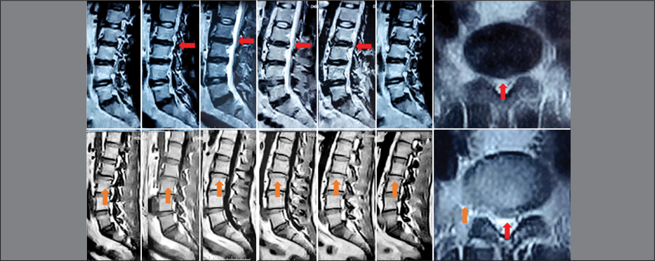
Magnetic resonance imaging (MRI) from December 7, 2023, showed initial L2-L3 spondylodiscitis with associated disc herniation (Figure 1).

MRI showing degeneration of the L2-L3, L3-L4, L4-L5, and L5-S1 intervertebral discs, a protruded L2-L3 herniation (red arrow), and blurring of the superior endplate of the L3 vertebral body (orange arrows), compatible with initial spondylodiscitis.
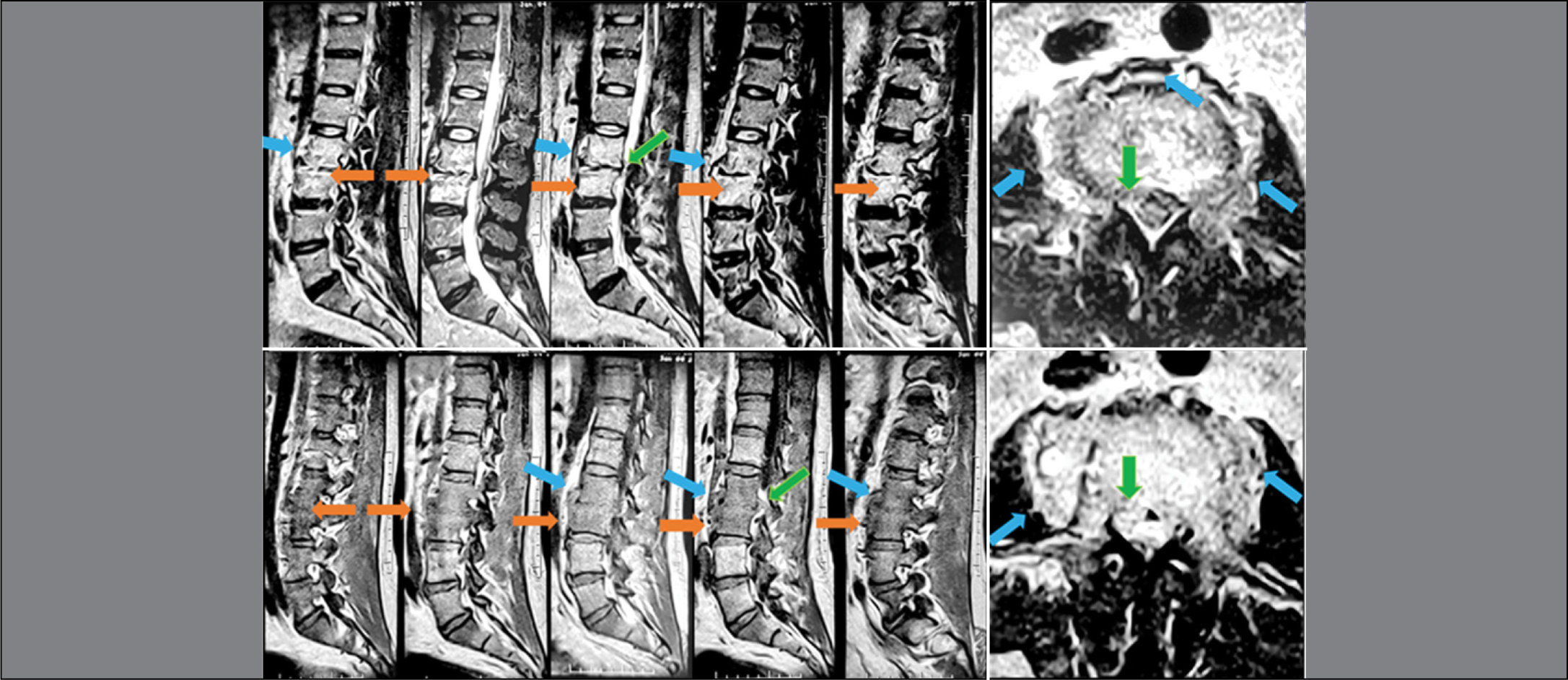
An MRI from January 4, 2024, shows L2-L3 spondylodiscitis with a lateral paravertebral abscess extending beyond both segments, an epidural abscess displacing the dural sac, and disc herniation at that level, without kyphosis. The scan shows very evident changes with significant disease progression compared to the previous study (Figure 2).
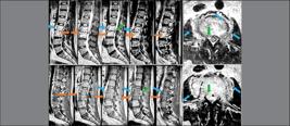
MRI showing degeneration of the L2-L3, L3-L4, L4-L5, and L5-S1 intervertebral discs, a protruded L2-L3 herniation, L3-L4 spondylodiscitis (orange arrows), an epidural abscess (green arrows) compressing the dural sac, and lateral and anterior paravertebral abscesses (green arrows).
Preoperative laboratory studies showed normal complete blood count, blood glucose, creatinine, coagulation profile, erythrocyte sedimentation rate, and total protein levels, but hypoalbuminemia of 3.0 g/dL (reference range: 3.5 to 5.4 g/dL). A rapid HIV test was negative.
Due to the neurological deficit, a right L2-L3 hemilaminectomy, epidural abscess drainage, L3-L4 disc biopsy, and L3-L4 vertebral body biopsy were performed, revealing firm, fibrous, inflammatory-infectious tissue compressing the dural sac.
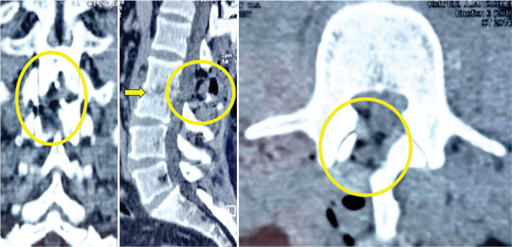
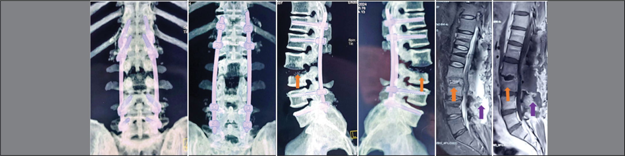
The patient continued to experience persistent lumbar pain. Postoperative computed tomography (CT) scans revealed significant vertebral bone destruction, exceeding the damage seen in the preoperative MRI (Figure 3). Another surgery was performed: L2-L3 laminectomy, T12-L1, L4, and L5 transpedicular fixation, and arthrodesis with an iliac crest autograft, achieving wide decompression and vertebral stabilization, as evidenced by the postoperative CT scan (Figure 4), which also shows significant bone involvement of the L2-L3 vertebral bodies and adequate decompression of the dural sac. The patient developed a deep seroma, which was identified in the postoperative MRI (Figure 4) and resolved with puncture and aspiration, resulting in complete remission.
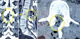
CT scan showing the area of minimally invasive decompression through right L3-L4 hemilaminectomy (yellow circles) and drainage of the epidural abscess. Bone involvement of the L3 and L4 vertebral bodies is evident (yellow arrow).

Postoperative CT scan and MRI showing the fixation system with pedicle screws in vertebrae T12, L1, L4, and L5, with lateral rods. There is evidence of further progression of the spondylodiscitis (orange arrows) and the deep seroma (purple arrows).
Cultures for common germs, yeasts, and fungi were negative. The Gram stain was negative. Ziehl-Neelsen staining did not detect acid-fast bacilli. Real-time PCR testing (Gen Xpert®) was positive for tuberculosis.
The histopathological study showed an active chronic granulomatous inflammatory process with epithelioid cell granulomas and Langhans giant cells.
Primary immunodeficiencies and HIV-related immunodeficiency were ruled out through several tests conducted by the immunology department. Liver and kidney function tests were normal. Vitamin levels, including vitamin D, were also normal.
The patient showed favorable progress, fully recovering his neurological functions and returning to his daily activities fifteen days after the second surgery. He is currently (October 2024) receiving antitubercular treatment with isoniazid, rifampicin, pyrazinamide, and ethambutol.
Although the vertebral bone involvement is significant, given his good clinical progression, no surgery for anterior vertebral support was considered.
DISCUSSION
This case involves rapid vertebral destruction due to vertebral tuberculosis, prompting us to conduct a non-systematic review of the literature. We identified the following factors as promoters of the progression of vertebral tuberculosis:
Age
Vertebral tuberculosis affects young patients more aggressively, as they have greater vascularization in the vertebral endplates.5 One of the reasons for vertebral deformity in children with spinal tuberculosis is the abundance of cartilaginous tissue in this region.6 Tuberculosis generally spreads hematogenously, traveling through the anterior and posterior spinal arteries of the vertebrae to finally reach the spongy bone region. According to Batson, through the valveless paravertebral venous plexus, the infection is transmitted to the lower and anterior parts of the vertebral body, facilitated by intra-abdominal and intrathoracic pressures. For these reasons, the most affected are children or young adults.5,7
HIV-Induced Immunosuppression
The risks of developing osteoarticular pathology associated with HIV infection are due to metabolic complications such as lipodystrophy, avascular necrosis, and decreased bone mineral density caused by both HIV and antiretroviral drugs. In 1987, Withringtonet al. reported the isolation of HIV from the synovial fluid of a patient with HIV-associated oligoarthritis, and in 2016, a group of authors published data on the presence of HIV RNA in pathologically altered tissues.8 These advances initiated studies into the potential inflammatory effects of HIV on the osteoarticular system. A study of HIV-infected patients who underwent surgery for confirmed tuberculous spondylitis and generalized tuberculosis detected HIV RNA in a focus of bone destruction, and similarly, polymorphological, microbiological, and genetic studies revealed the presence of Mycobacterium tuberculosis DNA. Consequently, it is inferred that HIV increases the likelihood of bone tissue invasion by Mycobacterium tuberculosis.9 HIV and Mycobacterium tuberculosis potentiate each other, and co-infection increases both the severity of tuberculosis symptoms and the likelihood of progressing from HIV to AIDS.5
The depletion of CD4+ T cells due to HIV plays a key role in the risk of co-infection with Mycobacterium tuberculosis. CD4+ T cells are essential for the host’s immunity to Mycobacterium tuberculosis infection, enhancing the effector functions of CD8+ T cells and preventing their exhaustion. The Th1 subtype of CD4+ T cells releases interferon-gamma (IFN-γ), a cytokine that activates inflammatory macrophages to initiate the phagocytosis of macrophages infected with Mycobacterium tuberculosis. The depletion of CD4+ Th1 cells and the lack of IFN-γ increase the incidence of initial Mycobacterium tuberculosis infection, the reactivation of latent disease, and the extrapulmonary manifestations seen in Pott’s disease. In turn, Mycobacterium tuberculosis increases the expression of CXCR4 receptors to promote HIV replication and increase viral load.5
HIV infection is the most significant independent risk factor for the progression of latent tuberculosis infection to active disease, increasing the likelihood of latent infection reactivation by 20 times. Patients who do not receive antiretroviral therapy and have a decreased CD4+ T cell count are prone to extrapulmonary tuberculosis due to the continuous immunosuppression caused by HIV.8
Vitamin B12 Deficiency
Cobalamin (vitamin B12) is a vitamin obtained solely from animal-derived foods, as humans are unable to synthesize it on their own. Therefore, individuals who do not consume animal products develop a deficiency of this vitamin.10 Vitamin B12 is essential for the metabolism of carbohydrates, fats, and proteins, the formation and regeneration of blood cells, as well as the maintenance of the central nervous system. It supports the proper functioning of the immune system by inducing cell replication, including the production of T lymphocytes. Its deficiency can impair defenses and promote the spread and progression of infections. This vitamin also stimulates osteoblastic and osteoclastic activity, meaning it regulates bone formation.11,12
The evolution of Mycobacterium tuberculosis is polymorphic, and it does not need to synthesize cobalamin, as its host provides this supplement. The bacterium merely sequesters vitamin B12 in the human body without expending energy, aiming to develop the disease successfully, as it requires this vitamin to synthesize methionine, a crucial amino acid for protein formation.13,14
Vitamin D Deficiency
Vitamin D activates and enhances innate immunity by promoting the fusion of the macrophage-phagolysosome complex, mediating the generation of reactive oxygen species (ROS), and minimizing the expression of the peroxisome proliferator-activated receptor to inhibit lipid metabolism in macrophages infected by Mycobacterium tuberculosis.15 Vitamin D promotes the elimination of Mycobacterium tuberculosis through macrophage-mediated destruction. The transcription of cathelicidin depends on adequate levels of vitamin D, and it has the ability to destroy mycobacterial membranes in the phagosome and lysosome of macrophages.16
Low levels of vitamin D are linked to infections such as tuberculosis and are important for preventing the symptomatic phase of this pathology. A study discovered that the polymorphism of the vitamin D receptor gene (VDR-FokI) is a susceptibility factor for spinal tuberculosis and is related to clinical severity. Therefore, low levels of this vitamin could compromise the body’s ability to fight Mycobacterium tuberculosis.17
Hypoalbuminemia
Defined as a serum concentration lower than 3.5 g/dL, hypoalbuminemia is considered a common representation of malnutrition. The higher incidence of postoperative complications in various orthopedic surgeries, ranging from spinal fusions to hip fractures, is closely related to hypoalbuminemia.18
In a study aimed at identifying the demographic characteristics and presentation of a population with spinal tuberculosis, 23 subjects were identified who, before treatment, exhibited hypoalbuminemia, elevated serum alkaline phosphatase, and increased C-reactive protein in their laboratory parameters.19
Patients with spinal tuberculosis are prone to hypoalbuminemia after surgery, which can easily lead to various complications and even adverse clinical outcomes. Preventing postoperative hypoalbuminemia in patients with spinal tuberculosis is of great clinical importance, and the preoperative albumin level in patients with vertebral tuberculosis may be a key factor in preventing postoperative hypoalbuminemia.18
Studies have shown that hypoalbuminemia is an independent risk factor for wound infection after spinal surgery, related to the occurrence of perioperative pneumonia, postoperative sepsis, myocardial infarction, and secondary revision surgery, both in spinal fusion and total hip arthroplasty. Therefore, it is of clinical importance to explore the risk factors for hypoproteinemia after surgery in patients with spinal tuberculosis. One study found that complicated pulmonary tuberculosis, preoperative serum albumin levels, and operation time are independent risk factors for postoperative hypoalbuminemia.18
Resistance to Antitubercular Drugs
The discovery of multidrug-resistant tuberculosis (MDR) and extensively drug-resistant tuberculosis (XDR) has had a major impact and become a significant challenge in the treatment of spinal tuberculosis.20 Tuberculosis can be classified as primary if the pathology occurred when the patient had not been exposed to any specific drug, and secondary if it occurred after exposure of a strain to chemotherapy. Non-lethal treatments induce mutations in Mycobacterium tuberculosis, and the variability of these mutations alters the level of drug resistance, regardless of being located in the same codon.21,22
A study that genotyped Mycobacterium tuberculosis strains of the Beijing and non-Beijing types using spoligotyping and drug resistance testing observed a higher incidence of multidrug-resistant tuberculosis associated with the Beijing type compared to the non-Beijing type. Drug resistance in the Beijing genotype was observed in descending order, with isoniazid resistance at 62.96%, rifampicin at 59.26%, streptomycin at 48.15%, ethambutol at 33.33%, ciprofloxacin at 18.52%, ofloxacin at 14.81%, capreomycin at 11.11%, and azithromycin with a low resistance rate of 3.7%. Highlighting the increasing drug resistance rates in Mycobacterium tuberculosis, it was noted that recurrent spinal tuberculosis is becoming more prominent.23
Another study determined that the greatest risk factor for postoperative recurrence in patients with spinal tuberculosis is rifampicin resistance, pointing out that due to incomplete elimination of the lesion, drug-resistant tubercle bacilli multiply at an accelerated rate and lead to the onset and recurrence of spinal tuberculosis, given the rich vascular supply of the vertebrae. Therefore, early diagnosis of rifampicin resistance and the initiation of appropriate chemotherapy are emphasized to prevent postoperative recurrence of spinal tuberculosis.24
To suspect drug-resistant spinal tuberculosis, clinical criteria include patients with spinal tuberculosis on first-line drug therapy for 5 months or more who exhibit one of the following findings: inadequate clinical and radiological response, development of recent osteoarticular tuberculosis lesions, worsening of spinal deformity, onset of draining sinuses, or dehiscence of the surgical wound.25
Vertebral tuberculosis can progress rapidly, especially in patients with risk factors that compromise their immune and nutritional responses. The case we presented showed a slow evolution, with poor response to an initial attempt at minimally invasive surgery, which necessitated a more aggressive surgical treatment. In this patient, hypoalbuminemia was identified as a key risk factor, while other pathologies potentially associated with the rapid progression of the disease were ruled out.
It is essential to consider the factors that modify the progression of vertebral tuberculosis, as their presence can accelerate bone destruction and compromise adjacent neurological structures. Upon identifying these factors, more aggressive therapeutic interventions, such as surgical decompression, stabilization, and solid fixation, should be implemented.
CONCLUSION
The analyzed case presented rapid progression of vertebral tuberculosis. Initially, minimally invasive treatment was insufficient and required more aggressive surgical management. Hypoalbuminemia was identified as a risk factor for its accelerated progression. After a non-systematic review of the literature, we determined that the risk factors for the rapid progression of vertebral tuberculosis are: young age (children), vitamin B12 and D deficiencies, immunosuppression due to diseases such as HIV, preoperative hypoalbuminemia, and resistance to antitubercular drugs. These factors must be considered when managing the treatment of patients with vertebral tuberculosis.
-
Study conducted by the Center of Neurological Diseases, La Paz/Santa Cruz, Bolivia.
REFERENCES
-
1 Organización Mundial de la Salud (OMS). Tuberculosis [Internet]. Ginebra: OMS; 2023 [access in 2024 5 sep 5]. Available from:https://www.who.int/es/news-room/fact-sheets/detail/tuberculosis
» https://www.who.int/es/news-room/fact-sheets/detail/tuberculosis - 2 Arias Deroncerés IJ, Puente Saní V, Lamotte Castillo JA, Ojeda Sánchez L. Tuberculosis vertebral (mal de Pott) e infección por el virus de la inmunodeficiencia humana. Medisan. 2011;15(12):1791–7.
-
3 Paz Cuéllar, K Factores de riesgo asociados a tuberculosis extrapulmonar en el programa de control de tuberculosis del Hospital Nacional Dos de Mayo en el periodo julio 2015 – junio 2016. [Internet]. Universidad Ricardo Palma - URP; 2017. [access in 2024 5 sep 5]. Available from: https://repositorio.urp.edu.pe/handle/20.500.14138/5190
» https://repositorio.urp.edu.pe/handle/20.500.14138/5190 -
4 Vergara-Amador E, Galván-Villamarin F, Piña-Quintero M. Tuberculosis Osteoarticular Primaria: Reaparición de una Patología Olvidada [Primary osteoarticular tuberculosis: the reappearance of a forgotten pathology]. Rev Salud Publica. 2007;9(3):465-70. Spanish. doi: 10.1590/s0124-00642007000300015
» 10.1590/s0124-00642007000300015 -
5 Glassman I, Nguyen KH, Giess J, Alcantara C, Booth M, Venketaraman V. Pathogenesis, Diagnostic Challenges, and Risk Factors of Pott’s Disease. Clin Pract. 2023;13(1):155-165. doi: 10.3390/clinpract13010014
» 10.3390/clinpract13010014 -
6 Jain AK, Sreenivasan R, Mukunth R, Dhammi IK. Tubercular spondylitis in children. Indian J Orthop. 2014;48(2):136-44. doi: 10.4103/0019-5413.128747
» 10.4103/0019-5413.128747 -
7 Disseminated tuberculosis [Internet]. University of Florida Health. 2023. [access in 2024 5 sep 5]. Available from: https://ufhealth.org/conditions-and-treatments/disseminated-tuberculosis
» https://ufhealth.org/conditions-and-treatments/disseminated-tuberculosis -
8 Li K, Liu B, Zhang Q. Recurrent Spinal Tuberculosis with HIV Infection After Surgery: A Rare Case of Recurrence and Drug Resistance. Infect Drug Resist. 2023;16:7827-7833. doi: 10.2147/IDR.S438184
» 10.2147/IDR.S438184 -
9 Peretsmanas EO, Tyulkova TE, Zubikov VS, Gerasimov IA, Kaminsky GD. Human Immunodeficiency Virus in the Focus of Bone Tissue Destruction in Patients With Aseptic Osteonecrosis of the Femoral Head: Two Case Reports. Traumatology and Orthopedics of Russia. 2023;29(3):94-101. doi:https://doi.org/10.17816/2311-2905-7989
» https://doi.org/10.17816/2311-2905-7989 -
10 Obeid R, Heil SG, Verhoeven MMA, van den Heuvel EGHM, de Groot LCPGM, Eussen SJPM. Vitamin B12 Intake From Animal Foods, Biomarkers, and Health Aspects. Front Nutr. 2019;6:93. doi: 10.3389/fnut.2019.00093
» 10.3389/fnut.2019.00093 -
11 Bach A, Ferreres Giménez I, Gloria Pueyo Alamán M, Ferreres Giménez Bayer Hispania I, Av Baix Llobregat S, Joan Despí S. Inmunonutrición y (su impacto en la) salud. Micronutrientes y factores debilitantes. Nutrición Hospitalaria. 2023;40(2):3–8. doi: 10.20960/nh.04945
» 10.20960/nh.04945 -
12 Campos-Pardos E, Uranga S, Picó A, Gómez AB, Gonzalo-Asensio J. Dependency on host vitamin B12 has shaped Mycobacterium tuberculosis Complex evolution. Nat Commun. 2024;15(1):2161. doi: 10.1038/s41467-024-46449-8
» 10.1038/s41467-024-46449-8 -
13 Mashabela GT, de Wet TJ, Warner DF. Mycobacterium tuberculosis Metabolism. Microbiol Spectr. 2019;7(4):10.1128/microbiolspec.gpp3-0067-2019. doi: 10.1128/microbiolspec.GPP3-0067-2019
» 10.1128/microbiolspec.gpp3-0067-2019» 10.1128/microbiolspec.GPP3-0067-2019 -
14 Young DB, Comas I, de Carvalho LPS. Phylogenetic analysis of vitamin B12-related metabolism in Mycobacterium tuberculosis. Front Mol Biosci. 2015;2. doi: 10.3389/fmolb.2015.00006
» 10.3389/fmolb.2015.00006 -
15 Tang L, Liu S, Bao YC, Gao RX, Han CF, Sun XC, et al. Study on the relationship between vitamin D deficiency and susceptibility to spinal tuberculosis. Int J Surg. 2017;44:99-103. doi: 10.1016/j.ijsu.2017.05.077
» 10.1016/j.ijsu.2017.05.077 - 16 Ramírez-Ramos CF, Salamanca-Montilla JF, Correa S, Torres-Restrepo JM, Ramírez-Méndez DA, Rivera-Marín JD, et al. Tuberculosis and Vitamin D: an Intriguing Relationship. Rev. Colomb. Endocrinol. Diabet. Metab. 2019;6(2).114-120.
-
17 Ajay C, Minish RK, Raghunath JK, Anant G, Kartik HS. Role of vitamin D in patients with Potts spine. Indian Journal of Orthopaedics Surgery. 2024;10(1):9–16. doi: 10.18231/j.ijos.2024.002
» 10.18231/j.ijos.2024.002 -
18 Jiang G, Zhu Y, Luo W, Zhang W, Qin W, Ou Y. What Level Should Preoperative Albumin of Thoracic and Lumbar Tuberculosis Patients Be Reached: A Case-Controlled Study. Front Nutr. 2022;9:740459. doi: 10.3389/fnut.2022.740459
» 10.3389/fnut.2022.740459 -
19 Cormican L, Hammal R, Messenger J, Milburn HJ. Current difficulties in the diagnosis and management of spinal tuberculosis. Postgrad Med J. 2006;82(963):46-51. doi: 10.1136/pgmj.2005.032862
» 10.1136/pgmj.2005.032862 -
20 Kumar V, Neradi D, Sherry B, Gaurav A, Dhatt SS. Tuberculosis of the spine and drug resistance: a review article. Neurosurg Rev. 2022;45(1):217-229. doi: 10.1007/s10143-021-01595-1
» 10.1007/s10143-021-01595-1 -
21 Soundararajan DCR, Rajasekaran RB, Rajasekaran S, Shetty AP, Kanna RM. Drug-resistant spinal tuberculosis - Current concepts, challenges, and controversies. J Clin Orthop Trauma. 2020t;11(5):863-870. doi: 10.1016/j.jcot.2020.07.028
» 10.1016/j.jcot.2020.07.028 -
22 Migliori GB, Tiberi S, Zumla A, Petersen E, Chakaya JM, Wejse C, et al. MDR/XDR-TB management of patients and contacts: Challenges facing the new decade. The 2020 clinical update by the Global Tuberculosis Network. Int J Infect Dis. 2020;92S:S15-S25. doi: 10.1016/j.ijid.2020.01.042
» 10.1016/j.ijid.2020.01.042 -
23 Liu Z, Li W, Zhang Y, Wu Y, Xiao X, Sun Z, et al. Analysis of Clinical Factors, Bacterial Genotyping, and Drug Resistance for Spinal Tuberculosis in South-Central China. Biomed Res Int. 2020;2020:9871390. doi: 10.1155/2020/9871390
» 10.1155/2020/9871390 -
24 Fan J, An J, Shu W, Tang K, Shang Y, Xue Y, et al. Impact of Xpert MTB/RIF on Outcomes of Adults Hospitalized With Spinal Tuberculosis: Findings From a Comparative Cohort in Beijing, China. Front Public Health. 2022;10:901504. doi: 10.3389/fpubh.2022.901504
» 10.3389/fpubh.2022.901504 -
25 Bhosale S, Prabhakar A, Srivastava S, Raj A, Purohit S, Marathe N. Pattern of Drug Resistance in Primary Spinal Tuberculosis: A Single-Center Study From India. Global Spine J. 2021;11(7):1070-1075. doi: 10.1177/2192568220941445
» 10.1177/2192568220941445