
 INTERLAMINAR ENDOSCOPIC DISCECTOMY: AN 80-CASE SERIES
ABSTRACT
Objective: This retrospective study describes the clinical and functional outcomes of 80 patients who underwent totally endoscopic inter-laminar surgery for lumbar disc herniation between May 2018 and May 2020 at a private healthcare facility. Methods: Medical records were analyzed to collect demographic data, pre- and postoperative pain levels using the Visual Analog Scale (VAS), functional disability according to the Oswestry Disability Index (ODI), and procedure-related complications. Results: Fifteen days after surgery, a significant improvement in pain and disability levels was observed (p < 0.001). Pain decreased significantly in the first 30 days, remaining stable between 60 and 90 days (p < 0.05) and 90 and 180 days (p = 0.82). Regarding functional disability, patients achieved almost complete improvement at 30 days, with no statistically significant variation at subsequent intervals of 60, 90, and 180 days (p = 0.4; p = 1; p = 1, respectively). Conclusion: Minimally invasive techniques, such as interlaminar endoscopic lumbar discectomy, have been consolidating as a trend in modern neurosurgical practice, as they allow for faster, more precise procedures with lower morbidity. Level of Evidence IV; Series of Cases.
Keywords:
Diskectomy; Intervertebral Disc Displacement; Intervertebral Disc Degeneration; Minimally Invasive Surgical Procedures; Retrospective Studies
Resumo:
Objetivo: Este estudo retrospectivo descreve os desfechos clínicos e funcionais de 80 pacientes submetidos à cirurgia endoscópica por via interlaminar para remoção de hérnia discal lombar, realizados entre maio de 2018 e maio de 2020 em um serviço de Saúde Suplementar. Métodos: Foi realizada análise de prontuários com coleta de dados demográficos, níveis de dor pré e pós-operatórios por meio da Escala Visual Analógica (EVA), grau de incapacidade funcional segundo o Oswestry Disability Index (ODI) e registro de complicações relacionadas ao procedimento. Resultados: Após 15 dias de pós-operatório, observouse melhora significativa nos níveis de dor e incapacidade (p < 0,001). A dor apresentou redução expressiva nos primeiros 30 dias, mantendo-se estável entre os períodos de 60 a 90 dias (p < 0,05) e de 90 a 180 dias (p = 0,82). Em relação à incapacidade funcional, os pacientes atingiram melhora quase completa aos 30 dias, sem variação estatisticamente significativa nos intervalos subsequentes de 60, 90 e 180 dias (p = 0,4; p = 1; p = 1, respectivamente). Conclusão: Técnicas minimamente invasivas, como a discectomia lombar endoscópica por via interlaminar, vêm se consolidando como tendência na prática neurocirúrgica moderna, por permitirem procedimentos mais rápidos, precisos e com menor morbidade. Nível de Evidência IV; Serie de Casos.
Descritores:
Discotomia; Deslocamento do Disco Intervertebral; Degeneração do Disco Intervertebral; Procedimentos Cirúrgicos Minima-mente Invasivos; Estudos Retrospectivos
Resumen:
Objetivo: Este estudio retrospectivo describe los resultados clínicos y funcionales de 80 pacientes sometidos a cirugía interlaminar endoscópica por hernia discal lumbar entre mayo de 2018 y mayo de 2020 en un centro de salud privado. Métodos: Se analizaron las historias clínicas para recopilar datos demográficos, niveles de dolor preoperatorio y posoperatorio mediante la Escala Visual Analógica (EVA), discapacidad funcional según el Índice de Discapacidad de Oswestry (ODI) y complicaciones relacionadas con el procedimiento. Resultados: Quince días después de la cirugía, se observó una mejoría significativa del dolor y la discapacidad (p < 0,001). El dolor mostró una reducción significativa en los primeros 30 días, manteniéndose estable entre los 60 y 90 días (p < 0,05) y entre los 90 y 180 días (p = 0,82). En cuanto a la discapacidad funcional, los pacientes lograron una mejoría casi completa a los 30 días, sin variación estadísticamente significativa en los intervalos posteriores de 60, 90 y 180 días (p = 0,4; p = 1; p = 1, respectivamente). Conclusión: Las técnicas mínimamente invasivas, como la discectomía lumbar endoscópica interlaminar, se han consolidado como tendencia en la práctica neuroquirúrgica moderna, ya que permiten procedimientos más rápidos, precisos y con menor morbilidad. Nivel de Evidencia IV; Serie de Casos.
Descriptores:
Discectomía; Desplazamiento del Disco Intervertebral; Degeneración del Disco Intervertebral; Procedimientos Quirúrgicos Mínimamente Invasivos; Estudios Retrospectivos
INTRODUCTION
The surgical treatment of lumbar disc hernia has undergone a remarkable evolution in recent decades, with the progressive transition from conventional open techniques to minimally invasive approaches. Initially, procedures such as broad laminectomy were widely employed, but associated with greater morbidity, extensive muscle dissection and iatrogenic segmental instability. Subsequently, techniques such as microdiscectomy and tubular approaches brought important advances in reducing surgical trauma, but still involved significant manipulation of paravertebral structures.
In this context, spinal endoscopy emerged as a powerful tool, offering direct access to the spinal canal with minimal tissue aggression and excellent intraoperative visualization. Among the possibilities, interlaminary endoscopic discectomy stands out as an effective alternative for lumbar hernia, especially in the lower lumbar levels, where the interlaminary space favors the introduction of the working canula, opening of the yellow ligament and access to the disc.
This study aims to evaluate the clinical and functional outcomes of a consecutive series of 80 patients with symptomatic lumbar disc hernia undergoing interlaminal endoscopic discectomy; and compare the results obtained between levels L4–L5 and L5–S1.
METHODOLOGY
This retrospective study analyzed 80 patients undergoing endoscopic lumbar discectomy via interlamination, in the period from May 2018 to May 2020, in a single specialized center. All patients presented with lumbar disc hernia with clinical-radiological correlation, including those with a sciatica and compatible findings in imaging. Patients with a diagnosis of lumbar stenosis, segmental instability or a history of previous spinal surgery were excluded.
Demographic data (age, gender), anatomical level operated, intensity of lumbar and radiculary pain evaluated by the Visual Analog Scale (VAS), and degree of functional disability by the Oswestry Disability Index (ODI) were collected.1,2 The evaluations were performed in the preoperative period and at 15, 30, 60, 90 and 180 days after the procedure. Complications related to this follow-up period were also recorded.
The surgical procedures followed standard protocol. Patients were placed in ventral decubitus on a radiotransparent table, with a slight flexion of hips and knees under general anesthesia. After the antiseptic and the preparation of the operating field, skin marking was performed with the help of fluoroscopy to identify the level to be addressed. Local anesthesia was applied at the entry point with 1% lidocaine without vasoconstrictor, followed by the introduction of a spinal needle into the interlaminal space. Confirmed the level, a paramedian cutaneous incision of approximately 8 mm was performed, about 1 cm from the middle line. Through blunt dissection and sequential dilation, the dilator was advanced and positioned against the lamina of the cranial vertebra at the affected level.
The working canula was then inserted on the bottom edge of the blade, guided by fluoroscopy. With the help of an endoscope with a working channel and a 30° angle optics, a partial removal of the yellow ligament and bone resection of the lateral recession with a high rotation drill were performed, to create a lateral space for the dural bag. This space allowed the controlled descent of the working canula with minimal manipulation of the hard-matter. After identification of the herniated disc, it was carefully removed using appropriate forceps under endoscopic dissection. The complete neural decompression was confirmed by direct visualization of the hard pulse and nerve root mobility. The hemostatic control was obtained with retractile bipolar and continuous irrigation with saline solution in gravity. The instru-mental was removed at the end of the procedure and the skin was closed with a single intradermal spot. There was no need for drainage.
The collected data was analyzed with the R software (version 4.2.2 for Windows). Initially, the distribution of the variables was evaluated by histograms, boxplots, QQ-plot charts and the Shapiro- -Wilk normality test. Since the variables VAS and ODI presented an abnormal distribution, non-parametric tests were applied. VAS was analyzed as a qualitative variable, by sequential time pairs (pre-operative vs 15 days, 15 vs 30 days, etc.), using the Wilcoxon test. The ODI, treated as a quantitative variable, was also evaluated by the Wilcoxon test for paired samples. Subsequently, the clinical performance was compared between patients operated at levels L4–L5 and L5–S1, applying the Wilcoxon-Mann-Whitney test for the VAS and ODI scales at the different evaluation points. In addition, the ODI scores were categorized in functional disability degrees (minimal, moderate, severe) and presented graphically as a percentage.
The study was approved by the Research Ethics Committee, with exemption from the Terms of Free and Informed Consent (CAAE): 63378822.5.0000.8667).
RESULTS
80 patients were evaluated, including 35 male (43.75%) and 45 female (56.25%), with ages between 18 and 84 years and a median of 39 years. As for the anatomical level operated, 40 cases were observed in L4–L5 (50%), 38 cases in L5–S1 (47.5%) and 1 case (1.25%) in L2–L3 and 1 (1.25%) in L3–L4 (Table 1).
Regarding the complications, a single case of durotomy (1.25%) was registered, managed conservatively with good clinical progress and no symptoms in follow-up. Three patients (3.75%) had recurrence of disc herniation at the same level operated and were submitted to reintervention by endoscopy, with complete resolution of symptoms in all cases. The longitudinal analyses revealed significant clinical and functional improvement as early as 15 days after surgery. There was an expressive reduction in the medians of VAS and ODI scores, with statistical significance (p < 0.001).
Regarding pain, patients showed progressive improvement in the first four weeks after surgery. Between the 60 and 90 day periods, VAS scores remained stable, with no statistically significant difference (p > 0.05), which was also observed between 90 and 180 days (p = 0.82) as shown in Figure 1.
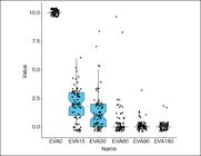
Regarding functional disability, measured by ODI, significant improvement was observed until 30 days after surgery. In subsequent periods (60, 90 and 180 days), scores remained stable, with no statistically significant difference (p = 0.4; p = 1; p = 1, respectively), as illustrated in Figure 2. The average time to return to work observed was 20.55 days.

In the comparative analysis between the two main anatomical levels operated (L4–L5 and L5–S1), no statistically significant differences in VAS and ODI scores were observed in any of the evaluated periods, indicating that the clinical and functional outcomes were similar, regardless of the level addressed (p > 0.05).
DISCUSSION
Lower back pain is one of the leading causes of global disability, affecting approximately 7.5% of the world population at some point in life.3 It is estimated that by the age of 30, almost half of adults have experienced at least one episode of significant lumbar pain. The lumbar disc hernia represents a frequent etiology in this scenario, being the main cause of sciatic pain and the most common indication for spine surgery, especially in men between the third and fourth decades of life. Surgery is mostly indicated in refractory cases to conservative treatment, which includes motor rehabilitation, weight control, analgesia and blockages.4
Historically, the surgical treatment of disc hernia began with the broad laminectomies described by Mixter and Barr in 1934, based on conventional posterior access with extensive removal of bone and ligament structures. Since then, the search for less invasive techniques has led to the development of procedures with lower morbidity. The introduction of microcirurgical discectomy and, more recently, endoscopic techniques, represents a milestone in the evolution of spine surgery. From the 1990s, with technological advances in optics and instrumentation, percutaneous endoscopic discectomy, notably with the works of Yeung, Foley and Smith, gained space as a minimally invasive, effective and reproducible alternative.5
Interlaminal endoscopic discectomy presents particular technical advantages in the lower levels of the lumbar spine, especially in L4-L5 and L5-S1, due to the greater amplitude of the interlaminal space. The technique allows direct access to the hernia with minimal manipulation of the dural bag, selective resection of the yellow ligament and the possibility of bone decompression of the lateral rejection, keeping the segmental stability. In addition to the reduction in hospitalization time, there is strong evidence of lower morbidity, less intraoperative bleeding and early functional return.6-8
In this study, the longitudinal analysis of VAS and ODI scores demonstrated significant clinical and functional improvement in the first weeks after surgery. The stability of these results between the periods of 60, 90 and 180 days reinforces the durability of the clinical effects of the endoscopic approach. These findings are compatible with the results described in international literature. Hua et al. (2018)9, for example, did not identify any clinically relevant differences in the outcomes of patients operated at levels L4-L5 and L5-S1, corroborating the data presented here, which also revealed no statistically significant difference between these anatomical levels.
The rate of reoperation observed (3.75%) was below the 5% described as an acceptable pattern for lumbar discectomias.4,10 Similarly, the incidence of durotomy was only 1.25%, lower than the average of 3.1% found in recent metaanalyses, with successful conservative management. These numbers reinforce the safety of the interlaminar technique when executed with appropriate selection criteria and technical rigor.
The average return time to work (20.5 days) was compatible with previous data, although this study did not stratify patients by occupation type, a recognizably important variable, since activities that require physical effort tend to require longer distances.11
Despite the positive results, this study presents limitations. The sample size (n=80) restricts the generalization of the findings. In addition, this is a retrospective analysis of a number of cases, without comparative control group with other techniques, which limits conclusive inferences about superiority of the endoscopic approach. Still, the presented data contribute to important clinical evidence on the efficacy and safety of interlaminal endoscopic discectomy, in particular at levels L4-L5 and L5-S1.
CONCLUSION
Interlaminal endoscopic discectomy presents itself as a safe and effective surgical technique for the treatment of lumbar disc hernia, with satisfactory clinical and functional results. The data demonstrated significant improvement in VAS pain scores and functional disability due to ODI in the first weeks after the procedure, with maintenance of results up to six months of follow-up. The low rate of complications and the possibility of endoscopic reintervention reinforce the role of the technique as a reliable alternative to conventional approaches.
The absence of statistically significant differences between operated levels (L4-L5 vs. L5-S1) reinforces the versatility of the technique, applicable to different levels of the lower lumbar spine. Despite the methodological limitations inherent in retrospective studies, the findings presented here contribute to the growing body of evidence that support the use of endoscopy as a relevant tool in the current scenario of spine surgery. Prospective clinical trials, with larger samples and prolonged follow-up, are needed to consolidate their long-term effectiveness.
-
Study conducted by the Clínica Atualli Spine Care. Rua Bela Cintra, 539, 2º andar, Consolação, São Paulo/SP, Brazil.
DATA AVAILABILITY DECLARATION
The contents underlying the research are available in the manuscript.
REFERENCES
-
1 Downie WW, Leatham PA, Rhind VM, Wright V, Branco JA, Anderson JA. Studies with pain rating scales. Ann Rheum Dis. 1987;37(4):378-81. doi: 10.1136/ard.37.4.378
» 10.1136/ard.37.4.378 -
2 Vigatto R, Alexandre NMC, Correa Filho HR. Development of a Brazilian portuguese version of the Oswestry Disability Index: cross-cultural adaptation, reliability, and validity. Spine (Phila Pa 1976). 2007;32(4):481-6. doi: 10.1097/01.brs.0000255075.11496.47
» 10.1097/01.brs.0000255075.11496.47 -
3 Wu A, March L, Zheng X, Huang J, Wang X, Zhao J, et al. Global low back pain prevalence and years lived with disability from 1990 to 2017: estimates from the Global Burden of Disease Study 2017. Ann Transl Med. 2020;8(6):299. doi: 10.21037/atm.2020.02.175
» 10.21037/atm.2020.02.175 - 4 Maroon JC. Current concepts in minimally invasive discectomy. Neurosurgery. 2002;51(5 Suppl):S137-S45.
-
5 Gadjradj PS, Rubinstein SM, Peul WC, Depauw PR, Vleggeert-Lankamp CL, Seiger A, et al. Full endoscopic versus open discectomy for sciatica: randomised controlled non-inferiority trial. BMJ. 2022;376:e065846. doi: 10.1136/bmj-2021-065846
» 10.1136/bmj-2021-065846 -
6 Assis RR, Defino HLA, Costa HRT, Dowling Á, Bergamaschi JPM. Surgery for lumbar disc herniation: open x minimally invasive technique. Coluna/Columna. 2021;20(1):47-9. doi: 10.1590/S1808-185120212001235649
» 10.1590/S1808-185120212001235649 -
7 Cong L, Zhu Y, Tu G. A meta-analysis of endoscopic discectomy versus open discectomy for symptomatic lumbar disk herniation. Eur Spine J. 2016;25(1):134-43. doi: 10.1007/s00586-015-3776-6
» 10.1007/s00586-015-3776-6 -
8 Gadjradj PS, Broulikova HM, van Dongen JM, Rubinstein SM, Depauw PR, Vleggeert C, et al. Cost-effectiveness of full endoscopic versus open discectomy for sciatica. Br J Sports Med. 2022;56(18):1018-25. doi: 10.1136/bjsports-2021-104808
» 10.1136/bjsports-2021-104808 -
9 Hua W, Tu J, Li S, Wu X, Zhang Y, Gao Y, et al. Full-endoscopic discectomy via the inter-laminar approach for disc herniation at L4-L5 and L5-S1: an observational study. Medicine (Baltimore). 2018;97(17):e0585. doi: 10.1097/MD.0000000000010585
» 10.1097/MD.0000000000010585 -
10 Desai A, Ball PA, Bekelis K, Lurie JD, Mirza SK, Tosteson TD, et al. Outcomes after incidental durotomy during first-time lumbar discectomy. J Neurosurg Spine. 2011;14(5):647-53. doi: 10.3171/2011.1.SPINE10426
» 10.3171/2011.1.SPINE10426 -
11 Song Z, Ran M, Luo J, Zhan K, Ye Y, Zheng J, et al. Follow-up results of microendoscopic discectomy compared to day surgery using percutaneous endoscopic lumbar discectomy for the treatment of lumbar disc herniation. BMC Musculoskelet Disord. 2021;22(1):160. doi: 10.1186/s12891-021-04038-6
» 10.1186/s12891-021-04038-6


