| Osteosarcoma |
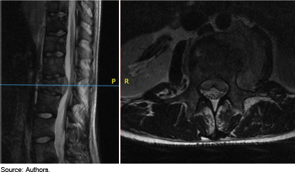
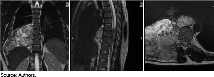
The most common primary malignant bone tumor. Spinal involvement is less frequent than in long bones |
Mixed lytic and blastic pattern (osteoid matrix). Periosteal reaction (“sunburst” or “Codman’s triangle”).1,3
|
| Primary Spinal Lymphoma |
Systemic B symptoms (fever, night sweats, weight loss), similar to ES. More common in older adults than in children. |
Diffuse marrow infiltration and lytic lesions. Less overt cortical bone destruction and a more homogeneous soft tissue. Definitive differentiation relies on histopathology and immunohistochemistry (CD45, CD20).1
|
| Metastatic Neuroblastoma |
The most common extracranial solid tumor in children. Metastatic lesions to the spine are typically lytic and can cause back pain and neurological deficits |
Differentiation often relies on the identification of the primary tumor, elevated urinary catecholamines (VMA, HVA), and specific immunohistochemical markers (e.g., synaptophysin, chromogranin) and molecular genetics (e.g., MYCN amplification) on biopsy, contrasting with the EWSR1 fusion characteristic of ES.3
|
| Eosinophilic Granuloma |
Can cause pain and even neurological symptoms due to vertebral collapse, but have a more favorable course and lack the systemic aggression of ES |
On radiographs, purely lytic, well-defined lesions. Common benign cause of “flat vertebra” (diffuse collapse of a vertebral body). MRI may show diffuse marrow signal changes without the large enhancing soft tissue component seen in ES.7
|
| Aneurysmal Bone Cyst |
Benign lesion that commonly affects the spine in children and adolescents. Pain, spinal deformity, and neurological symptoms due to mass effect. |
Expansile, osteolytic lesion with multiple fluid-fluid levels on MRI, reflecting blood products within cystic loculations, a pathognomonic feature not seen in ES.7
|
| Osteoblastoma |
Benign bone-forming tumor, typically affecting the posterior elements of the spine in children and young adults. Back pain is common. |
Well-circumscribed, often with a sclerotic rim on radiographs and CT, and may have a central nidus. Tend to be less aggressive and infiltrative than ES. The presence of osteoid matrix on biopsy is benign, unlike the malignant osteoid of osteosarcoma.1
|
| Spondylodiscitis and Vertebral Osteomyelitis |
Back pain is often accompanied by fever and elevated inflammatory markers (ESR, CRP). The presence of acute inflammatory markers and, sometimes, positive blood cultures or disc aspirates, aids in diagnosis |
On MRI, spondylodiscitis typically shows narrowing and enhancement of the disc space, along with T2 hyperintensity and enhancement of adjacent vertebral endplates. (Figure 3) While ES can cause diffuse marrow signal changes, disc space involvement is rare in ES, and the soft tissue mass in ES is usually more prominent and less centered on the disc.3
|
| Spinal Tuberculosis (Pott’s Disease) |
Remains a significant differential in endemic areas. Slow, progressive destruction of the vertebral bodies (often anteriorly), with relative sparing of the posterior elements initially, leading to severe kyphotic deformity (Figure 3). Systemic symptoms (night sweats, weight loss) |
diffuse marrow signal changes, extensive paraspinal abscesses (cold abscesses), and involvement of multiple contiguous vertebral bodies and intervening discs. This multifocal, multi-level involvement with significant disc pathology is less typical of ES, which usually affects a single or limited number of vertebrae.3
|

 Tumor de Ewing: Diagnóstico Diferencial e Bases para o Tratamento Oncológico e Cirúrgico
 Thumbnail
 Thumbnail
 Thumbnail
 Thumbnail
 Thumbnail
 Thumbnail
 Thumbnail