 DIFFERENT CERVICAL LAMINOPLASTY TECHNIQUES - UPDATE AND SYSTEMATIC REVIEW
ABSTRACT
This review aims to update the cervical laminoplasty techniques described in the literature and perform a systematic review of the open-door (ODL) and French-door (FDL) laminoplasties. A literature review was conducted on the Pubmed and Scielo platforms. This study was separated into two phases. The first section reviewed the laminoplasty techniques described in the literature. In this phase, a literature review was carried out using the descriptor “cervical laminoplasty”, employing “clinical trial”, “meta-analysis”, “review,” and “systematic review” as filters. The second section was a systematic review of the two most-studied techniques, the open-door and French-door laminoplasties. In this phase, a review was carried out using the descriptors “cervical laminoplasty”, “open-door” and “French-door” or “double-door”, employing “meta-analysis” and “review” as filters. In both phases, no limits were defined on the period. The first laminoplasty was described in 1973 and, since then, there have been, at least, six more techniques described that involve opening the spinal canal. Other studies report slight modifications in the type of spine access or details related to the osteotomy technique. Five systematic reviews were performed comparing the ODL and FDL. Only two of them analyzed common variables, and their results were divergent. Both ODL and FDL achieve favorable clinical and radiological results for the patient, and it is not yet possible to say whether one is better than the other. Level of Evidence II; Therapeutic Studies.
Keywords:
Laminoplasty; Cervix Uteri; Myelopathy; Spinal Stenosis; Spondylosis
RESUMO
O objetivo desta revisão é atualizar as técnicas de laminoplastia cervical descritas na literatura e realizar uma revisão sistemática das laminoplastias open-door (ODL) e French-door (FDL). Foi realizada uma revisão de literatura nas plataformas Pubmed e Scielo. Este estudo foi separado em duas fases. A primeira seção revisou as técnicas de laminoplastia descritas na literatura. Nessa fase, foi realizada uma revisão da literatura utilizando o descritor “laminoplastia cervical”, empregando como filtros “ensaio clínico”, “meta-análise”, “revisão” e “revisão sistemática”. A segunda seção foi uma revisão sistemática das duas técnicas mais estudadas, as laminoplastias de porta aberta e de porta francesa. Nessa fase, foi realizada uma revisão utilizando os descritores “laminoplastia cervical”, “open-door” e “French-door” ou “double-door”, empregando como filtros “meta-análise” e “revisão”. Em ambas as fases, não foram definidos limites para o período. A primeira laminoplastia foi descrita em 1973 e, desde então, houve, pelo menos, mais seis técnicas descritas que envolvem a abertura do canal vertebral. Outros estudos relatam pequenas modificações no tipo de acesso à coluna ou detalhes relacionados à técnica de osteotomia. Foram encontradas cinco revisões sistemáticas comparando o ODL e a FDL. Apenas duas delas analisaram variáveis em comuns e seus resultados foram divergentes. Tanto o ODL quanto o FDL alcançam resultados clínicos e radiológicos favoráveis para o paciente, e ainda não é possível dizer qual é melhor. Nível de Evidência II; Estudos Terapêuticos.
Descritores:
Laminoplastia; Colo do Útero; Mielopatia; Estenose Espinal; Espondilose
RESUMEN
El propósito de esta revisión es actualizar las técnicas de laminoplastia cervical descriptas en la literatura y realizar una revisión sistemática de las laminoplastias open-door (ODL) y French-door (FDL). Se realizó una revisión bibliográfica en las plataformas Pubmed y Scielo. Este estudio se dividió en dos fases. En el primer apartado se revisaron las técnicas de laminoplastia descriptas en la literatura. En esta fase se realizó una revisión bibliográfica utilizando el descriptor “laminoplastia cervical”, empleando como filtros “ensayo clínico”, “metaanálisis”, “revisión” y “revisión sistemática”. La segunda sección consistió en una revisión sistemática de las dos técnicas más estudiadas, la laminoplastia open-door y la laminoplastia French-door. En esta fase se realizó una revisión utilizando los descriptores “laminoplastia cervical”, “open-door” y “French-door” o “double-door”, empleando como filtros “metaanálisis” y “revisión”. En ambas fases no se definieron límites en el período. La primera laminoplastia se describió en 1973 y, desde entonces, se han descripto, al menos, seis técnicas más que implican la apertura del canal medular. Otros estudios reportan ligeras modificaciones en el tipo de acceso de la columna vertebral o detalles relacionados con la técnica de osteotomía. Se encontraron cinco revisiones sistemáticas comparando el ODL y el FDL. Solo dos de ellos analizaron las variables comunes y sus resultados fueron divergentes. Tanto la ODL como la FDL consiguen resultados clínicos y radiológicos favorables para el paciente, y aún no es posible decir si una es mejor que la otra. Nivel de Evidencia II; Estudios Terapêuticos.
Descriptores:
Laminoplastia; Cuello del Útero; Mielopatía; Estenosis Espinal; Espondilosis
INTRODUCTION
Surgical approaches to the cervical spinal canal can be performed either by the anterior route, through discectomy and/or corpectomy, both associated with fusion, or posterior access by isolated spinal canal decompression, laminoplasty, or laminectomy with fusion.1-3 The posterior approach ends up being selected for cases of spinal canal stenosis and multi-level degenerative diseases, especially in cases where cervical lordosis is preserved.1,3,4
Laminectomy has been the main technique used in these cases, but there have been some complications, such as instability, kyphosis, and the possibility of neurological deterioration.4-6 In the 1970s, Orthopedic surgeons developed a way of enlarging the diameter of the spinal canal while preserving the posterior arch of the vertebrae to prevent such complications: laminoplasty.7,8 Since then, laminoplasty has been accepted as one of the standard techniques for posterior decompression of the cervical spinal canal.3,9-11
Various laminoplasty techniques have been described throughout the years for improved cervical mobility preservation, decreased aggression on the muscles, less postoperative neck pain, and, consequently, a better quality of life for patients.12-19
The most common laminoplasties are the open-door and French-door. Both are safe and improve the myelopathy’s symptoms, but it is difficult to ascertain which is better.20-24
Therefore, this study aimed to update the laminoplasty techniques described in the literature and perform a systematic review of open-door and French-door laminoplasty.
METHODS
This study was divided into two phases. The first section reviewed the laminoplasty techniques described in the literature, and the second section was a systematic review of the two most studied techniques: open-door and French-door laminoplasties.
First phase
In April 2024, a literature review was carried out on the PubMed and Scielo platforms, using the descriptor “cervical laminoplasty” and employing “clinical trial,” “meta-analysis,” “review,” and “systematic review” as filters.
Initially, 66 articles were selected that included something related to types of laminoplasty techniques in their titles. The article abstracts were read, and those that dealt with a review of the subject or described a new type of laminoplasty or technical variation of open-door or French-door were chosen.
In the end, 20 articles were used to review the literature on the different types of laminoplasty.
Second Phase
A literature review was carried out on the PubMed and Scielo platforms, using the descriptors “cervical laminoplasty”, “open-door”, and “French-door” or “double-door”, employing “meta-analysis” and “review” as filters, in April 2024. No limits were defined on the period.
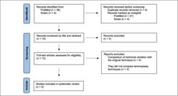
Initially, 36 articles were found. After reading the article abstracts, all narrative reviews were excluded, and only meta-analyses were used. This review complied with the Preferred Reported Items for Systematic Reviews and Meta-Analysis (PRISMA) guideline. (Figure 1)
RESULTS
Five meta-analyses were found comparing open-door and French-door laminoplasties. All meta-analyses used the PRISMA methodology to choose the articles, and only randomized controlled trials and retrospective analyses were included. All meta-analyses were classified as level II of evidence (Table 1). Table 2 summarizes the findings from each article, and Table 3 compiles the most frequently found in the meta-analyses.
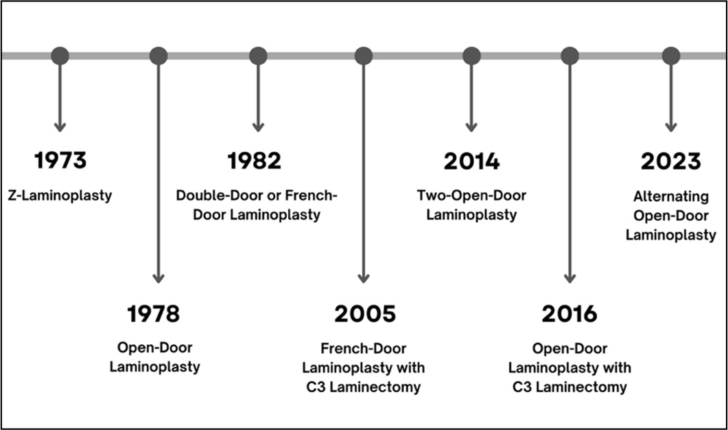
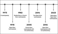
Figure 2 summarizes the cervical laminoplasty timeline. Seven types of laminoplasty have been identified since 1973. Next, the authors describe the main characteristics of the techniques.
DISCUSSION
The first laminoplasty
Oyama described the first laminoplasty in 1973 as a “Z-shaped expansive laminoplasty.” The laminae were cut in a “Z” shape, elevated, and sutured in the midline (Figure 3). The posterior spinal canal was reconstructed to avoid scar formation over the dural surface, preventing the invasion of scar tissue. All patients who underwent this technique improved clinically in the postoperative period.25
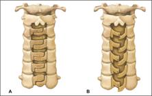
Z-laminoplasty. (A) Laminotomies are formed in each lamina. (B) Split laminae are elevated and fixed.
According to the author’s opinion of this review, this technique appears to be difficult to perform, which may have motivated the development of other types of laminoplasty years later.
Open-door laminoplasty
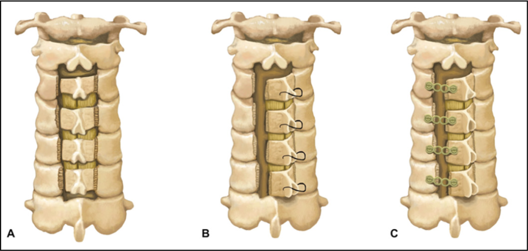
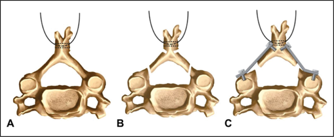
In 1978, Hirabayashi developed an epoch-making laminoplasty. This open-door expansive laminoplasty consists of a total laminotomy on one side and a hinge made on the opposite side by drilling the outer cortex of the laminae (Figure 4). Several levels of the spinal cord could be decompressed simultaneously, earlier mobilization of patients could be achieved, post-operative kyphotic deformity of the cervical spine could be avoided, and mobility of the cervical region maintained.7,8
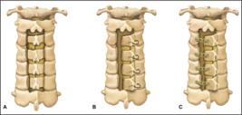
Open-door laminoplasty. (A) Total laminotomy on one side and a hinge made on the opposite side. (B) Laminae are kept open with sutures on the facet capsule on the hinge side or with (C) titanium plates.
During the 4.5-year follow-up period, most patients achieved favorable outcomes in this study, with a 66% increase in the JOA scale score. On average, the spinal canal’s diameter increased by 5 mm. Two patients showed worsening in the postoperative period, and the laminoplasty did not remain open. After these cases, the authors began to sew sutures between the ligamentum flavum and the deep muscles around the facets on the hinge side, which was enough to maintain spinal canal decompression.7
Subsequently, studies have shown that fastening the laminae with titanium plates is more effective in preventing post-operative complications, such as neurological damage and axial pain.23,26
French-door laminoplasty
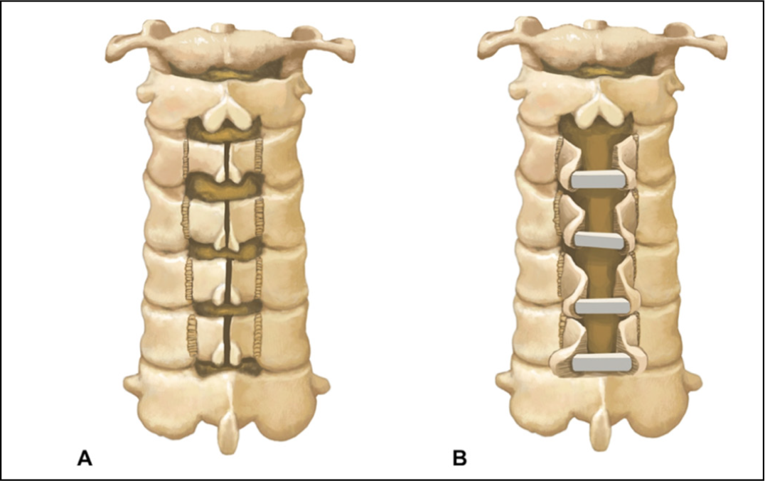
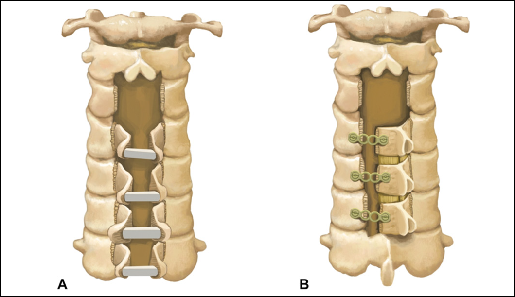
French-door laminoplasty, also described as double-door, consists of performing a longitudinal osteotomy of the spinous processes, dividing it in half along the midline, and creating the hinge on the laminae bilaterally (Figure 5). The spacers can be autologous, homologous, or synthetic bone grafts. They are placed between the flaps of the spinous processes to keep the spinal canal open. The final design of the laminoplasty resembles a French window, hence its name. Kurokawa described this technique in 1982.27
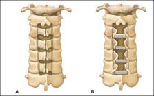
The French-door laminoplasty. (A) Longitudinal osteotomy of the spinous processes and the hinge in the laminae bilaterally. (B) Laminae are elevated bilaterally.
The author defends the great advantage of French-door laminoplasty, which allows symmetrical decompression on both sides of the spinal canal.27 One of the disadvantages of this technique is the increased chance of dural laceration and spinal cord injury, especially in extreme cases of canal stenosis, such as in patients with ossification of the posterior longitudinal ligament.11
Some authors consider this laminoplasty to be technically difficult due to the sectioning of the spinous process. Tomita et al. (1998)28 described using a Gigli saw to section the spinous processes and considered the method safe and effective, as they did not observe any dural injury in any single case.
Open-Door vs French-Door: Which one is best?
Nakashima et al. (2014)29 compared the two laminoplasty techniques (Open-door and French-door) in a randomized study involving 92 patients with cervical spondylotic myelopathy. It concluded that there was no difference regarding neurological recovery or perioperative complications. However, the average reduction in post-operative cervical lordosis was significantly greater in patients who underwent open-door laminoplasty. Therefore, they suggest that French-door laminoplasty is preferable to open-door laminoplasty for maintaining cervical alignment.
Five meta-analyses were found comparing the two techniques in the literature review. Wang et al. (2015)20 considered the open-door laminoplasty group to achieve better post-operative recovery. However, Luo et al. (2018)21 regarded the canal expansion as superior to the open-door technique; thus, it has not yet been possible to conclude that either of these procedures is more effective than the other. Other authors who revealed that open-door laminoplasty presented more adverse events than French-door laminoplasty deemed French-door laminoplasty to be more effective and safer.23 Chen et al. (2021)24 suggested that both methods could achieve good postoperative results. However, they argue that French-door laminoplasty is superior because this group experienced less intraoperative blood loss and axial pain. Wiguna et al. (2019)22 suggested that open-door laminoplasty improved functional outcomes and recovery rates compared to French-door laminoplasty.
After analyzing these five studies, the authors of this review observed that open-door laminoplasty appears to be superior in three meta-analyses. However, only two studies analyzed common variables, and their results were divergent. Therefore, it is still difficult to define whether one technique is superior to the other.
The lateral recess cannot be forgotten when decompression is performed, and, in the authors’ opinion, French-door laminoplasty does not seem to promote decompression of this portion of the vertebral canal since the laminae are open in the midline. So, open-door laminoplasty may be more efficient considering decompression involving both the midline and the lateral recess. A radiological study of the spinal canal before and after decompression can be interesting to evaluate and try to clarify this doubt.
Another factor that must be considered when choosing the technique is the surgeon’s familiarity with it. Cases where the surgeon is proficient in the applied technique will have a better clinical outcome.
Open-door laminoplasty variations
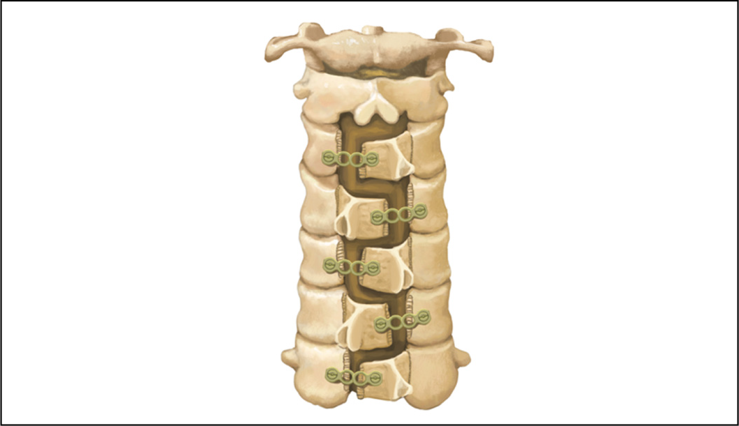
Arantes Júnior et al. (2014)30 idealized the two-open-door technique, where an opening is made in the C3 and C4 laminae on one side and C5 and C6 on the opposite side (Figure 6). This makes it possible to perform bilateral C4-C5 foraminotomy, avoiding C5 root neuropraxia. It also promotes three arthrodesis points between 1) the C3 lamina and the C2 spinous process, 2) the C4 and C5 laminae, and 3) the C6 lamina to the C7 spinous process. Their study included 86 patients in a five-year follow-up period. The spinal canal increased in diameter (from 11 mm to 17 mm); there were no cases of instability and 88% functional improvement on the Nurick scale.
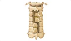
The great advantage of this technique is the possibility of performing bilateral foraminotomy. However, since there are arthrodesis points, cervical mobility may decrease over time and worsen patients’ quality of life.
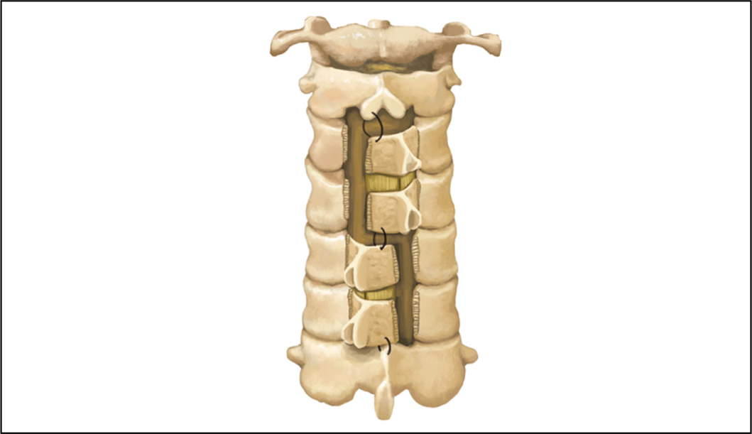
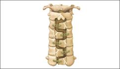
Huang et al. (2023)31 also described a variation of the open-door technique similar to Arantes’s. He approached from C3 to C7 and made alternating openings in C3, C5, and C7, as the openings of the laminae were on the left and in C4 and C6 on the right (Figure 7). These authors performed this procedure on a single 74-year-old patient, and there was an improvement in the mJOA score (preoperative score of 8 pts./14th postoperative day: 12 points/after five years of the segment: 14 points), neck pain was absent, and preserved cervical stability. The idea of performing alternating decompression may promote a wider decompression of the spinal canal, but as there are increased cutting areas of the blades, surgical morbidity may increase. Therefore, the authors believe that to validate this technique, it is necessary to increase the number of operated cases and, preferably, to develop a study comparing it with the previously established techniques.
Suh et al. (2016)13 developed a variation of open-door laminoplasty in which the cranial portion of the posterior arch of each vertebra was resected in a wedge shape. That was to avoid impact between neighboring laminae, avoid segmental fusion, and reduce the incidence of postoperative neck pain. In a randomized study, the authors compared this technique to Hirabayashi’s and concluded that wedge resection provided decreased postoperative neck pain and increased preservation of cervical mobility.
Laminoplasty associated with C3 laminectomy
The literature reports that cervical mobility may decrease after open-door laminoplasty, mainly due to interlaminar fusion between the C2-C3 and/or C3-C4 segments. This may cause limited mobility and interfere with quality of life.32
Takeuchi et al. (2005)33 performed C3 laminectomy associated with French-door laminoplasty at the other levels based on this principle (Figure 8a). They showed that this variation in the technique decreases the incidence of postoperative neck pain. Chen et al. (2021)34 compared the clinical results of C3 laminectomy to French-door laminoplasty versus classic French-door laminoplasty and observed that C3 laminectomy could reduce operating time, maintain cervical mobility, and reduce the incidence of postoperative neck pain.
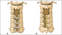
Lee et al. (2016)12 performed C3 laminectomy associated with laminoplasty at the other levels (Figure 8b). The study included 59 patients, retrospectively, with a three-year follow-up period. Fourteen patients underwent C3 laminectomy + open-door laminoplasty at the other levels. None of these cases displayed any sign of fusion between C2-C3 and/or C3-C4, while 42.2% of the patients who underwent laminoplasty, including C3, incurred interlaminar bone fusion. Therefore, the authors concluded that C3 laminectomy associated with open-door laminoplasty prevented interlaminar bone fusion in the segments. This resulted in better preservation of cervical mobility without the development of focal kyphosis or instability.
Muscle injury
Some technical details have been studied to preserve the paravertebral muscles to reduce postoperative axial pain. Meta-analysis studies have proven a significant reduction in the incidence and severity of axial symptoms and an improvement in cervical mobility, especially by preserving the muscle attachment in the spinous processes of C2 and C7.15,19 Lin et al. (2023)17 developed a technical variation of the French-door which preserved the muscle insertion in the spinous process of C2 and C7 and performed laminectomy of C3 and laminoplasty in C4, C5, and C6. The authors compared this technique to the traditional French-door technique and observed spinal cord decompression and neurological recovery like in the conventional technique. Additionally, allowing a lower incidence of postoperative neck pain and improved angle of movement of the cervical spine without causing instability.
A description of a minimally invasive technique of open-door laminoplasty is included. Benglis et al. (2008)35 conducted a study on six cadavers using tubular access through two small incisions at the C4 and C6 levels. It is possible to access the other levels and proceed to laminoplasty by moving the cranial tube caudally; the great advantage of this technique is decreased muscle lesions as they are not detached from the midline.
Wang et al. (2021)16 developed a bilateral laminotomy technique associated with longitudinal separation of the spinous process, preserving part of the paravertebral musculature. Subsequently, the lamina was fastened to the lateral mass of the same vertebra, thus enlarging the vertebral canal. This method was applied only in the posterior arch of C6, and in the other segments, open-door laminoplasty was performed. These authors observed a significant increase in the spinal canal area. There were not any complications, and there was a postoperative functional improvement, but postoperative neck pain had not been evaluated in the study.
It is already evident that muscle preservation benefits the patient, so this should always be considered during laminoplasty, which is the intention of the technique used.
Endoscopic laminoplasty
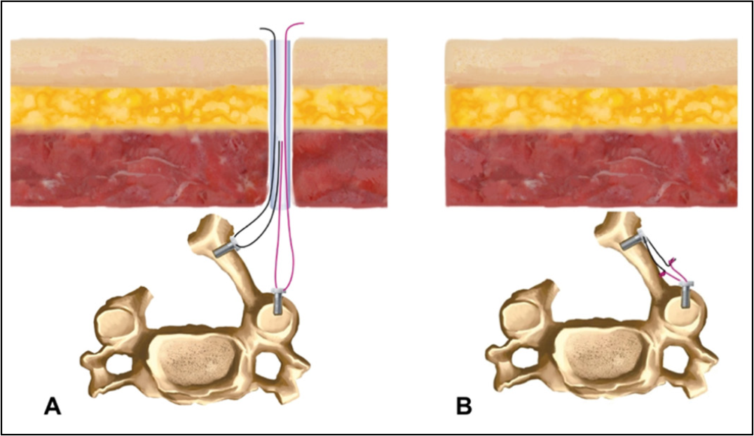
New laminoplasty techniques have been described, with promising results due to the advent and expansion of endoscopic spine surgery.14,18 Zhang et al. (2016)14 operated on 45 patients employing an endoscopic laminoplasty technique and observed clinical improvement based on the mJOA scale. The vertebral canal increased the diameter from 1 to 3 mm, and there was no collapse of the laminoplasty. They performed bilateral endoscopic laminotomy associated with traction of the posterior arch of the vertebra, by decompressing the vertebral canal. Then, the laminae were fastened to the lateral mass by titanium plates (Figure 9). Zhu et al. (2022)18 reported the case of a patient with stenosis of the cervical vertebral canal from C3 to C6 who applied a biportal endoscopic technique and underwent open-door laminoplasty, including laminae fastening by suturing to prevent their closure (Figure 10). There was clinical improvement in the immediate postoperative period, and an MRI of the cervical spine showed effective decompression of the spinal cord.

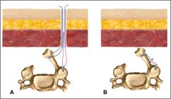
Undoubtedly, the endoscopic technique tends to become a much more common practice, but the learning curve will be long, and the benefit for the patient should be indisputable.
CONCLUSIONS
Several technical variations of cervical laminoplasty exist. All approaches appear to be safe for the patient, and there are few complications. Reserving muscle insertion in the spinous processes of C2 and C7 allows for decreased postoperative neck pain, regardless of the technique.
Both open-door and French-door laminoplasty yield favorable clinical and radiological results for the patient, and it is not yet possible to say whether one is superior to the other. Undoubtedly, the surgeon’s familiarity with the surgical technique is a criterion that must be considered when defining which approach to use.
-
Study conducted by the Faculdade Evangélica Mackenzie, Curitiba, PR, Brazil.
REFERENCES
-
1 Benzel EC, Lancon J, Kesterson L, Hadden T. Cervical laminectomy and dentate ligament section for cervical spondylotic myelopathy. J Spinal Disord. 1991;4(3):286-95. doi: 10.1097/00002517-199109000-00005.
» https://doi.org/10.1097/00002517-199109000-00005. -
2 Fehlings MG, Wilson JR, Kopjar B, Yoon ST, Arnold PM, Massicotte EM, et al. Efficacy and safety of surgical decompression in patients with cervical spondylotic myelopathy results of the aospine north america prospective multi-center study. J Bone Joint Surg Am. 2013;95(18):1651-8. doi: 10.2106/JBJS.L.00589.
» https://doi.org/10.2106/JBJS.L.00589. -
3 Zhong H, Xu C, Wang R, Wu X, Wu H, Sun B, et al. Anterior cervical discectomy and fusion, open-door laminoplasty, or laminectomy with fusion: Which is the better treatment for four-level cervical spondylotic myelopathy?. Front Surg. 2023;9:1065103. doi: 10.3389/fsurg.2022.1065103.
» https://doi.org/10.3389/fsurg.2022.1065103. -
4 Naderi S, Benzel EC, Baldwin NG. Cervical spondylotic myelopathy: surgical decision making. Neurosurg Focus. 1996;1(6):e1. doi: 10.3171/foc.1996.1.6.1.
» https://doi.org/10.3171/foc.1996.1.6.1. -
5 Buell TJ, Buchholz AL, Quinn JC, Shaffrey CI, Smith JS. Importance of sagittal alignment of the cervical spine in the management of degenerative cervical myelopathy. Neurosurg Clin N Am. 2018;29(1):69-82. doi: 10.1016/j.nec.2017.09.004.
» https://doi.org/10.1016/j.nec.2017.09.004. -
6 Alam I, Sharma R, Borkar S, Goda R, Katiyar V, Kale S. Factors predicting loss of cervical lordosis following cervical laminoplasty: A critical review. J Craniovertebr Junction Spine. 2020;11(3):163-8. doi: 10.4103/jcvjs.JCVJS_70_20.
» https://doi.org/10.4103/jcvjs.JCVJS_70_20. -
7 Hirabayashi K, Watanabe K, Wakano K, Suzuki N, Satomi K, Ishii Y. Expansive open-door laminoplasty for cervical spinal stenotic myelopathy. Spine (Phila Pa 1976). 1983;8(7):693-9. doi: 10.1097/00007632-198310000-00003.
» https://doi.org/10.1097/00007632-198310000-00003. -
8 Hirabayashi K, Satomi K. Operative procedure and results of expansive open-door laminoplasty. Spine (Phila Pa 1976). 1988;13(7):870-6. doi: 10.1097/00007632-198807000-00032.
» https://doi.org/10.1097/00007632-198807000-00032. -
9 Bakhsheshian J, Mehta VA, Liu JC. Current diagnosis and management of cervical spondylotic myelopathy. Global Spine J. 2017;7(6):572-86. doi: 10.1177/2192568217699208.
» https://doi.org/10.1177/2192568217699208. -
10 Wilson JR, Tetreault LA, Kim J, Shamji MF, Harrop JS, Mroz T, et al. State of the art in degenerative cervical myelopathy: An update on current clinical evidence. Clin Neurosurg. 2017;80(3S):S33-S45. doi: 10.1093/neuros/nyw083.
» https://doi.org/10.1093/neuros/nyw083. -
11 Weinberg DS, Rhee JM. Cervical laminoplasty: Indication, technique, complications. J Spine Surg. 2020;6(1):290-301. doi: 10.21037/jss.2020.01.05.
» https://doi.org/10.21037/jss.2020.01.05. -
12 Lee DH, Cho JH, Hwang CJ, Lee CS, Cho SK, Ha JK. Can C3 laminectomy reduce interlaminar bony fusion and preserve the range of motion after cervical laminoplasty?. Spine (Phila Pa 1976). 2016;41(24):1884-90. doi: 10.1097/BRS.0000000000001852.
» https://doi.org/10.1097/BRS.0000000000001852. -
13 Suh BG, Ahn MW, Kim HJ, Yeom JS, Lee GW. Wedge-shaped resection of the posterior bony arch during open door laminoplasty to prevent postoperative motion limitation. Spine (Phila Pa 1976). 2017;42(3):143-50. doi: 10.1097/BRS.0000000000001689.
» https://doi.org/10.1097/BRS.0000000000001689. -
14 Zhang C, Li D, Wang C, Yan X. Cervical endoscopic laminoplasty for cervical myelopathy. Spine (Phila Pa 1976). 2016;41(Suppl 19):B44-B51. doi: 10.1097/BRS.0000000000001816.
» https://doi.org/10.1097/BRS.0000000000001816. -
15 Qi Q, Chen Y, Ling Z, Hu H, Ma J, Guo W, et al. Modified laminoplasty preserving the posterior deep extensor insertion into C2 improves clinical and radiologic results compared with conventional laminoplasty: A meta-analysis. World Neurosurg. 2018;111:157-65. doi: 10.1016/j.wneu.2017.12.098.
» https://doi.org/10.1016/j.wneu.2017.12.098. -
16 Wang H, Zhang L. Expansion of spinal canal with lift-open laminoplasty: A new method for compression cervical myelopathy. Orthop Surg. 2021;13(5):1673-81. doi: 10.1111/os.13026.
» https://doi.org/10.1111/os.13026. -
17 Lin W, Song J, Zhang Y, Yao S, Yi M, Yao M, et al. Comparison of clinical outcomes of modified laminoplasty with preservation of muscle group inserted into C2 and C7 spinous processes versus conventional C3-C7 laminoplasty: a prospective, randomized, controlled, noninferiority trial. Int J Surg. 2023;109(4):905-12. doi: 10.1097/JS9.0000000000000358.
» https://doi.org/10.1097/JS9.0000000000000358. -
18 Zhu C, Wang J, Cheng W, Wang D, Pan H, Zhang W. Case Report: Bilateral biportal endoscopic open-door laminoplasty with the use of suture anchors: A technical report and literature review. Front Surg. 2022;9:913456. doi: 10.3389/fsurg.2022.913456.
» https://doi.org/10.3389/fsurg.2022.913456. -
19 Qu L, Li Z, Wang X, Yuan L, Li C. Axial symptoms after conventional and modified laminoplasty: A meta-analysis. World Neurosurg. 2023;180:112-22. doi: 10.1016/j.wneu.2023.09.086.
» https://doi.org/10.1016/j.wneu.2023.09.086. -
20 Wang L, Wang Y, Yu B, Li Z, Liu X. Open-door versus French-door laminoplasty for the treatment of cervical multilevel compressive myelopathy. J Clin Neurosci. 2015;22(3):450-5. doi: 10.1016/j.jocn.2014.08.022.
» https://doi.org/10.1016/j.jocn.2014.08.022. -
21 Luo W, Li Y, Zhao J, Gu R, Jiang R, Lin F. Open-versus French-Door laminoplasty for the treatment of cervical multilevel compressive myelopathy: A meta-analysis. World Neurosurg. 2018;117:129-36. doi: 10.1016/j.wneu.2018.06.026.
» https://doi.org/10.1016/j.wneu.2018.06.026. -
22 Wiguna IGLNAA, Magetsari R, Noor Z, Suyitno S, Nindrea RD. Comparative effectiveness and functional outcome of open-door versus french-door laminoplasty for multilevel cervical myelopathy: A meta-analysis. Open Access Maced J Med Sci. 2019;7(19):3348-52. doi: 10.3889/oamjms.2019.739.
» https://doi.org/10.3889/oamjms.2019.739. -
23 Ma JX, Han XZ, Wang XY. Comparison of single versus double door posterior cervical laminoplasty for patients with cervical spondylotic myelopathy: A systematic review and meta-analysis. Medicine (United States). 2020;99(25):E20538. doi: 10.1097/MD.0000000000020538.
» https://doi.org/10.1097/MD.0000000000020538. -
24 Chen T, Zhang X, Meng F, Zhang T, Zhao Y, Yan J, et al. Open-door versus French-door laminoplasty for patients with multisegmental cervical spondylotic myelopathy: A systematic review and meta-analysis. World Neurosurg. 2021;155:82-93. doi: 10.1016/j.wneu.2021.08.032.
» https://doi.org/10.1016/j.wneu.2021.08.032. -
25 Hirano Y, Ohara Y, Mizuno J, Itoh Y. History and evolution of laminoplasty. Neurosurg Clin N Am. 2018;29(1):107-13. doi: 10.1016/j.nec.2017.09.019.
» https://doi.org/10.1016/j.nec.2017.09.019. -
26 Mo Z, Li D, Zhang R, Chang M, Yang B, Tang S. Comparison of three fixation modalities for unilateral open-door cervical laminoplasty: a systematic review and network meta-analysis. Neurosurg Rev. 2020;43(3):813-23. doi: 10.1007/s10143-018-1035-0.
» https://doi.org/10.1007/s10143-018-1035-0. -
27 Kurokawa R, Kim P. Cervical laminoplasty: The history and the future. Neurol Med Chir (Tokyo). 2015;55(7):529-39. doi: 10.2176/nmc.ra.2014-0387.
» https://doi.org/10.2176/nmc.ra.2014-0387. -
28 Tomita K, Kawahara N, Toribatake Y, Heller JG. Expansive midline T-saw laminoplasty (modified spinous process-splitting) for the management of cervical myelopathy. Spine (Phila Pa 1976). 1998;23(1):32-7. doi: 10.1097/00007632-199801010-00007.
» https://doi.org/10.1097/00007632-199801010-00007. -
29 Nakashima H, Kato F, Yukawa Y, Imagama S, Ito K, Machino M, et al. Comparative effectiveness of open-door laminoplasty versus French-door laminoplasty in cervical compressive myelopathy. Spine (Phila Pa 1976). 2014;39(8):642-7. doi: 10.1097/BRS.0000000000000252.
» https://doi.org/10.1097/BRS.0000000000000252. -
30 Arantes Júnior AA, da Silva Junior GA, Malheiros JA, Gonçalves FF, Magaldi M, Santiago E, et al. A new expansive two-open-doors laminoplasty for multilevel cervical spondylotic myelopathy: Technical report and follow-up results. Arq Neuropsiquiatr. 2014;72(1):49-54. doi: 10.1590/0004-282X20130240.
» https://doi.org/10.1590/0004-282X20130240. -
31 Huang X, Liu D, Yang Y, Qiu H, Ma Z, Lei W, et al. A novel surgical technique for cervical laminoplasty in patients with multilevel cervical spondylotic myelopathy: A case report and literature review. Front Surg. 2023;10:1078138. doi: 10.3389/fsurg.2023.1078138.
» https://doi.org/10.3389/fsurg.2023.1078138. -
32 Iizuka H, Iizuka Y, Nakagawa Y, Nakashima T, Toda N, Shimegi A, et al. Interlaminar bony fusion after cervical laminoplasty: its characteristics and relationship with clinical results. Spine (Phila Pa 1976). 2006;31(6):644-7. doi: 10.1097/01.brs.0000203707.79269.6a.
» https://doi.org/10.1097/01.brs.0000203707.79269.6a. -
33 Takeuchi K, Yokoyama T, Aburakawa S, Saito A, Numasawa T, Iwasaki T, et al. Axial symptoms after cervical laminoplasty with C3 laminectomy compared with conventional C3-C7 laminoplasty: a modified laminoplasty preserving the semispinalis cervicis inserted into axis. Spine (Phila Pa 1976). 2005;30(22):2544-9. doi: 10.1097/01.brs.0000186332.66490.ba
» https://doi.org/10.1097/01.brs.0000186332.66490.ba -
34 Chen T, Zhang X, Meng F, Yan J, Xu G, Zhao W. Is laminoplasty or laminectomy the best strategy for C3 segment in French-door laminoplasty? A systematic review and meta-analysis. J Orthop Surg Res. 2021;16(1):557. doi: 10.1186/s13018-021-02596-y.
» https://doi.org/10.1186/s13018-021-02596-y. -
35 Benglis DM, Guest JD, Wang MY. Clinical feasibility of minimally invasive cervical laminoplasty. Neurosurg Focus. 2008;25(2):E3. doi: 10.3171/FOC/2008/25/8/E3.
» https://doi.org/10.3171/FOC/2008/25/8/E3.
Edited by
-
Reviewed by:
Aluízio Augusto Arantes