 MODIFIED THREE-DIMENSIONAL CLASSIFICATION OF THE ADOLESCENT IDIOPATHIC SCOLIOSIS
ABSTRACT
Objective: This study aims to present the modifications to the Modified Three-Dimensional Classification (MTC) and show the results of its use in the classification of radiographic images of the spine of four hundred and four patients who underwent surgery for Adolescent Idiopathic Scoliosis (AIS).
Method: MTC was used to classify the radiographic images of the spine of four hundred and four patients who had undergone surgery for AIS. A panoramic X-ray of the spine was performed in the orthostatic position, in the lateral and anterior pos-teroanterior views, neutral and with forced inclination to the right and left. The results of the classification variables and the relevant patient data were collected from the medical records and a biostatistical study was carried out. The MTC is made up of three factors: quantitative, localizing and sagittal.
Results: A higher incidence of double curve scoliosis was observed. The main curve predominated in the distal thoracic region. The sagittal plane showed a higher incidence of normal angle. There was a higher incidence of hyperlordosis in females. There was a predominance of hypokyphosis in younger individuals. The results tables show that these predominances occurred with significant differences.
Conclusion: The study of the MTC variables applied to the classification of patients undergoing AIS surgery showed a three-dimensional scope, and therefore concludes a truly three-dimensional classification. Level of Evidence III; Retrospective Study.
Keywords:
Scoliosis; Idiopathic; Classification; Tridimensional
Resumo:
Objetivo: Apresentar as modificações da Classificação Tridimensional Modificada (CTM), e mostrar os resultados do seu emprego na classificação das imagens radiográficas da coluna vertebral, de quatrocentos e quatro pacientes operados de Escoliose Idiopática do Adolescente (EIA).
Método: Usou-se a CTM para classificar as imagens radiográficas da coluna vertebral, de quatrocentos e quatro pacientes operados de EIA. Para tal, realizou-se Rx panorâmica da coluna, na posição ortostática, nas incidências em perfil e póstero anterior, neutro e com inclinação forçada para direita e esquerda. Colheu-se os resultados das variáveis da classificação e os dados pertinentes dos pacientes nos prontuários, realizou-se o estudo bioestatístico. A CTM é composta de três fatores: quantitativo, localizador e sagital.
Resultados: Observou-se maior incidência da escoliose de dupla curva. Predominância da curva principal na região torácica distal. O plano sagital apresentou maior incidência de ângulo normal. Maior presença de hiperlordose no sexo feminino. E predomínio de hipocifose nos indivíduos mais jovens. Pode-se visualizar nas tabelas dos resultados, que essas predominâncias, ocorreram com diferenças significativas.
Conclusão: O estudo das variáveis da CTM aplicada na classificação de pacientes operados de EIA, mostrou ser de âmbito tridimensional, portanto, conclui ser uma classificação realmente tridimensional. Nível de Evidência III; Estudo Retrospectivo.
Descritores:
Escoliose ; Idiopática; Classificação; Tridimensional
Resumen:
Objetivo: Presentar las modificaciones de la Clasificación Tridimensional Modificada (CTM) y mostrar los resultados de su utilización en la clasificación de imágenes radiográficas de la columna vertebral de cuatrocientos cuatro pacientes operados de Escoliosis Idiopática del Adolescente (EIA).
Método: Se utilizó la CTM para clasificar las imágenes radiográficas de la columna vertebral de cuatrocientos cuatro pacientes operados de AIS. Para ello, se realizó una radiografía panorámica de la columna vertebral en posición ortostática, en las vistas lateral y posteroanterior, neutra y con inclinación forzada a derecha e izquierda. Los resultados de las variables de clasificación y los datos relevantes del paciente se recogieron de las historias clínicas y se realizó un estudio bioestadístico. La CTM se compone de tres factores: cuantitativo, localizador y sagital.
Resultados: Se observó una mayor incidencia de escoliosis de doble curva. La curva principal predominaba en la región torácica distal. El plano sagital mostró mayor incidencia de ángulo normal. Mayor presencia de hiperlordosis en el sexo femenino. Y un predomínio de la hipocifosis en los indivíduos más jóvenes. Las tablas de resultados muestran que estos predomínios ocurrieron con diferencias significativas.
Conclusión: El estudio de las variables de la CTM aplicadas a la clasificación de los pacientes sometidos a cirugía de EIA demostró que se trata de una clasificación tridimensional. Nivel de Evidencia III; Estudio Retrospectivo.
Descriptores:
Escoliosis; Idiopática; Clasificación; Tridimensional
INTRODUCTION
The AIS is a three-dimensional deformity of the spine that encompasses the curvature of the coronal plane and rotation on the axial axis, with maximum rotational and translational deviations occurring at the apex of the curve.1 The classification by King et al. was first published in 1983, evaluates the deformity in the coronal plane, describes five types of thoracic curves, and recommends levels of vertebral segments to be included in the arthrodesis.2
The Lenke et al. classification (LC) was developed in 2001 and has three components: Six types of curves (1 to 6); the lumbar column modifier (A, B or C); and the sagittal thoracic modifier (−, N or +).3 The authors emphasize that the corrective surgical strategy for AIS may need to incorporate spinopelvic parameters in the classification to achieve a balanced spine, requiring minimal energy expenditure.4
Coonrad et al. reviewed two thousand cases of idiopathic scoliosis and described 21 types of curves. The author indicated 98.7% and 100% for inter-observer and intra-observer reliability, respectively.5 Kadoury et al. (2012) seek to understand how to classify and quantify three-dimensionally in (3D) the deformities of the spine. They studied 170 patients with AIS, Lenke type 1, when the results showed that multiple representation can potentially be useful for the classification of spinal pathologies in 3D, such as AIS, and serve as a tool to understand the progression of deformities in longitudinal studies.6 Qiu et al. defined the PUMC classification system in 2005. This method is useful for determining the surgical approach and defining the related fusion levels, and there are 3 main categories such as Type I (single curve), Type II (double curve), and Type III (triple curve) with a total of 13 subtypes.7
Comparing the PUMC and Lenke classification systems, Qiu et al. stated that the PUMC classification system is relatively simpler and the inter- and intra-observer variability causes less confusion in surgical planning.8 In 2018, Garcia et al. created the Three-Dimensional Classification (CT), composed of three components: the first called the quantitative factor, which is divided into three types according to the number of curves; the second, the locator factor, highlights the main curve, and the third, sagittal factor, which globally assesses the sagittal plane.9
OBJECTIVE
Present the modifications of the Modified Three-Dimensional Classification (MTC), and show the results of its application in the classification of radiographic images of the spine, of four hundred and four patients operated on for Adolescent Idiopathic Scoliosis (AIS).
MATERIAL AND METHOD
Project approved by Scientific Committee. The study had the knowledge free, clarified, and signed by all participants. In 2023, four hundred and four medical records of patients with AIS were studied, all of whom underwent surgical correction using transpedicular fixation in our service, without any specific selection, and they presented common characteristics of the disease in question. All complete and available records have been included. 83 cases that did not meet the requirements were excluded.
Complementary changes were made to the Three-Dimensional Classification, such as the inclusion of Sacral Incidence in the measurement of the sagittal plane. Residual curves are considered to be all those equal to or greater than 10°. The column was divided into three segments: (A), called cervicothoracic (from cervical to T5), (B), distal thoracic (from T5 to T12), and (C), lumbar, (from L1 to the sacrum). Radiological images from the pre-operative phase of the case in question were used, and radiographs taken in panoramic view of the spine were evaluated, with the patient standing, in the posteroanterior (PA) incidences, profile, and in PA with forced inclination to the right and left.
This MTC study used three specific assessments, referring to the Coronal and Sagittal planes. Three Factors are considered: Quantitative Factor, the Locator Factor, and the Sagittal Factor. The quantitative factor considers: Type I (single curve), Type II (double curve), and Type III (triple curve). The Locator Factor is related to the apex of the main curve in relation to the studied segment of the column. It was divided into three segments: A, B and C. It is considered (A) when the apex of the main curve is located in the cervico-thoracic region. The type (B) when the apex of the curve is more structurally located in the distal thoracic column. E (C) when the apex of the curvature of greater dimension is observed in the lumbar column. Type 2B is subdivided into 2BT when the compensatory curve is located in the cervicothoracic region. It considers 2BL when the compensatory curve is located in the lumbar column.
The sagittal factor of the MTC evaluates the entire sagittal plane of the spine using the Cobb method. The entire extent of the cyphotic area was measured, from the upper terminal vertebra to the lower one. Hypocifosis is considered when the angle is less than 25°, normal when the angle is between 25° and 45°, and hypercifosis above 45°. Lumbar lordosis is measured from its beginning to S1. Hypolordosis is considered (−) with Cobb below 40°, Normal (N) from 40 to 60°, and Hyperlordosis (+) with Cobb above 60°. The measurement of the Sacral Incidence in degrees has been completed. From 30° to 50° (N) normal, below would be (−) hypo and above corresponds to (+) hyper. A statistical study was conducted on the incidence of the MTC variables, describing characteristics and scales in patients with AIS and relating personal characteristics to the scales, as well as the scales to each other. For the analyses, IBM-SPSS for Windows version 22.0 was used, and for data tabulation, Microsoft Excel 2013 was used. The tests were conducted with a significance level of 5%.10,11
RESULTS
Table 1. Description of the characteristics evaluated in all patients.
Table 2. Description of the scales according to the patients’ sex and the results of the association tests.
Description of the scales according to the sex of the patients and the results of the association tests.
Table 2 shows that in female patients the frequency of hyper (+) lumbar sagittal factor was statistically higher than in male patients (p = 0.033).
According to Table 3, there was no statistically significant association between the patients’ color and the evaluated scales (p > 0.05).
Description of the scales according to the patients’ color and the results of the association tests.
Table 4 shows that there was a statistically significant average difference in the age of patients according to the thoracic sagittal factor (p = 0.009), with the patient with this factor of hypokyphosis being younger than the patient with this normal factor (p = 0.007).
Table 5 shows that there was a statistically significant association of all other scales with the quantitative factor (p < 0.05), with higher quantitative values being associated with lower expression in the sagittal factors, and patients with factor 2 presented statistically more of locator factor C (p < 0.001).
Description of the other scales according to the quantitative factor and the results of the association tests.
According to Table 6, there was no statistically significant association of the locator factor with the sagittal factors (p > 0.05).
Description of the other scales according to the locator factor and the results of the association tests.
Table 7 shows that the thoracic sagittal factor was statistically associated with the lumbar sagittal factor in a direct manner (p < 0.001).
Description of the other scales according to thoracic sagittal factor and the results of the association tests.
According to Table 8, there was a statistically significant association of the lumbar sagittal factor with the sacral sagittal factor, with a direct association between the scales; people who were more hypoexpressive in one parameter were more hypoexpressive in the other parameter, and people who were more hyperexpressive in one parameter were more hyperexpressive in the other (p < 0.001).
Description of the other scales according to lumbar sagittal factor and the results of the association tests.
DISCUSSION
The authors of this article have been using the MTC, therefore, this methodology was compared to that of Lenke (LC)3 as it is considered three-dimensional and widely used, which covers only the six most frequent types of the ten described by the Classification discussed in this article.
In cases with three curvatures, if the main curve is in the cervicothoracic or lumbar segment, it does not fit into the LC. If the patient has two curves located in the thoracic spine, with the main one in the cervicothoracic region, it is also not contemplated. In cases with only one structured curvature also in the cervicothoracic column, it again does not align with the referred Classification.
The lumbar column modifier of the LC, identified as A, B, and C, is based on the relationship between the central vertical sacral line and the apex of the lumbar curve; therefore, since types 1 and 2 of this classification do not present a structured curve in the lumbar column, this modifier does not apply. Types 5 and 6 are invariably considered C. Therefore, the Lumbar Modifier is only relevant in types 3 and 4.
The radiological visualization of the T5 vertebra in the lateral view is undoubtedly more difficult due to its overlap with the scapulae, especially in cases of thoracic hypolordosis. Therefore, the option to use T5 as a reference to evaluate the alignment of the sagittal plane, as standardized in the LC, is concerning.
The MTC emphasizes a comprehensive assessment of the sagittal plane, including the measurement of thoracic kyphosis, lumbar lordosis, and sacral incidence, starting the measurement at the terminal vertebrae of the curvatures, which in our experience seems easier and safer.
The information about the alignment of the sagittal plane derived only from the measurement of the kyphosis obtained from T5 to T12 (seven vertebrae), remembering that the spine is composed of thirty-three vertebrae and the sagittal plane is composed of four structured curves. Therefore, the use of the LC does not warrant being considered a three-dimensional analysis of spinal deformities. Your assessment of the curves and sagittal balance is quite similar to the classifications of King et al2 and Coonrad et al5, which focus on the coronal plane.
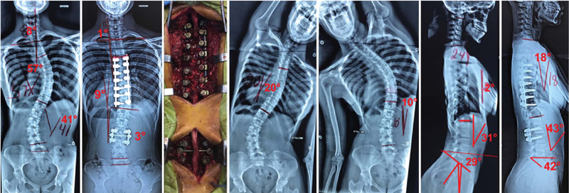
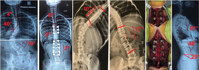
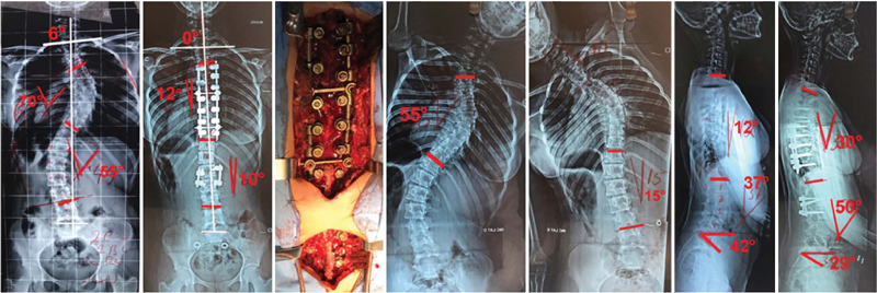
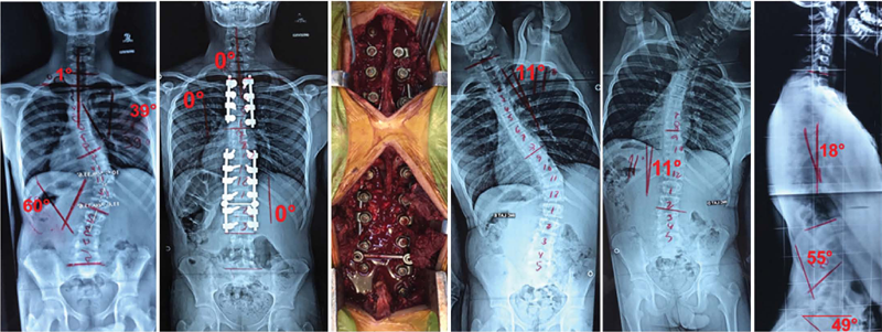
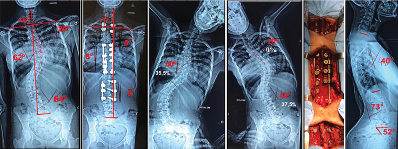
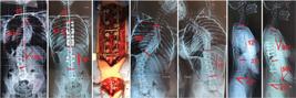
For support in the surgical correction of scoliosis, MTC values the full assessment of the coronal and sagittal planes, getting closer to the Kadoury Classification System6 and PUMC,8 focusing on its importance in surgical indications and in the design of scoliosis fixations, see figures 1 2 3 4 5 6.
17-year-old female patient, AIS, classified in the MTC as type 2 BL --- residual curves of 20° and 10°, above 10°, therefore, the two curves were fixed. Sacroclavicular Angle (SCA)12 corrected from 8° to 1°.
16-year-old female client, classified in the MTC as type 2 BT N N N, residual curves of 22° and 27°, residual curves above 10°, therefore, the two curves were fixed. The SCA corrected itself from 3° to 1°.
12-year-old girl, female, classified in the MTC as type 2 C N N -, residual curves of 10° and 17°, residual curves above 10°, therefore, surgery was indicated, both curves were addressed. The SCA corrected itself from 4° to 0°.
13-year-old female is classified in the MTC as type 2 BL - N N, residual curves of 55° and 15°, residual curves above 10°, therefore, both curves were addressed. The SCA corrected itself from 6° to 0°.
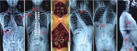
14-year-old boy, male, is classified in the MTC as type 2 BT - N N, residual curves of 11° and 11°, are above 10°, therefore, both were addressed. The SCA corrected itself from 1° to 0°.
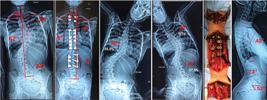
15-year-old female client is classified in the MTC as type 3 C N + +, residual curves of 20°, 40°, and 40° are above 10°, therefore, all three curvatures were addressed. The SCA corrected itself from 6° to −1°.
In this study, a higher incidence of double curve scoliosis was observed. Predominance of the main curve in the distal thoracic region. The sagittal plane showed a higher incidence of normal angle. Greater presence of hyperlordosis in females. It is the predominance of hypokyphosis in younger individuals. It can be seen in the result tables that these predominances occurred with significant differences, see tables 1 2 3 4 5 6 7 8. Findings that we observe in our daily lives in the treatment of scoliosis.
The importance of considering the fixation of the residual secondary curves, and the appreciation of the approach aims to reduce the trunk imbalance rate, believing and not concluding, that they are the main cause of these complications. Their presence, associated with the existence of one or more neutral vertebrae, can be addressed by Multiple Fixation12, with less loss of mobility of the spine. Undoubtedly, the MTC requires further tests and studies in the three-dimensional assessment of the spine.
CONCLUSIONS
The study of the variables of the MTC applied in the classification of patients operated on for AIS showed to be of a three-dimensional scope, therefore, it concludes to be a truly three-dimensional classification.
REFERENCES
- 1 Bernhardt M, Bridwell KH. Segmental analysis of the sagittal plane alignment of the normal thoracic and lumbar spines and thoracolumbar junction. Spine. 1989;14(7):717-21.
- 2 King HA, Moe JH, Bradford DS, Winter RB. The selection of fusion levels in thoracic idiopathic scoliosis. J Bone Joint Surg Am. 1983;65(9):1302-13.
- 3 Lenke LG, Betz RR, Harms J, Bridwell KH, Clements DH, Lowe TG, et al. Adolescent idiopathic scoliosis: a new classification to determine extent of spinal arthrodesis. J Bone Joint Surg Am. 2001;83-A(8):1169-81.
- 4 Coonrad RW, Murrell GA, Motley G, Lytle E, Hey LA. A logical coronal pattern classification of 2.000 consecutive idiopathic scoliosis cases based on the scoliosis research society-defined apical vertebra. Spine. 1998;23(12):1380-91.
-
5 Kadoury S, Labelle H. Classification of three-dimensional thoracic deformities in adolescent idiopathic scoliosis based on a multivariate analysis. Eur Spine J. 2012;21(1):40-9. doi: 10.1007/s00586-011-2004-2.
» https://doi.org/10.1007/s00586-011-2004-2 - 6 Qiu G, Zhang J, Wang Y, Xu H, Zhang J, Weng X, et al. A new Operative classification of idiopathic scoliosis: A peking union medical college method. Spine. 2005;30(12):1419-26.
- 7 Qiu G, Li Q, Wang Y, Yu B, Qian J, Yu K, et al. Comparison of reliability between the PUMC and Lenke classification systems for classifying adolescent idiopathic scoliosis. Spine (Phila Pa 1976). 2008;33(22):E836-42.
-
8 Garcia EB, Garcia LF, Giesbrecht ST, Vasconcelos LG, Garcia EB Jr, Camarinha MF, et at. Adolescent idiopathic scoliosis and similar conditions: three-dimensional classification. Coluna/Columna 2018;17(4):286-92. doi: 10.1590/S1808-185120181704191818.
» https://doi.org/10.1590/S1808-185120181704191818 - 9 Kirkwood BR, Sterne JAC. Essential medical statistics. 2nd ed. Massachusetts: Blackwell Science; 2006. p. 502.
- 10 Mccullagh P, Nelder JA. Generalized linear models. 2nd ed. New York: Chapman and Hall; 1989. p. 511.
- 11 Garcia EB, Payão GB, Garcia LF, Garcia EB Jr, Camarinha MF, Gonçalves RG, et al. Adolescent idiopathic Scoliosis and Similar Conditions: New Tool to Measure the Coronal Plane. Coluna/Columna. 2018;17(4):281-5.
- 12 Garcia EB, Garcia LF, Garcia EB Jr, Camarinha MF, Gonçalves RG, Garcia EB, et al. Escoliose idiopática do adolescente e similares: fixação curta, apical, única ou múltipla. Coluna/Columna. 2018;17(4):275-80.