 Arrhythmogenic cardiomyopathy in a cat
ABSTRACT:
Arrhythmogenic cardiomyopathy is a rare cardiac disease in cats, characterized by ventricular myocyte atrophy and replacement by fibrofatty tissue, predisposing to potentially fatal arrhythmias and heart failure. This report described a 12-year-old mixed-breed male cat with recurrent episodes of syncope, manifested by sudden weakness and altered consciousness. Physical examination revealed hypothermia, hypotension, and an irregular cardiac rhythm. Diagnostic evaluations confirmed chronic kidney disease and cardiomegaly, with echocardiographic findings compatible with arrhythmogenic cardiomyopathy. Despite treatment with pimobendan and clopidogrel, the heart failure progressed, culminating in refractory hypotension and the need for hospitalization. The hemodynamic instability and lack of response to therapy indicated a poor prognosis and led to the decision for euthanasia. Postmortem histopathological analysis confirmed the presence of biventricular arrhythmogenic cardiomyopathy. This case highlighted the importance of early diagnosis of feline arrhythmogenic cardiomyopathy, as advanced stages often follow a refractory course despite therapeutic interventions. Histopathological confirmation remains essential, particularly in atypical clinical presentations such as recurrent syncope in patients without overt signs of congestive heart failure.
Key words:
cardiac arrhythmia; cardiomyopathy; cats; heart failure; syncope
RESUMO:
A cardiomiopatia arritmogênica é uma doença cardíaca rara em gatos, sendo caracterizada por atrofia dos miócitos ventriculares e substituição por tecido fibrogorduroso, predispondo a arritmias potencialmente fatais e a insuficiência cardíaca. Este estudo relata o caso de um gato macho, sem raça definida, de 12 anos, com episódios recorrentes de síncope, caracterizados por fraqueza súbita e alteração no nível de consciência. O exame físico revelou hipotermia, hipotensão e ritmo cardíaco irregular. As avaliações diagnósticas confirmaram doença renal crônica e cardiomegalia, com evidências ecocardiográficas compatíveis com cardiomiopatia arritmogênica. Contudo, apesar do tratamento com pimobendam e clopidogrel, a insuficiência cardíaca progrediu, culminando em hipotensão refratária e a necessidade de hospitalização. Além disso, a instabilidade hemodinâmica e a ausência de resposta ao tratamento indicavam prognóstico ruim, o que culminou na decisão de eutanásia. Posteriormente, a análise histopatológica pós-morte confirmou a presença de cardiomiopatia arritmogênica com comprometimento biventricular. Desta maneira, destaca-se a importância do diagnóstico precoce da cardiomiopatia arritmogênica felina, uma vez que, nos estágios avançados, a doença tende a apresentar curso refratário mesmo diante de intervenções terapêuticas. Logo, a confirmação histopatológica permanece fundamental, especialmente diante de apresentações clínicas atípicas, como síncope recorrente, em pacientes sem sinais evidentes de insuficiência cardíaca congestiva.
Palavras-chave:
arritmia cardíaca; cardiomiopatia; gato; insuficiência cardíaca; síncope
INTRODUCTION
Arrhythmogenic cardiomyopathy (ACM) is a rare and underdiagnosed myocardial disease in cats, characterized by the progressive replacement of cardiac myocytes with fibrofatty tissue, predominantly affecting the right ventricle (RIVAS et al., 2023). While originally described in humans as arrhythmogenic right ventricular cardiomyopathy, the feline counterpart remains poorly understood, with few documented cases and no established diagnostic criteria (KITTLESON & CÔTÉ, 2021; RIVAS et al., 2023). In humans, ACM is most frequently associated with inherited mutations in genes encoding desmosomal proteins, which are essential for cell-to-cell adhesion and myocardial integrity. Over 50% of affected individuals have identifiable mutations; although, other genetic variants and epigenetic factors likely contribute to disease expression (SHAIKH et al., 2025). In dogs, particularly Boxers, a deletion in the striatin gene has been linked to the disease, further supporting the role of desmosomal dysfunction in its pathogenesis (MEURS, 2017). Despite these interspecies similarities, the molecular basis of ACM in cats remains undefined, underscoring the need for further research into its genetic and structural underpinnings in the feline population.
Since it was first reported in cats in 2000, additional isolated cases of ACM have been documented, consistently describing fibrofatty myocardial replacement associated with varying degrees of right and left ventricular involvement, as well as diverse clinical manifestations of congestive heart failure (FOX et al., 2000; HARVEY et al., 2005; CIARAMELLA et al., 2009; BACKSCHAT et al., 2016; FERASIN et al., 2020).
According to the 2020 ACVIM consensus statement on feline cardiomyopathies, classification should rely on morphofunctional assessment and included arrhythmias as part of phenotypic characterization (FUENTES et al., 2020). However, ACM is not currently listed among the major cardiomyopathy categories, likely reflecting the limited number of well-documented feline cases and the absence of standardized diagnostic criteria. This omission may contribute to underdiagnosis or misclassification, especially in cats presenting with arrhythmias or syncope without overt signs of heart failure.
The objective of this case report is to document a rare manifestation of ACM in a domestic shorthair cat, initially presenting with recurrent syncope rather than overt signs of congestive heart failure. This clinical profile differs from most previously documented cases and highlights the diagnostic complexity of ACM, especially in the absence of volume overload. By integrating clinical, electrocardiographic, and echocardiographic findings, this report underscores the value of a multimodal diagnostic approach to support the suspicion of arrhythmogenic cardiomyopathy in vivo, while histopathology remains essential for definitive postmortem confirmation. Additionally, it contributes to the limited veterinary literature by highlighting the importance of early recognition, differential diagnosis from other cardiomyopathies, and prompt therapeutic intervention in feline patients presenting with unexplained syncope or arrhythmias.
Case description
A 12-year-old, intact male domestic shorthair cat (black and white coat), weighing 2.8 kg, and with a body condition score of 3/9, was presented to the Veterinary Hospital with a history of two sudden and brief episodes of generalized weakness and altered consciousness after feeding the day prior to consultation. According to the owner, similar episodes had occurred six months earlier, with three events reported. The cat had no significant medical history beyond these episodes. Lethargy was noted, though appetite and water intake remained normal, with no other observed abnormalities. There were no notable familial or genetic histories reported.
Physical examination revealed an alert animal with hypothermia (rectal temperature of 37 °C) an irregular cardiac rhythm. Systemic arterial pressure (SAP) of 88 mmHg measured indirectly using a continuous-wave Doppler ultrasonographic device. Hematologic analysis revealed leukopenia (5,400/ µL; reference: 5,500-19,500/ µL) secondary to lymphopenia (1,080/ µL; reference: 1,500-7,000/ µL), with no abnormalities in red cell parameters. Serum biochemistry showed increased serum levels of urea (82 mg/dL; reference: 42.8-64.2 mg/dL) and creatinine (2.15 mg/dL; reference: 0.8-1.8 mg/dL). Urinalysis indicated a specific gravity of 1.020 (reference: ≥ 1.035) and a urine protein-to-creatinine ratio of 2.63 (reference: < 0.2). Reference intervals were provided by the local laboratory.
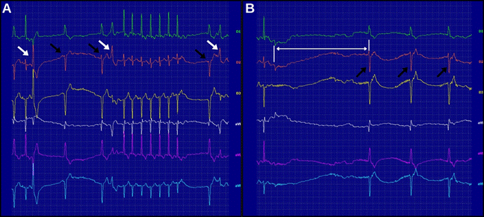
Abdominal ultrasound findings revealed bilateral renal atrophy, increased cortical echogenicity, altered corticomedullary ratio, and a heterogeneous medullary pattern, findings consistent with chronic kidney disease. Thoracic radiographs identified cardiomegaly, and a mild bronchial wall thickening and mineral opacities suggestive of chronic bronchopathy. Electrocardiography (ECG) performed using a 6-channel automated ECG device, revealed heart rate variability (49-211 bpm over a 5-minute interval), atrial fibrillation with a low ventricular response, aberrant conduction with left-axis deviation, and intermittent escape rhythm from the left ventricle, with a maximum pause of 3.15 seconds. Additionally, premature ventricular complexes (PVCs) originating from the right ventricle were identified (Figure 1).
Electrocardiographic tracings of the reported patient in frontal lead, with 2N sensitivity and a paper speed of 25 mm/s, demonstrating atrial fibrillation with an irregular rhythm. Panel (A) shows ventricular rate variability ranging from 50 bpm to 200 bpm, with ventricular escape beats originating from the left ventricle (black arrow) and premature ventricular complexes originating from the right ventricle (white arrow). Panel (B) highlights a pronounced ventricular depolarization pause lasting 3.15 seconds (white arrow), resulting in a transient heart rate of 26 bpm, followed by a ventricular escape rhythm (black arrows).
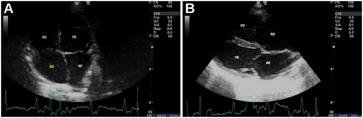
Echocardiographic evaluation revealed marked structural and functional abnormalities involving both atria and ventricles. Left atrial remodeling was characterized by an increased left atrium-to-aortic root ratio (LA/Ao) of 2.22 (reference: ≤ 1.50) (MACHADO et al., 2024). Indicators of atrial dysfunction included a markedly reduced left atrial fractional shortening (LAFS) of 12.66% (reference: 28.2 - 30.6% in M-mode short-axis view) (MACHADO et al., 2024) and a decreased left atrial emptying velocity of 0.17 m/s (reference: > 0.25 m/s) (SCHOBER & MAERZ, 2006), suggesting impaired contractility and an increased risk of thromboembolic events. The right atrium and ventricle appeared subjectively enlarged (Figure 2), and right ventricular systolic dysfunction was evidenced by reduced motion of the right ventricular free wall and a decreased tricuspid annular plane systolic excursion (TAPSE) of 5.7 mm (reference: 8.27-9.77 mm) (SPALLA et al., 2017). Left ventricular systolic function was also impaired, as demonstrated by a fractional shortening of 23.38% (reference: 40.9-52.2) (MACHADO et al., 2024); although, there was no ventricular remodeling. An irregular rhythm with atrioventricular valve flutter was noted, compatible with atrial fibrillation, likely secondary to atrial enlargement and fibrosis. Valvular assessment revealed mild mitral and moderate tricuspid insufficiency, both presumably functional in nature. Taken together, these findings are indicative of advanced structural and functional cardiac remodeling involving both sides of the heart. The pattern of right and left ventricular systolic impairment, right atrial and ventricle enlargement, rhythm disturbance, and associated valvular regurgitation raises strong suspicion of arrhythmogenic cardiomyopathy with biventricular involvement; although, differential diagnoses such as nonspecific cardiomyopathy or myocarditis cannot be excluded.
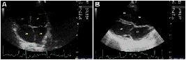
Echocardiographic assessment of right atrial and ventricular enlargement. Longitudinal B-mode echocardiographic images simultaneously recorded with electrocardiographic monitoring of the reported patient, showing marked enlargement of the right atrium (RA) and right ventricle (RV) on subjective assessment when compared to the left atrium (LA) and left ventricle (LV), in addition to an evident irregular rhythm and polymorphic electrocardiographic waves. Panel (A) displays an apical four-chamber view obtained from the left caudal parasternal window, and panel (B) shows a longitudinal four-chamber view from the right parasternal window.
Treatment was started with oral administration of pimobendan (0.25 mg/kg BID), clopidogrel (¼ of a 75 mg tablet, SID), and prednisolone (1 mg/kg SID for seven days, tapering to 0.5 mg/kg SID for reassessment). At follow-up, 20 days later, the owner reported four episodes of collapse on the day of consultation, coinciding with incomplete administration of pimobendan. The cat was admitted due to hypotension (SAP of 69 mmHg) and hypothermia (36.9 °C).
During hospitalization, pimobendan therapy was reinstituted and the patient’s SAP remained unstable, fluctuating around 80 mmHg. Repeat echocardiographic assessment showed persistently reduced left ventricular systolic function, with fractional shortening ranging from 23.26-39.29%. This marked variability was most likely associated with atrial fibrillation and its irregular ventricular response, which can result in beat-to-beat changes in diastolic filling and stroke volume. To manage SAP and ventricular systolic function, a continuous infusion of dobutamine was started at 2.5 µg/kg/min and titrated up to 7.5 µg/kg/min, resulting in a temporary stabilization of a SAP at 88 mmHg. However, a subsequent decline in SAP was observed, requiring an increase in dobutamine to 10 µg/kg/min and the addition of norepinephrine at 0.3 µg/kg/min. This combination stabilized SAP at approximately 102 mmHg for 15 hours. However, SAP eventually declined, and ascites developed. Despite these efforts, the patient showed a poor clinical response, and after 9 days of hospitalization, the owner elected euthanasia.
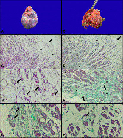
Postmortem examination revealed ascites, pleural effusion, and subcutaneous edema. The heart appeared globose, with ventricular dilation and thinning of the walls, as well as mild eccentric hypertrophy of the left ventricle, all subjectively assessed during gross examination (Figure 3A-B). Histopathological analysis of the cardiac tissue, stained with hematoxylin and eosin (H&E) and Gomori’s trichrome, revealed multifocal atrophy and degeneration of cardiomyocytes, associated with fibroadipose tissue infiltration between myocardial fibers, extending from the epicardium to the endocardium and affecting both ventricles (Figure 3C-H). Gomori’s trichrome staining confirmed the deposition of fibrous tissue. Clinical and morphologic findings confirmed the diagnosis of arrhythmogenic right and left ventricular cardiomyopathy.
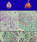
Arrhythmogenic right ventricular cardiomyopathy. (A) Gross appearance showing a globoid heart with rounded borders. (B) Opening of the right ventricle revealing thinning of the ventricular wall and multifocal pale areas within the wall (asterisk). (C) Photomicrograph showing fibroadipose tissue infiltration originating from the epicardium (arrow) and extending toward the endocardium (asterisk). Hematoxylin and eosin (H&E) stain, 4x. (D) Fibroadipose tissue infiltration with the same distribution pattern, originating from the epicardium (arrow) and extending toward the endocardium (asterisk). Gomori’s trichrome stain, 4x. (E) Cardiomyocytes exhibiting multifocal atrophy (arrow), H&E, 10x. (F) Replacement of cardiac muscle by fibrous (asterisk) and adipose (arrow) tissue. Gomori’s trichrome stain, 10x. (G, H) Higher magnification view showing adipocytes interspersed among cardiomyocytes (arrow). Gomori’s trichrome stain, 40x.
This case illustrates the diagnostic complexity of feline cardiomyopathies, particularly when clinical and imaging findings are initially nonspecific. At presentation, three differential diagnoses were considered: myocarditis, due to the acute onset of decompensation, bradyarrhythmia, and hypotension, which justified the empirical use of corticosteroids; arrhythmogenic cardiomyopathy (ACM), based on the presence of marked right atrial and ventricular enlargement and atrial fibrillation with a slow ventricular response, even in the absence of overt signs of congestion; and nonspecific cardiomyopathy (NCM), initially proposed due to the absence of imaging features characteristic of hypertrophic, dilated, or restrictive phenotypes (FUENTES et al., 2020; KITTLESON & CÔTÉ, 2021).
This provisional diagnosis highlighted the limitations of phenotype-based classification systems, particularly in early or atypical stages. As the condition progressed, the patient developed persistent arrhythmias, worsening systolic dysfunction of both ventricles, and clinical signs of biventricular failure. These findings strengthened the suspicion of ACM, which was ultimately confirmed postmortem. Histopathological analysis revealed fibrofatty infiltration of right and left ventricles, hallmark features of arrhythmogenic cardiomyopathy that retrospectively substantiated the clinical impression (BILHALVA et al., 2020; FUENTES et al., 2020; KITTLESON & CÔTÉ, 2021).
Although, ACM remains poorly characterized and has been reported in cats ranging from 1 to 20 years of age, it is generally believed to affect predominantly middle-aged to older animals (FOX et al., 2000; HARVEY et al., 2005; CIARAMELLA et al., 2009; FERASIN et al., 2020). The age of the present patient was therefore consistent with the typical demographic profile. This reinforces the importance of maintaining a high index of suspicion for ACM in cats of similar age, particularly when early clinical or imaging findings are subtle, as timely recognition of these nonspecific signs may be critical for prognostication and therapeutic planning in what is likely a chronically evolving condition.
Prednisolone was initially administered as a short-term anti-inflammatory therapy, based on the possibility of an underlying myocardial inflammatory process (KITTLESON & CÔTÉ, 2021). It was also intended to mitigate a suspected bronchial condition, as respiratory complications can further compromise cardiac function (REINERO et al., 2020). Although empirical, this intervention was considered clinically reasonable. However, due to the known risk of corticosteroid-induced fluid retention in cardiac patients, the drug was discontinued as the patient’s condition deteriorated.
Pimobendan and clopidogrel were selected as part of the initial therapeutic strategy to support myocardial function and reduce thromboembolic risk in a patient presenting with atrial fibrillation, biatrial enlargement, and systolic dysfunction, despite the absence of overt signs of congestive heart failure. Pimobendan is a calcium-sensitizing inodilator that improves contractility without significantly increasing myocardial oxygen consumption (BOYLE & LEECH, 2012). Its inotropic effects result from both calcium sensitization and PDE3 inhibition, and its active metabolite may also enhance inotropy through A1-adenosine receptor antagonism (BOYLE & LEECH, 2012). In addition, its balanced vasodilatory action reduces preload and afterload, which may improve cardiac output but can aggravate hypotension in unstable patients (GORDON et al., 2012). While its use in feline cardiomyopathies, including ACM, is not well established, it is considered a reasonable off-label option in cases with systolic impairment, supported by extrapolation from canine protocols and clinical reports (KITTLESON & CÔTÉ, 2021). In the present case, initial clinical deterioration may have been precipitated by the withdrawal of pimobendan by the caregiver, suggesting a potential role in stabilizing myocardial function during the early disease phase. However, in a later stage, its vasodilatory properties may have contributed to the difficulty in restoring arterial pressure, even in the presence of other inotropic and vasoactive agents such as dobutamine and norepinephrine.
Clopidogrel, a thienopyridine-class antiplatelet agent, acts by irreversibly inhibiting the platelet P2Y₁₂ receptor, preventing ADP-mediated platelet aggregation, and has been shown to be superior to aspirin in preventing arterial thromboembolism recurrence in cats (HOGAN et al., 2015). Although its specific role in feline ACM has not been evaluated, its use in this context was extrapolated from evidence in HCM and other thrombosis-prone feline populations (BLAIS et al., 2019). Ultimately, while the combination of pimobendan and clopidogrel was rationalized based on the patient’s cardiovascular profile and associated risks, it remains unclear whether either agent modifies the clinical course or survival in feline ACM. This highlighted the need for prospective, disease-specific studies to guide treatment in this underrecognized cardiomyopathy.
Additionally, this patient presented with azotemia and proteinuria, accompanied by ultrasonographic findings consistent with chronic kidney disease (CKD), suggesting preexisting chronic renal dysfunction. Nevertheless, reduced cardiac output and systemic hypotension likely contributed to a superimposed pre-renal component, further compromising renal perfusion and glomerular filtration pressure (MCCALLUM & TESTANI, 2023). This may have intensified proteinuria, which could reflect both chronic glomerular injury and acute hemodynamic alterations, including microthrombotic events associated with advanced cardiomyopathy. This pathophysiological interaction is consistent with mechanisms described in the cardiorenal syndrome literature (MCCALLUM & TESTANI, 2023). Although, these abnormalities are not specific to arrhythmogenic cardiomyopathy, they provided valuable insight into the extent of multisystem involvement and reinforced the complex interplay between cardiac and renal dysfunctions in this case.
Despite escalating supportive measures, including inotropes and vasopressors, the cat experienced rapid clinical deterioration and ultimately succumbed to refractory hypotension. This fulminant course underscores the aggressive nature of ACM and illustrates how early, seemingly nonspecific findings, such as isolated atrial fibrillation, atrial dysfunction, and transient syncope, may precede severe biventricular failure and circulatory collapse. Notably, diuretics were not administered during hospitalization, as there were no clear clinical or imaging signs of volume overload at initial evaluation. Fluid translocation and overt congestion became apparent only in the terminal stages, by which time the patient was already in a state of refractory hemodynamic instability.
In summary, this case highlighted the diagnostic challenges of feline ACM and the dynamic progression of myocardial disease. The integration of clinical assessment, electrocardiographic, echocardiographic, and histopathological findings was essential to establishing a definitive diagnosis. Clinical evaluation played a pivotal role in identifying subtle early signs, guiding differential diagnoses, and informing therapeutic decisions throughout the disease course. This report reinforces the urgent need for validated ante-mortem diagnostic criteria and evidence-based management strategies for this often fatal and underdiagnosed condition in cats.
ACKNOWLEDGMENTS
The authors would like to thank the Coordenação de Aperfeiçoamento de Pessoal de Nível Superior (CAPES), Brasil - Finance code 001.
REFERENCES
-
BACKSCHAT, P. S. et al. Arrhythmogenic right ventricular cardiomiopathy in a feline: case report. Arquivo Brasileiro de Medicina Veterinária e Zootecnia, v.68, n.5, p.1112-1116, oct. 2016. Available from: <Available from: https://www.scielo.br/j/abmvz/a/zd6gBjsJs6fV7xps6vLTV9p/?format=html⟨=pt >. Accessed: Mar. 20, 2020. doi: 10.1590/1678-4162-8616.
» https://doi.org/10.1590/1678-4162-8616.» https://www.scielo.br/j/abmvz/a/zd6gBjsJs6fV7xps6vLTV9p/?format=html⟨=pt -
BILHALVA, M. et al. Cardiomiopatia arritmogênica do ventrículo direito em felinos: revisão. Pubvet, v.14, n.07, 4 aug. 2020. Available from: <Available from: https://ojs.pubvet.com.br/index.php/revista/article/view/401 >. Accessed: Jul. 03, 2022. doi: 10.31533/pubvet.v14n7a611.1-5.
» https://doi.org/10.31533/pubvet.v14n7a611.1-5.» https://ojs.pubvet.com.br/index.php/revista/article/view/401 -
BLAIS, M. et al. Consensus on the rational use of antithrombotics in veterinary critical care (CURATIVE): domain 3 - defining antithrombotic protocols. Journal of Veterinary Emergency and Critical Care, v. 29, n. 1, p.60-74, 17 jan. 2019. Available from: <Available from: https://pubmed.ncbi.nlm.nih.gov/30654416/ >. Accessed: Dec. 08, 2020. doi: 10.1111/vec.12795.
» https://doi.org/10.1111/vec.12795.» https://pubmed.ncbi.nlm.nih.gov/30654416/ -
BOYLE, K. L.; LEECH, E. A review of the pharmacology and clinical uses of pimobendan. Journal of Veterinary Emergency and Critical Care, v.22, n.4, p.398-408, 28 aug. 2012. Available from: <Available from: https://pubmed.ncbi.nlm.nih.gov/22928748/ >. Accessed: Aug. 14, 2015. doi: 10.1111/j.1476-4431.2012.00768.x.
» https://doi.org/10.1111/j.1476-4431.2012.00768.x.» https://pubmed.ncbi.nlm.nih.gov/22928748/ -
CIARAMELLA, P. et al. Arrhythmogenic right ventricular cardiomyopathy associated with severe left ventricular involvement in a cat. Journal of Veterinary Cardiology, v.11, n.1, p.41-45, jun. 2009. Available from: <Available from: https://pubmed.ncbi.nlm.nih.gov/19473902/ >. Accessed: Jun. 14, 2023. doi: 10.1016/j.jvc.2009.02.007.
» https://doi.org/10.1016/j.jvc.2009.02.007.» https://pubmed.ncbi.nlm.nih.gov/19473902/ -
FERASIN, L. et al. Twenty-four-hour ambulatory (Holter) electrocardiographic findings in 13 cats with non-hypertrophic cardiomyopathy. The Veterinary Journal, v.264, p.105537, oct. 2020. Available from: <Available from: https://pubmed.ncbi.nlm.nih.gov/33012440/ >. Accessed: Sept. 06, 2024. doi: 10.1016/j.tvjl.2020.105537.
» https://doi.org/10.1016/j.tvjl.2020.105537.» https://pubmed.ncbi.nlm.nih.gov/33012440/ -
FOX, P. R. et al. Spontaneously occurring arrhythmogenic right ventricular cardiomyopathy in the domestic cat. Circulation, v.102, n.15, p.1863-1870, 10 oct. 2000. Available from: <Available from: https://www.ahajournals.org/doi/10.1161/01.CIR.102.15.1863 >. Accessed: Oct. 10, 2023. doi: 10.1161/01.CIR.102.15.1863.
» https://doi.org/10.1161/01.CIR.102.15.1863.» https://www.ahajournals.org/doi/10.1161/01.CIR.102.15.1863 -
GORDON, S. G. et al. Effect of oral administration of pimobendan in cats with heart failure. Journal of the American Veterinary Medical Association, v.241, n.1, p.89-94, 1 jul. 2012. Available from: <Available from: https://pubmed.ncbi.nlm.nih.gov/22720992/ >. Accessed: Jul. 01, 2023. doi: 10.2460/javma.241.1.89.
» https://doi.org/10.2460/javma.241.1.89.» https://pubmed.ncbi.nlm.nih.gov/22720992/ -
HARVEY, A. M. et al. Arrhythmogenic right ventricular cardiomyopathy in two cats. Journal of Small Animal Practice, v.46, n.3, p.151-156, mar. 2005. Available from: <Available from: https://pubmed.ncbi.nlm.nih.gov/15789811/ >. Accessed: Mar. 20, 2024. doi: 10.1111/j.1748-5827.2005.tb00306.x.
» https://doi.org/10.1111/j.1748-5827.2005.tb00306.x.» https://pubmed.ncbi.nlm.nih.gov/15789811/ -
HOGAN, D. F. et al. Secondary prevention of cardiogenic arterial thromboembolism in the cat: the double-blind, randomized, positive-controlled feline arterial thromboembolism; clopidogrel vs. aspirin trial (FAT CAT). Journal of Veterinary Cardiology, v.17, p.S306-S317, dec. 2015. Available from: <Available from: https://pubmed.ncbi.nlm.nih.gov/26776588/ >. Accessed: Dec. 12, 2024. doi: 10.1016/j.jvc.2015.10.004.
» https://doi.org/10.1016/j.jvc.2015.10.004.» https://pubmed.ncbi.nlm.nih.gov/26776588/ -
KITTLESON, M. D.; CÔTÉ, E. The feline cardiomyopathies: 3. cardiomyopathies other than HCM. Journal of Feline Medicine and Surgery, v.23, n.11, p.1053-1067, 25 nov. 2021. Available from: <Available from: https://pubmed.ncbi.nlm.nih.gov/34693805/ >. Accessed: Nov. 14, 2023. doi: 10.1177/1098612X211030218.
» https://doi.org/10.1177/1098612X211030218.» https://pubmed.ncbi.nlm.nih.gov/34693805/ -
FUENTES, V. L. et al. ACVIM consensus statement guidelines for the classification, diagnosis, and management of cardiomyopathies in cats. Journal of Veterinary Internal Medicine, v.34, n.3, p.1062-1077, 3 may, 2020. Available from: <Available from: https://onlinelibrary.wiley.com/doi/10.1111/jvim.15745 >. Accessed: Apr. 03, 2024. doi: 10.1111/jvim.15745.
» https://doi.org/10.1111/jvim.15745.» https://onlinelibrary.wiley.com/doi/10.1111/jvim.15745 -
MACHADO, A. et al. Left atrial fractional shortening in cats: a comparison between two echocardiographic views. Journal of Veterinary Cardiology, v.55, p.38-47, oct. 2024. Available from: <Available from: https://www.sciencedirect.com/science/article/pii/S1760273424000717 >. Accessed: Oct. 16, 2023. doi: 10.1016/j.jvc.2024.08.002.
» https://doi.org/10.1016/j.jvc.2024.08.002.» https://www.sciencedirect.com/science/article/pii/S1760273424000717 -
MCCALLUM, W.; TESTANI, J. M. Updates in cardiorenal syndrome. Medical Clinics of North America, v.107, n.4, p.763-780, jul. 2023. Available from: <Available from: https://pubmed.ncbi.nlm.nih.gov/37258013/ >. Accessed: Jul. 25, 2024. doi: 10.1016/j.mcna.2023.03.011.
» https://doi.org/10.1016/j.mcna.2023.03.011.» https://pubmed.ncbi.nlm.nih.gov/37258013/ -
MEURS, K. M. Arrhythmogenic right ventricular cardiomyopathy in the boxer dog. Veterinary Clinics of North America: Small Animal Practice, v.47, n.5, p.1103-1111, sept. 2017. Available from: <Available from: https://pubmed.ncbi.nlm.nih.gov/28647112/ >. Accessed: Jun. 21, 2024. doi: 10.1016/j.cvsm.2017.04.007.
» https://doi.org/10.1016/j.cvsm.2017.04.007.» https://pubmed.ncbi.nlm.nih.gov/28647112/ -
REINERO, C. et al. ACVIM consensus statement guidelines for the diagnosis, classification, treatment, and monitoring of pulmonary hypertension in dogs. Journal of Veterinary Internal Medicine, v.34, n.2, p.549-573, 1 mar. 2020. Available from: <Available from: https://pubmed.ncbi.nlm.nih.gov/32065428/ >. Accessed: Mar. 20, 2024. doi: 10.1111/jvim.15725.
» https://doi.org/10.1111/jvim.15725.» https://pubmed.ncbi.nlm.nih.gov/32065428/ -
RIVAS, V. N. et al. The role of personalized medicine in companion animal cardiology. Veterinary Clinics of North America: Small Animal Practice, v.53, n.6, p.1255-1276, nov. 2023. Available from: <Available from: https://pmc.ncbi.nlm.nih.gov/articles/PMC11184409/ >. Accessed: Jul. 07, 2023. doi: 10.1016/j.cvsm.2023.05.016.
» https://doi.org/10.1016/j.cvsm.2023.05.016.» https://pmc.ncbi.nlm.nih.gov/articles/PMC11184409/ -
SCHOBER, K. E.; MAERZ, I. Assessment of left atrial appendage flow velocity and its relation to spontaneous echocardiographic contrast in 89 cats with myocardial disease. Journal of Veterinary Internal Medicine, v.20, n.1, p.120-130, 28 jan. 2006. Available from: <Available from: https://pubmed.ncbi.nlm.nih.gov/16496931/ >. Accessed: Mar. 20, 2024. doi: 10.1892/0891-6640(2006)20[120:aolaaf]2.0.co;2.
» https://doi.org/10.1892/0891-6640(2006)20[120:aolaaf]2.0.co;2.» https://pubmed.ncbi.nlm.nih.gov/16496931/ -
SHAIKH, T. et al. Arrhythmogenic right ventricular cardiomyopathy: a comprehensive review. Journal of Cardiovascular Development and Disease, v.12, n.2, p.71, 13 feb. 2025. Available from: <Available from: https://pubmed.ncbi.nlm.nih.gov/39997505/ >. Accessed: Feb. 13, 2025. doi: 10.3390/jcdd12020071.
» https://doi.org/10.3390/jcdd12020071.» https://pubmed.ncbi.nlm.nih.gov/39997505/ -
SPALLA, I. et al. Mitral annular plane systolic excursion and tricuspid annular plane systolic excursion in cats with hypertrophic cardiomyopathy. Journal of Veterinary Internal Medicine, v.31, n.3, p.691-699, 30 may, 2017. Available from: <Available from: https://pubmed.ncbi.nlm.nih.gov/28370313/ >. Accessed: May, 22, 2023. doi: 10.1111/jvim.14697.
» https://doi.org/10.1111/jvim.14697.» https://pubmed.ncbi.nlm.nih.gov/28370313/
-
CR-2025-0199.R1
-
DATA AVAILABILITY STATEMENT
All data pertaining this study can be obtained through contacting the author for correspondence at talita.raposo@uvv.br.
-
DECLARATION OF USE OF ARTIFICIAL INTELLIGENCE
This manuscript employed artificial intelligence assistance through ChatGPT (OpenAI) to translate the original text from Brazilian Portuguese to English. The authors carefully reviewed the translated text to ensure its accuracy and fidelity.
Edited by
-
ASSOCIATE EDITOR:
Rudi Weiblen (0000-0002-1737-9817)
All data pertaining this study can be obtained through contacting the author for correspondence at talita.raposo@uvv.br.