 Platynosomosis in the municipality of Santa Maria, RS, Brazil - diagnosis by cholecystocentesis
ABSTRACT:
Platynosomosis is the main liver and bile duct verminosis in domestic cats. In Brazil, the cases are concentrated in the southeast and northeast regions, with few reports in the south of the country. A four-year-old male feline, mixed breed, with a history of jaundice, hyporexia and abdominal distension, was referred for clinical care. The leukogram showed leukocytosis due to eosinophilia and serum biochemistry showed a marked increase in liver extravasation enzymes. On ultrasound examination, there was marked hepatomegaly and the presence of dilated, tortuous bile ducts, as well as severe fullness of the gallbladder. Coproparasitological examination proved to be inconclusive. The feline then underwent cholecystocentesis in order to determine the etiology of the condition. The cytological examination of the bile fluid revealed parasite structures with a morphology consistent with operculate eggs of Platynosomum spp. The diagnostic conclusion allowed the establishment of appropriate therapy in a timely manner, which enabled the resolution of the problem. This research reported the first case of feline platynosomosis in the municipality of Santa Maria with clinical cure.
Key words:
Platynosomum spp.; trematoda; jaundice; cholecystocentesis
RESUMO:
Dá-se o nome de Platinosomose à principal verminose hepática e de vias biliares do gato doméstico. No Brasil, a casuística se concentra nas regiões sudeste e nordeste, com poucos relatos desta doença no sul do país. Um felino macho, sem raça definida, de quatro anos de idade e apresentando histórico de icterícia, hiporexia e dilatação abdominal, foi encaminhado para atendimento clínico. O leucograma evidenciou leucocitose por eosinofilia e a bioquímica sérica marcou aumento de enzimas de extravasamento hepático. No exame ultrassonográfico havia acentuada hepatomegalia e presença de ductos biliares dilatados, tortuosos, bem como severa repleção da vesícula biliar. O exame coproparasitológico mostrou-se inconclusivo. O felino, então, foi submetido ao procedimento de colecistocentese no intuito de determinar a etiologia do agravo. O exame citológico do fluido biliar obtido revelou estruturas parasitárias de morfologia condizente com ovos operculados de Platinossomum spp. A conclusão diagnóstica permitiu o emprego terapêutico em tempo hábil, o que possibilitou a resolução do agravo. Esse trabalho relata o primeiro caso de platinosomose felina no município de Santa Maria com cura clínica.
Palavras-chave:
Platynosomum spp.; trematoda; icterícia; colestase.
Trematodes of the genus Platynosomum are significant causes of hepatic and bile duct verminosis in domestic cats (Feliscatus) (TAYLOR et al., 2017). They have a worldwide distribution, with reports from Brazil concentrated in the southeast and northeast regions (SILVA et al., 2001; BRAGA et al., 2016). Their life cycle requires the presence of at least two intermediate hosts (snails and terrestrial isopods), as well as a paratenic host (anoles of the genus Anolis or toads of the genus Bufo) (EISENBRAUN et al., 2020). Felines are definitive hosts and acquire the trematode through predation of parasitized reptiles or amphibians (TAYLOR et al., 2017).
Feline platynosomosis is typically asymptomatic or potentially severe when associated with clinical signs of lethargy, weight loss, anorexia, vomiting, ascites, abdominal pain, jaundice, and hepatic failure (MICHAELSEN et al., 2012). Diagnosis is usually obtained through coproparasitological examination or cholecystectomy (HENRIQUE et al., 2018), the inclusion of investigative tools in the diagnostic routine is essential to ensure greater analytical sensitivity than coproparasitological methods and lower risk compared to surgical exploration (SAVARY-BATAILLE et al., 2003; EISENBRAUN et al., 2020). Ultrasonography-guided cholecystocentesis may serve this purpose (SAVARY-BATAILLE et al., 2003), though its application in the context of feline platynosomosis remains underutilized, requiring further case reports and clinical trials to validate and encourage its use (KÖSTER et al., 2016).
This study reported the first case of feline platynosomosis in Santa Maria/RS, with the etiological diagnosis obtained via ultrasonography-guided cholecystocentesis followed by cytological analysis of the obtained material and clinical cure of the patient.
A male, mixed-breed, intact four-year old cat, weighing 2.9 kg, was referred to the clinical service at the Universidade Federal de Santa Maria (UFSM). According to the owner, the cat had been missing from home for 30 days and was found three weeks ago. At the time of rescue, the cat was thin, with moderate abdominal distension, reduced appetite, and yellowish skin coloration. Over the weeks, the clinical condition worsened, prompting the search for veterinary care.
The cat was alert but unresponsive, with a low body condition score (2/5), moderately bristled and dull fur, with some fleas present, markedly yellowed skin and mucous membranes (Figure 1A and Figure 1B), moderate dehydration (6-8%), characterized by a skin tenting of 4 seconds, slight enophthalmia; and a capillary refill time of 3 seconds. No abnormalities were noted on auscultation, and the body temperature was normal. The cat was promptly referred to the hospitalization service at HVU-UFSM for dehydration correction and further diagnostic tests.
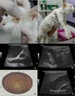
Cat, showing marked yellowing of the conjunctival mucosa and third eyelid (A) and moderate abdominal distension and diffuse yellowing of the skin (B). Gallbladder and extrahepatic bile ducts with thick and irregular walls, markedly distended by anechoic content with presence of gravity-dependent echogenic particles (C). Ultrasound-guided cholecystocentesis for sample collection; note the insertion of the needle (*) in the cranioventral margin of the gallbladder, which extends into the lumen (D). Biliary effusion containing an operculated egg (E) morphologically consistent with Platynosomum spp. (X100). Gallbladder and extrahepatic ducts in the last ultrasound examination, with slightly thickened walls and moderately distended by anechoic content with echogenic particles, showing improvement in relation to the first examination (F).
The erythrogram revealed an increase in total plasma proteins (11.2, reference value (RV): 5.9 to 7.5), and the leukogram showed leukocytosis (19,800/μL, RV: 5,100 to 16,200/μL) due to eosinophilia (2,376/μL, RV: 100 to 1,800/μL) and basophilia (792/μL, RV: 0 to 200/μL), with jaundice plasma (3 crosses). Increases in serum activities of ALT (770.00 IU/L, RV: 28.00 to 109.00 IU/L), AST (196.8 IU/L, RV: 17.0 to 48.0 IU/L), ALP (1,033.0 IU/L, RV: 11.0 to 49.0 IU/L), and GGT (30.6 mg/dL, RV: 0.0 to 2.0 mg/dL) were observed in the biochemical analysis. On abdominal ultrasound examination, the liver was enlarged (hepatomegaly), and the bile ducts were markedly dilated with thickened walls. The gallbladder appeared markedly distended, with a hyperechoic, thickened, and irregular wall, and contained echogenic particles in suspension, sometimes small hyperechoic filamentous structures were observed (Figure 1C).
In light of the new findings, fecal samples (two samples collected one day apart) were submitted for coproparasitological analysis (Willis-Mollay), which did not yield conclusive results. Subsequently, ultrasonography-guided cholecystocentesis was performed. The cat’s permissive temperament allowed the procedure to be carried out without sedation. For the procedure, hair from the ventral abdominal region of the feline was shaved, followed by antisepsis with 70% alcohol. The liver and bile duct were delineated with the transducer. Subsequently, ultrasound-guided percutaneous cholecystocentesis was performed using a 22-gauge needle and a 5 mL syringe (Figure 1D), resulting in the collection of 2 mL of amber-colored, low-viscosity bile fluid, which was sent for laboratory analysis. With the aid of light microscopy at a magnification of 100x, after prior centrifugation of the sample, occasional operculated eggs morphologically consistent with Platynosomum spp. were identified in the precipitate of the bile fluid by direct analysis (Figure 1E).
Treatment was initiated with oral fenbendazole at a dose of 50 mg/kg every 24 hours for three days, and praziquantel at a dose of 5.75 mg/kg orally as a single dose. It was recommended to the animal’s owner to repeat this treatment protocol after ten weeks (SHELL et al., 2015). The cat was discharged two days after the start of treatment, as it was found to be clinically healthy (hydrated and had normal appetite). Upon return, 30 days after the start of therapy, there was no longer jaundice and no abdominal distension observed on physical examination. The cat weighed 3.2 kg, was hydrated, alert, and interactive. Both the hemogram (erythrogram and leukogram) and serum biochemistry (ALT, AST, ALP, GGT, urea, and creatinine) were within their respective reference ranges. In the latest abdominal ultrasound, the previous findings remained, with a moderate reduction in their intensity (Figure 1F).
The conclusive diagnosis of platynosomosis in a cat was obtained through the association of clinical, imaging, and laboratory findings. The etiology of the condition is sometimes limited to the visualization of operculated trematode eggs through coproparasitological examination. However, this investigative tool’s analytical sensitivity is directly related to the intensity of cholestasis and the type of analysis employed (WILLARD & FOSSUM, 2005; TAYLOR et al., 2017). Partial or complete obstructive processes in the bile ducts reduce the availability of eggs in the feces, often resulting in false negative results as seen in this case (JORGE et al., 2020). It is also reported that the availability of eggs in bile is much higher when compared to fecal count for the diagnosis of platynosomosis (KÖSTER et al., 2016). Furthermore, routine analytical techniques such as Willis-Mollay are not suitable for identifying operculated eggs of Platynosomum spp. (TAYLOR et al., 2017). Centrifugal fecal flotation techniques using Sheather’s sugar solution (ROCHA et al., 2014), which have demonstrated high analytical sensitivity for detecting trematodes (EISENBRAUN et al., 2020), are preferred.
Ultrasound-guided cholecystocentesis procedures are commonly performed with the assistance of anesthesia (KÖSTER et al., 2016). However, it is essential to consider the temperament and physical condition of the animal during the procedure (SAVARY-BATAILLE et al., 2003). In severely debilitated patients, sedation may pose risks that outweigh the benefits of the examination itself (WILLARD & FOSSUM, 2005).
Most cats affected by platynosomosis are asymptomatic (BRAGA et al., 2016). However, the severity of jaundice, associated with morphological changes in the liver and gallbladder, along with a myriad of laboratory alterations, in addition to clinical findings of vomiting, dehydration, anorexia, and abdominal pain, allow characterization of the parasitism in the cat in question as intense and potentially severe, with unlikely possibility of spontaneous resolution (MICHAELSEN et al., 2012; WATSON et al., 2019).
The jaundice present in cases of Platynosomum spp. parasitism in cats is primarily due to hepatic and post-hepatic obstructive processes and direct injury to the liver (BRAGA et al., 2016). There is also speculation about the possibility of the development of intrahepatic and extrahepatic constrictive fibrosis resulting from the movement of trematodes within the bile ducts, which could lead to momentary episodes of bile flow obstruction (MARTIN et al., 2003). This event could characterize partial or transient cholestasis and thus justify the presence of ductal cystic dilations and severe gallbladder distension. Furthermore, in chronic obstructions, the common bile duct can remain dilated even after the cause of obstruction has been alleviated (D´ANJOU & PENNINCK, 2015), which justifies the persistence of ultrasound findings weeks after clinical cure of the patient.
Exploratory laparotomy followed by cholecystectomy is a common approach in the management of feline patients affected by platynosomosis, often performed when coproparasitological analysis fails (HENRIQUE et al., 2018). However, it is an inherently risky procedure and is often performed on animals that are already severely ill, substantially increasing the risk of perioperative and postoperative death (SAVARY-BATAILLE et al., 2003). The procedure should be performed when there is evidence of cholestasis with imminent risk of gallbladder rupture (HENRIQUE et al., 2018). The prior performance of ultrasonography-guided cholecystocentesis, as in the present case, resulted in early diagnosis of the parasitosis and avoided the need for surgical intervention.
Feline platynosomosis; therefore, is a verminosis present in the municipality of Santa Maria-RS and should be included among the differential diagnoses for diseases/conditions that cause jaundice in the species. Ultrasonography-guided cholecystocentesis is a minimally invasive diagnostic tool that potentially offers greater analytical precision in cases of partial or complete biliary obstruction and should be considered whenever coproparasitological examination yields negative results.
ACKNOWLEDGEMENTS
The authors are thankful for Conselho Nacional de Desenvolvimento Científico e Tecnológico (CNPq) and Coordenação de Aperfeiçoamento de Pessoal de Nível Superior (CAPES) for supporting this study, which was financed in part by CAPES. Brazil - Finance code 001.
REFERENCES
-
BRAGA, R. R. et al. Prevalence of Platynosomumfastosum infection in free roaming cats in northeastern Brazil: Fluke burden and grading of lesions. Veterinary Parasitology, v.227, p.20-25, 2016. Available from: <Available from: https://doi.org/10.1016/j.vetpar.2016.07.021 >. Accessed: Feb. 10, 2024. doi: 10.1016/j.vetpar.2016.07.021.
» https://doi.org/10.1016/j.vetpar.2016.07.021.» https://doi.org/10.1016/j.vetpar.2016.07.021 - D´ANJOU, M.; PENNINCK, D. Liver. In: Atlas of Small Animal Ultrasonography, Iowa: Wiley Blackwell, 2015. cap.6, p.183-238.
-
EISENBRAUN, H. et al. Comparison of fecal analysis methods for the detection of Platynosomumfastosum in naturally infected cats. Journal of Feline Medicine and Surgery, v.4, n.22, p.395-398, 2020. Available from: <Available from: https://doi.org/10.1177/1098612X19848173 >. Accessed: Feb. 10, 2024. doi: 10.1177/1098612X19848173.
» https://doi.org/10.1177/1098612X19848173.» https://doi.org/10.1177/1098612X19848173 -
HENRIQUE, F. V. et al. Cholecystectomy associated to duodenal enterotomy in the infestation by Platynosomum sp. In a domestic cat. Veterinaria e Zootecnia, v.25, n.2, p.2-4, 2018. Available from: <Available from: https://doi.org/10.35172/rvz.2018.v25.75 >. Accessed: Feb. 10, 2024. doi: 10.35172/rvz.2018.v25.75.
» https://doi.org/10.35172/rvz.2018.v25.75.» https://doi.org/10.35172/rvz.2018.v25.75 -
JORGE, A. L. T. A. et al. Cholecystoduodenostomy for treatment of biliary obstruction secondary to feline platinossomosis. Acta Scientiae Veterinariae, v.48, p.498, 2020. Available from: <Available from: https://doi.org/10.22456/1679-9216.100612 >. Accessed: Feb. 10, 2024. doi: 10.22456/1679-9216.100612.
» https://doi.org/10.22456/1679-9216.100612.» https://doi.org/10.22456/1679-9216.100612 - MARTIN, R. A. et al. Liver and biliary system. In: Textbook of Small Animal Surgery, Philadelphia: Elsevier Science, 2003, p.708-724.
-
KÖSTER, L. et al. Percutaneous ultrasound-guided cholecystocentesis and Bile Analysis for the Detection of Platynosomum spp.-Induced Cholangitis in Cats. Journal of internal medicine veterinary, v.30, n.3, p.787-793, 2016. Available from: <Available from: https://doi.org/10.1111/jvim.13943 >. Accessed: Jun. 10, 2024. doi: 10.1111/jvim.13943.
» https://doi.org/10.1111/jvim.13943.» https://doi.org/10.1111/jvim.13943 -
MICHAELSEN, R. et al. Platynosomumconcinnum (Trematoda: Dicrocoeliidae) em gato doméstico da cidade de Porto Alegre, Rio Grande do Sul, Brasil. Veterinária em Foco, v.10, n.1, p.53-60, 2012. Available from: <Available from: http://www.periodicos.ulbra.br/index.php/veterinaria/article/view/1167/883 >. Accessed: Feb. 10, 2024.
» http://www.periodicos.ulbra.br/index.php/veterinaria/article/view/1167/883 -
ROCHA, N. O. et al. Comparison of two coproparasitological techniques for the detection of Platynosomum sp. infection in cats. VeterinaryParasitology, v.204, n.3-4, p.392-395, 2014. Available from: <Available from: https://doi.org/10.1016/j.vetpar.2014.04.022 >. Accessed: May, 18, 2024. doi: 10.1016/j.vetpar.2014.04.022.
» https://doi.org/10.1016/j.vetpar.2014.04.022.» https://doi.org/10.1016/j.vetpar.2014.04.022 -
SAVARY-BATAILLE, K. C. M. et al. Percutaneous ultrasound-guided cholecystocentesis in healthy cats. Journal of Veterinary Internal Medicine, Filadelfia, v.17, p.298-303, 2003. Available from: <Available from: https://doi.org/10.1111/j.1939-1676.2003.tb02451.x >. Accessed: Feb. 10, 2024. doi: 10.1111/j.1939-1676.2003.tb02451.x.
» https://doi.org/10.1111/j.1939-1676.2003.tb02451.x.» https://doi.org/10.1111/j.1939-1676.2003.tb02451.x -
SHELL, L. et al. Praziquantel treatment for Platynosomum species infection of a domestic cat on St Kitts, West Indies. Journal of Feline Medicine and SurgeryOpen Reports, Londres, v.1, n.1, p.1-4, 2015. Available from: <Available from: https://doi.org/10.1177/2055116915589834 >. Accessed: Feb. 10, 2024. doi: 10.1177/2055116915589834.
» https://doi.org/10.1177/2055116915589834.» https://doi.org/10.1177/2055116915589834 -
SILVA, H. C. et al. Helminthic phaune of dogs and cats of some municipalities of São Paulo State. Semina Ciências Agrárias, v.22, n.1, p.67-71, 2001. Available from: <Available from: https://doi.org/10.5433/1679-0359.2001v22n1p67 >. Accessed: Feb. 10, 2024. doi: 10.5433/1679-0359.2001v22n1p67.
» https://doi.org/10.5433/1679-0359.2001v22n1p67.» https://doi.org/10.5433/1679-0359.2001v22n1p67 - TAYLOR, M. A. Helmintologia Veterinária - Filo Platyhelminthes - Classe Trematoda. In: Parasitologia Veterinária, Rio de Janeiro: Guanabara Koogan, 4 ed, p.74-75, 2017.
- WATSON, P. J. Hepatobiliary diseases in cat. In: Small Animal Internal Medicine, St. Louis, Missouri: Elsevier Mosby, 6 ed, p.561-583, 2019.
- WILLARD, M. D.; FOSSUM, T. W. Diseases of the Gallbladder and Extrahepatic Biliary System. In: Textbook of veterinary internal medicine: diseases of the dog and cat, Missouri: Elsevier Saunders, p.1478-1482, 2005.
-
CR-2024-0194.R1
-
BIOETHICS AND BIOSECURITY COMMITTEE APPROVAL
We authors of the article entitled “Platynosomosis in the municipality of Santa Maria, RS, Brazil - diagnosis by cholecystocentesis” declared, for all due purposes, the project that gave rise to the present data of the same has not been submitted for evaluation to the Ethics Committee of the University of Santa Maria (UFSM), but we are aware of the content of the Brazilian resolutions of the National Council for Control of Animal Experimentation - CONCEA <http://www.mct.gov.br/index.php/content/view/310553.html> if it involves animals. Thus, the authors assume full responsibility for the presented data and are available for possible questions, should they be required by the competent authorities.
Edited by
-
Editor
Rudi Weiblen (0000-0002-1737-9817)