 Characterization of a novel MSH2 variant in Lynch syndrome: clinical data and complementary bioinformatics assessment
ABSTRACT
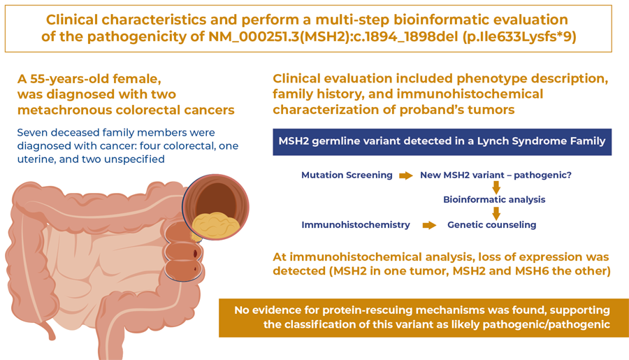
Objective To describe the clinical characteristics and perform a multi-step bioinformatics evaluation of the pathogenicity of NM_000251.3(MSH2):c.1894_1898del (p.Ile633Lysfs*9), an MSH2 germline variant detected in a family with Lynch syndrome.
Methods Clinical evaluation included description of phenotype, family history, and immunohistochemical characterization of the proband’s tumors. For pathogenicity classification according to the American College of Genetics and Genomics/Association for Molecular Pathology (ACMG/APA) criteria, bioinformatics analyses included: (i) literature and database screening, searching for the variant allele frequency, case reports, or functional studies, including ClinVar, VarSome, Ensembl, PubMed, EVA, and ABraOM; (ii) prediction of variant impacts using ExPASy Translate, Pfam, and Modeller 9.24; and, (iii) mechanisms that could mitigate the effects of the variant included alternative splicing and exon skipping (UniProt and GTex) and nonsense-mediated decay (NMD; MutationTaster2021).
Results The proband, a 55-year-old female, was diagnosed with two metachronous colorectal cancers. Immunohistochemical analysis showed loss of expression ( MSH2 in one tumor, and MSH2 and MSH6 in the other). Seven deceased family members were diagnosed with cancer (four colorectal, one uterine, and two unspecified). This variant caused a stop codon in MSH2 exon 12 of 16. When translated, the protein loses 294 C-terminal residues, which may prompt protein degradation. If the mutated protein escapes degradation, dimerization and DNA-binding domains will be present. Therefore, negative dominance effects were possible. No isoforms ending in exon 12 have been identified in the literature or in RNA splicing databases. A stop codon before the last exon-exon boundary indicated the occurrence of NMD.
Conclusion No evidence of protein-rescuing mechanisms was found, supporting the classification of this variant as likely pathogenic/pathogenic.
Lynch syndrome; Neoplastic syndromes; Hereditary; Colorectal neoplasms; DNA mismatch repair; MutS homolog 2 protein
Highlights
Family identified with recurrent colorectal cancer, suggestive of Lynch syndrome.
A new variant was detected, NM_000251.3(MSH2):c.1894 _1898del (p.Ile633Lysfs*9).
The variant causes a frameshift mutation leading to a premature stop codon, suggesting pathogenicity.
Using bioinformatics tools, we investigated possible escape mechanisms, such as the presence of isoforms without the mutated exon.Location of the variant suggests that it causes non-sense mediated decay, which is reinforced by immunohistochemistry findings showing absence of MSH2 protein expression.
Detailed assessment allows this variant to be classified as probably pathogenic/pathogenic.
Data from the article was extracted from the thesis by author Anisse Marques Chami presented to the Faculdade de Medicina of the Universidade Estadual Paulista “Júlio de Mesquita Filho ”, Botucatu Campus, to obtain the title of Doctor from the Graduate Program in Obstetrics and Gynecology, Concentration Area: Oncology, in 2022.
In Brief
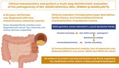
Familial cancer is a complex condition requiring genetic counseling. Screening for genetic variants is necessary in addition to clinical diagnosis. An identified genetic variant must be classified according to its potential pathogenicity. We identified a novel variant in the MSH2 gene, in a family with clinical manifestations compatible with Lynch Syndrome. Bioinformatics analysis showed that the variant created a frameshift mutation, leading to a premature stop codon. No compensatory mechanism was identified, confirming that this variant as likely pathogenic.
INTRODUCTION
Lynch syndrome (LS; OMIM 120435) is amongst the most common hereditary cancer predispositions.( 1 - 3 )It includes gastrointestinal, gynecological, skin, urinary tract, and central nervous system cancers.( 2 ) Colorectal and endometrial cancers are the most frequent in LS, and it has been estimated that 3% of all colorectal cancers and 3% of endometrial cancers are associated with LS.( 4 , 5 ) Lynch syndrome is an autosomal dominant condition with incomplete penetrance and heterogeneous clinical manifestations caused by deleterious germline variants in the post-replicative mismatch repair (MMR) genes MLH1 (OMIM 120436) , MSH2 (OMIM 609309) , MSH6 (OMIM 600678) , and PMS2 (OMIM 600259). In addition, germline EPCAM microdeletions or epimutations can influence MSH2 expression and cause LS.( 2 ) Cancer risk varies according to the mutated genes. Pathogenic variants in MLH1 and MSH2 present higher penetrance, with the risk of colorectal cancer exceeding 50%, whereas PMS2 and MSH6 pathogenic variants present lower penetrance.( 6 ) Among other functions, MMR proteins correct DNA replication errors, reduce mutation rates, control the fidelity of meiotic recombination, block recombination between non-homologous sequences, and help deal with environmental mutagens by mediating DNA damage-induced apoptosis.( 7 - 9 )
In recent decades, knowledge of the genotype-phenotype correlations for each LS gene has allowed the development of risk reduction management protocols for patients with pathogenic variants in MMR genes.( 6 ) Complete anamnesis (including family history), tumor pathology, microsatellite instability analysis, and immunohistochemistry of MMR proteins are crucial for guiding the diagnosis and management of LS. The Revised Bethesda Guidelines and Amsterdam (I and II) clinical criteria enable the identification of high-risk patients or families.( 10 - 12 ) Prediction models, such as the PREdiction Model for gene Mutations (PREMM5), also help screen individuals who may be mutation carriers.( 13 ) Furthermore, the inclusion of immunohistochemistry and/or microsatellite instability analysis in Universal Testing for all colorectal and endometrial cancers is currently recommended to improve the identification rate of individuals at risk of LS.( 14 ) However, as LS is defined by the presence of a pathogenic variant in one of the MMR genes,( 2 ) genetic-molecular studies are fundamental for identifying germline variants and establishing their phenotypic impacts.
Several mutations in MMR genes have already been described in LS and are shared in the main international variants databases, such as InSiGHT, VarSome, and Human Genetic Variation Database (HGVD).( 15 - 17 ) The description of mutations and associated phenotypes assists in the pursuit of better care by identifying specific population or family risk groups.( 14 ) It is fundamental to accurately characterize the pathogenic potential of newly identified genetic variants, and the strategies required vary according to each variant. Recently, we identified a new variant, NM_000251.3(MSH2):c.1894_1898del (p.Ile633Lysfs*9), in a Brazilian family with LS. In VarSome, this variant was classified as pathogenic according to the American College of Medical Genetics and Genomics and the Association for Molecular Pathology (ACMG-AMP) criteria.( 18 ) Nonetheless, no clinical description has been associated with this variant and no investigation has been conducted thus far, to evaluate the mechanisms underlying its pathogenicity.
OBJECTIVE
In this study, we aimed to describe the phenotype associated with NM_000251.3(MSH2):c.1894_1898del (p.Ile633Lysfs*9) and present a step-by-step bioinformatics analysis for characterization of its pathogenetic mechanisms.
METHODS
The proband was referred for genetic counseling, considering LS as a diagnostic hypothesis. The proband was a 55-year-old female, diagnosed with metachronous colorectal cancer in the previous year. The first tumor was in the sigmoid colon and the second was diagnosed 12 months later, in the cecum. Both tumors were pathologically described as moderately differentiated invasive adenocarcinomas. The anatomopathological records did not describe mucinous, signet ring cell histologic features, medullary growth patterns, or infiltrating lymphocytes in the tumor. Immunohistochemical analysis of MMR proteins showed loss of MSH2 and MSH6 expression in the first tumor, and loss of MSH2 expression only in the second tumor. At cancer staging, both tumors were classified as initial, without signs of invasion or spread. Consequently, both tumors were resected, and no further oncological treatment was required.
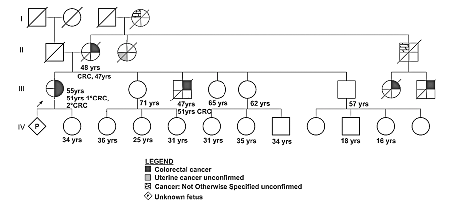
Family history is relevant to colorectal cancer. The proband’s grandmother had gynecological/uterine cancer. Unfortunately, the proband was unable to provide additional details on family history or add confirmatory reports regarding the type of tumor for distant family members. The family came from Teofilo Otoni, a city in northern Minas Gerais State in southeastern Brazil. Family history met the Bethesda criteria for LS. Definitive evidence for the Amsterdam II criteria was not obtained because all other affected relatives were already deceased, and we did not have access to their histological records ( Figure 1 ). Nonetheless, LS diagnosis was supported by immunohistochemical analysis of the proband’s tumors, evidencing the loss of expression of MSH2 in one tumor and MSH2 and MSH6 in the other.
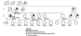
Proband pedigree showing familial segregation of tumors, age of onset, and cancer types. Age in years refers to the current age of the person or age at death. A well-defined age at diagnosis was obtained for III.1 (51-year-old), II.2 (47-year-old), and III.3 (46-year-old)
After pre-test genetic counseling, mutation screening was conducted in the peripheral blood using a next-generation sequencing (NGS)-based multi-gene panel comprising all MMR genes associated with LS, including EPCAM. Mutation screening was performed in a CLIA-approved laboratory (on April 2, 2018). No copy number variation analyses were performed. A novel germline variant was identified, NM_000251.3(MSH2):c.1894_1898del (p.Ile633Lysfs*9), which was heterozygous. In this study, we describe the analysis of the functional impact and potential pathogenicity of this variant by adopting a multistep approach to investigate the possible effects, at the RNA and protein levels.
This bioinformatics analysis included the following steps: 1) Previous descriptions of this variant were searched in ClinVar,( 19 ) Database of Genomic Variants,( 20 ) Ensembl,( 21 ) OMIM, PubMed,( 22 ) European Variation Archive,( 23 ) GeneCard,( 24 ) InSIGHT DNA Variant Database,( 14 ) VarSome, and ABraOM;( 25 ) 2) Variant impacts were predicted using the Variant Effect Predictor (VEP);( 21 ) 3) CDS sequence containing NM_000251.3(MSH2):c.1894_1898del was produced by editing the FASTA format sequence of the MSH2 (NM_000251.3) CDS, downloaded from the National Center for Biotechnology Information (NCBI);( 26 ) 4) This RNA sequence was translated to generate the protein sequence containing NM_000251.3(MSH2):c.1894_1898del (p.Ile633Lysfs*9) using ExPASy Translate;( 27 ) 5) MSH2 protein isoforms were searched in the UNIPROT database.( 28 ) Initially, two MSH2 protein isoforms were identified (P43246-1 and P43246-2) and compared using Basic Local Alignment Search Tool proteins (BLASTP).( 29 ) Isoforms were also predicted using the Variant Effect Predictor (VEP) and GTEx;( 30 ) 6) Nonsense-mediated decay (NMD) was evaluated using Mutation Taster 2021;( 31 ) 7) Evidence for exon skipping was searched in CDS and protein databases as well as in the literature; 8) Information on MSH2 protein domains was obtained using Pfam;( 32 ) 9) Impact of this variant on the MSH2 3D-structure was evaluated by homology modeling using Modeller 9.24,( 33 ) with eight models (2O8B, 2O8C, 2O8D, 2O8F, 3THW, 3THX, 3THY, and 3THZ) obtained from PDB.( 34 ) Ten models of NM_000251.3(MSH2):c.1894_1898del (p.Ile633Lysfs*9) were generated. Among them, the five models presenting the lowest Discrete Optimizer Protein Energy (DOPE) values were selected to produce Ramachandran plots in PDBsum( 35 ) and estimate Z-scores using the PROSA-Web( 36 ) (Protein Structure Analyses) software. Best models were selected based on higher PROCHECK/Z-score. Once validated, the model with the highest PROCHECK/Z-score was selected for comparison of the 3D-structures (MSH2 wild-type versus NM_000251.3(MSH2):c.1894_1898del (p.Ile633Lysfs*9)) using PyMOL software.( 37 )
This project was approved by the Ethics in Research Committee of the Universidade Federal de Minas Gerais under the protocols CAAE: 01758418.0.0000.5149; # 3.134.990 and CAAE: 48770621.6.0000.5149; # 4.865.051.
❚RESULTS
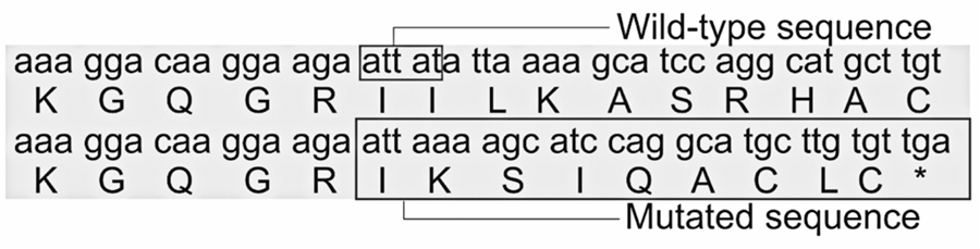
NM_000251.3(MSH2):c.1894_1898del (p.Ile633Lysfs*9) was not identified in any of the databases searched. The MSH2 CDS containing the five-nucleotide deletion induced by this variant and the resulting protein sequence predicted using ExPASy Translate are provided in Table 1S , Supplementary Material. The wild-type MSH2 protein contains 934 amino acids, and this deletion resulted in a reduced protein length to 640 amino acids. Therefore, 294 amino acids at the C-terminus were lost. The five-nucleotide deletion affected codons 632 and 633. After the frameshift mutation (fsm), amino acid 632, an isoleucine residue, did not change. The first change observed in the protein sequence was in amino acid residue 633, which was a substitution of isoleucine with lysine. A premature stop codon was present at the 9th position ( Figure 2 ). We used the NM_000251.3(MSH2):c.1894_1898del (p.Ile633Lysfs*9)-predicted protein sequence to search the NCBI database, using BLASTP. No match for the sequence was identified after the frameshift mutation, supporting the idea that this variant has not been previously reported.
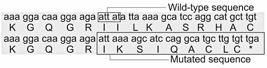
Comparative alignment of MSH2 mRNA and protein, wild-type vs . NM_000251.3(MSH2):c.1894_1898del (p.Ile633Lysfs*9) variant. The 5-nucleotide deletion is shown within a box in the wild-type mRNA sequence. Observe that the first codon created after the 5-nucleotide deletion is an ATT and, therefore, the first amino acid in the mutated segment is still an isoleucine (position 632). The sequence changes from codon 633 onwards, to include a stop codon at position 641
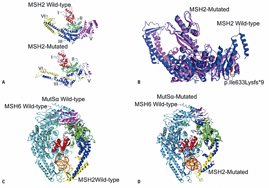
Next, we evaluated the effect of NM_000251.3(MSH2):c.1894_1898del (p.Ile633Lysfs*9) on MSH2 protein domains. MSH2 contains 33 protein domains and characteristic molecular features ( Table 2S , Supplementary Material). This variant disrupts exon 12 and leads to the deletion of exons 13 to 16. NM_000251.3(MSH2):c.1894_1898del (p.Ile633Lysfs*9) is located in the loop between MutS domains III/IV and V, causing the loss of domain V, a highly conserved ATPase domain that provides energy for the first steps of MMR.( 38 ) The 3D-model of the wild-type MSH2 protein selected using Modeller was 2O8B. Visual inspection of the comparative 3D-models of the wild-type and mutated proteins suggested that this variant affects MSH2 and MSH6 protein interactions ( Figure 3 ).
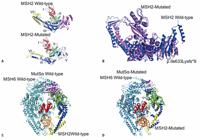
A) Comparison between MSH2 wild-type and NM_000251.3(MSH2):c.1894_1898del p.Ile633Lysfs*9 proteins. MutS I domain is represented in red; MutS II domains in green; MutS III domain in blue; MutS IV domain in yellow; and, MutS V domain in magenta; B) Overlapping blue wild-type MSH2 protein and magenta mutated MSH2 protein. The portion of the protein that is affected by the frame-shift mutation is indicated in red; C) Wild-type MutS alpha complex. Blue contains wild-type MSH2 protein and orange contains wild-type MSH6 protein; D) Mutated MutS alpha complex. Blue shows the mutated MSH2 protein and orange, the wild-type MSH6 protein
In addition, we evaluated the impact of this variant on different MSH2 RNA isoforms and overlapping genes and investigated the mechanisms that may reduce the impact of the variant, such as alternative splicing, exon skipping, and NMD. MSH2 is located in the 2p21-p16.3 region, CHR2:47403067-47634501 (GRCh38 p.13). Typically, MSH2 is composed of 16 exons; however, this gene contains 46 possible exons. MSH2 mRNA undergoes alternative splicing. One difficulty in predicting the impact of the MSH2 variant is that the number of CDS and protein isoforms, as well as their identification numbers, vary according to the source consulted. For example, nine isoforms have been described in Ensembl, and 10 isoforms have been described in GTEx. The CDS and MSH2 protein isoforms linking different nomenclatures are shown in Table 1S . The two canonical MSH2 protein isoforms (P43246-1 and P43246-2) were similarly affected by the NM_000251.3(MSH2):c.1894_1898del (p.Ile633Lysfs*9) variant. P43246-1 was 66 amino acids longer than P43246-2. This difference is due to alternative splicing of the first exon, which encodes a flexible loop at the N-terminus of the protein and does not change the MutS domains. Therefore, the 294 amino acid deletion induced by NM_000251.3(MSH2):c.1894_1898del (p.Ile633Lysfs*9) starts at residue 640 in P43246-1 and residue 576 in the P43246-2 isoform. However, the sequences and, consequently, the variant effects of both isoforms were the same in this region. Additional information on the impact of this variant on different isoforms is provided in Table 1S , Supplementary Material.
There are two additional genes in this region, MSH2-OT1 and KCNK2 . MSH2-OT1 and KCNK12 overlap with the long MSH2 isoforms. None of these neighboring genes/isoforms were affected by the NM_000251.3(MSH2):c.1894_1898del (p.Ile633Lysfs*9) variant.
Alternative splicing, which can rescue a mutated allele by generating a shorter but functional protein, has also been investigated.( 39 ) No MSH2 protein isoform without exon 12 but preserving NH2- and COOH-ends was supported (GTex, accessed May 29, 2025). To date, no such isoform has been reported in either the RNA or protein databases. The predicted impact of NM_000251.3(MSH2):c.1894_1898del (p.Ile633Lysfs*9) on different MSH2 RNA and protein isoforms is presented in Table 2S , Supplementary Material. Considering the immunohistochemical findings, showing loss of MSH2 protein expression in one tumor and that of MSH2 and MSH6 in the other, we investigated the hypothesis of NMD. The premature termination codon inserted as a consequence of the frameshift mutation occurs in the 12th out of 16 exons, before the last exon-exon boundary (NM_000251.3). Consequently, Mutation Taster predicts that this variant leads to NMD.
DISCUSSION
In the present study, a patient presenting the LS phenotype with a newly identified germline variant was investigated clinically, immunohistochemically, and at the molecular level. NM_000251.3(MSH2):c.1894_1898del (p.Ile633Lysfs*9) was not found in the aforementioned variant databases or in studies describing Brazilian samples published to date.( 40 , 41 ) In VarSome, a similar fsm in the same region, MSH2(ENST00000233146.7):c.1899_1900del p.(Leu634LysfsTer9), is listed as pathogenic; however, the associated phenotypes and molecular mechanisms are not provided. Here, we discuss two aspects: the complexity of establishing a diagnosis of LS in small families and the difficulty of classifying germline variants in relation to their pathogenicity.
First, one of the dilemmas in assisting people with mutations that predispose them to an inherited disease but do not necessarily determine the disease, is how to guide them appropriately through the decision-making process. The main questions concern medical support and balancing the risks and benefits of the available risk-reducing procedures. Some risk-reducing procedures are highly invasive, and the decision-making process requires a solid scientific basis as well as common sense. Since LS is a heterogeneous disease, optimized management may be limited, depending on how well documented and complete the family data is, in order to apply current guidelines.( 42 ) Complete family data is frequently difficult to obtain, particularly the actual number of affected relatives, information on the type of neoplasia, age of onset, and tumor documentation. In addition, families tend to be small, and patients may have restrictions on contacting family members; thus, improving the quality of the data may be difficult. Second, we consider the evidence for MSH2 loss of function and NM_000251.3(MSH2):c.1894_1898del (p.Ile633Lysfs*9) contribution to LS in this family. Loss of MSH2 expression was implicated by immunohistochemistry, in the tumors of the proband. Identification of NM_000251.3(MSH2):c.1894_1898del (p.Ile633Lysfs*9) confirmed the diagnosis of LS. Frame-shift mutations are automatically classified as pathogenic by most software used in variant pathogenicity classification, and most fsms are probably or certainly pathogenic. However, compensatory cellular mechanisms function at both the RNA and protein levels. At the RNA level, damage-reducing mechanisms include NMD, alternative splicing, and exon skipping.( 43 , 44 ) At the protein level, some mechanisms are damaging, such as dominant-negative effects, a phenomenon that occurs in dimeric or multimeric proteins in which the entry of a mutated subunit into the complex induces its precipitation.( 45 ) Consequently, the mutated allele reduces the function of the normal allele. However, dominant-negative effects are not observed when the variant affects the protein surface, which interacts with other proteins and prevents it from entering the complexes.( 46 ) Therefore, the impact of fsms should be carefully evaluated.
At the RNA level, we searched for evidence of alternative splicing, exon skipping, or NMD. Isoforms described in MSH2 in both healthy and colon cancer cell lines include the deletion of some exons;( 47 ) however, no evidence of alternative splicing or exon skipping resulting in an isoform without exon 12 was found in literature or any database. This variant fulfills the typical requirements for NMD, that is, the creation of a stop codon before the last exon-exon boundary, and consequently, distant from the typical termination of translation.( 48 ) The loss of MSH2 expression observed by immunohistochemistry provides additional evidence of NMD. However, another possible loss-of-function mechanism has emerged. Protein variants causing loss of the C-terminus are frequently driven to degradation, a mechanism that may also explain the loss of MSH2 expression in the proband tumor, as detected by immunohistochemistry.( 49 )
Despite the evidence for NMD or protein degradation due to the lack of a C-terminus, we investigated the consequences of NM_000251.3(MSH2):c.1894_1898del (p.Ile633Lysfs*9) at the protein level. In presence of this mutation, MSH2 protein loses 294 C-terminal amino acid residues. NM_000251.3(MSH2):c.1894_1898del (p.Ile633Lysfs*9) is expected to cause the loss of MutS domain V and, consequently, the loss of the interaction with ADP/ATP and the ATPase activity needed for mismatch recognition and DNA binding, at the very beginning of the MMR.( 3 , 41 , 50 ) MSH2 interacts with MSH6 and MSH3 to form MutSalpha and MutSbeta, respectively, via MutS domains I and IV. These domains are also involved in DNA binding. MSH2 MutS domains II and III act as connectors between MutS domains V, I, and IV.( 3 , 41 , 50 ) At first sight, it appears that the loss of MutS domain V may not prevent the mutated MSH2 protein monomer from entering the MutSalpha or MutSbeta complexes. An additional description of the interaction mechanisms of the MutS complexes involved in MMR is provided in Table 3S , Supplementary Material. We modeled protein complexes containing MSH2 to understand how they may be affected by the presence of NM_000251.3(MSH2):c.1894_1898del (p.Ile633Lysfs*9) ( Figure 3 ). The results suggest that this variant prevents MSH2 from interacting with MSH6 to form the MutSalpha complex. No evidence of the ability of MSH2 or MSH3 to form MutSbeta was found.
After considering the overall results, we found no evidence that NM_000251.3(MSH2):c.1894_1898del (p.Ile633Lysfs*9) is a non-pathogenic variant. The strengths of the present study are as follows. 1) This variant was not found in any of the population databases. It has been included in VarSome, but without additional information; 2) It causes the loss of MutS domain V, and therefore, the ADP binding ability of the MSH2 protein; 3) There is no evidence that the effects of this variant may be ameliorated by physiological alternative splicing or exon skipping. The large number of exon 12 pathogenic variants in InSIGHT suggests that no such rescue mechanisms are possible; 4) There is evidence of NMD; and, 5). In case this mRNA escapes NMD, the resulting protein may possibly enter MutSalpha complexes; however, these complexes would be less compact than those established by the normal allele and probably non-functional because of the loss of the MSH2 ADP binding site. Regarding limitations of the study , no in vitro analyses were performed to validate or exclude the mechanisms identified in silico . However, according to the ACMG criteria, in vitro or in vivo analyses are not formally required when loss of function is a highly probable consequence of a genetic variant (e.g., an fsm). NM_000251.3(MSH2):c.1894_1898del (p.Ile633Lysfs*9) fulfills the criteria for PVS1 (null variant - nonsense, frameshift, canonical +/- 1 or splice sites, initiation codon, and single or multiple exon deletion). It also fulfills the PM2 criterion (absent from controls or present with extremely low frequency, if recessive, in the Exome Sequencing Project,( 51 ) 1000 Genomes Project,( 52 ) or Exome Aggregation Consortium,( 53 ) which supports its classification as pathogenic. The additional bioinformatics analysis described here provided no evidence of rescue mechanisms acting to alleviate the effects of NM_000251.3(MSH2):c.1894_1898del (p.Ile633Lysfs*9). The bioinformatic analysis strategy proposed in this study was limited by the absence of validation by analysis of similar variants, in other genes.
CONCLUSION
In the present study, we used several different bioinformatics tools to evaluate the NM_000251.3(MSH2):c.1894_1898del (p.Ile633Lysfs*9) germline variant identified in a patient with Lynch syndrome. No evidence was identified for a compensatory mechanism that may reduce the impact of NM_000251.3(MSH2):c.1894_1898del (p.Ile633Lysfs*9). In conclusion, the evidence reported in this study indicates that NM_000251.3(MSH2):c.1894_1898del p.Ile633Lysfs*9 is a likely pathogenic variant that can explain the occurrence of Lynch syndrome in this family.
DATA AVAILABILITY:
Data are available to reviewers upon request.
ACKNOWLEDGMENTS
We thank the patient and the patient’s family for their participation in this study.
Funding Thalia Rodrigues de Souza Zózimo and Carolina Guimarães Ramos Matosinho were awarded a fellowship from the Fundação de Amparo à Pesquisa do Estado de Minas Gerais (FAPEMIG) and Coordenação de Pessoal de Nível Superior (CAPES), respectively; Maria Raquel Santos Carvalho was awarded a fellowship from the Conselho Nacional de Desenvolvimento Científico e Tecnológico (CNPq, grant numbers 312068/2015-8 and 312405/2018-9); Pró-Reitoria de Pesquisa de Universidade Federal de Minas Gerais (PRPq/UFMG). Agnaldo Lopes da Silva Filho was awarded Fundação de Amparo à Pesquisa do Estado de Minas Gerais (FAPEMIG) grant numbers APQ-02373-17 and APQ-02856-23, and Letícia da Conceição Braga was awarded FAPEMIG grant number APQ-01292-21. This study was also supported by the FAPEMIG Minas Gerais Network for Cancer Translational Research (RED-00110-23).
REFERENCES
- 1 McKusick VA. Mendelian inheritance in man: a catalog of human genes and genetic disorders. JHU Press; 1998.
- 2 Lynch HT, Snyder CL, Shaw TG, Heinen CD, Hitchins MP. Milestones of Lynch syndrome: 1895-2015. Nat Rev Cancer. 2015;15(3):181-94.
- 3 Gupta S, Gellert M, Yang W. Mechanism of mismatch recognition revealed by human MutSß bound to unpaired DNA loops. Nat Struct Mol Biol. 2011;19(1):72-8.
- 4 Biller LH, Syngal S, Yurgelun MB. Recent advances in Lynch syndrome. Fam Cancer. 2019;18(2):211-9.
- 5 Ryan NA, Glaire MA, Blake D, Cabrera-Dandy M, Evans DG, Crosbie EJ. The proportion of endometrial cancers associated with Lynch syndrome: a systematic review of the literature and meta-analysis. Genet Med. 2019;21(10):2167-80.
- 6 Dominguez-Valentin M, Sampson JR, Seppälä TT, Ten Broeke SW, Plazzer JP, Nakken S, et al. Cancer risks by gene, age, and gender in 6350 carriers of pathogenic mismatch repair variants: findings from the Prospective Lynch Syndrome Database. Genet Med. 2020;22(1):15-25.
- 7 Crouse GF. Non-canonical actions of mismatch repair. DNA Repair (Amst). 2016;38:102-9.
- 8 Toft NJ, Winton DJ, Kelly J, Howard LA, Dekker M, te Riele H, et al. Msh2 status modulates both apoptosis and mutation frequency in the murine small intestine. Proc Natl Acad Sci USA. 1999;96(7):3911-5.
- 9 Li MM, Chao E, Esplin ED, Miller DT, Nathanson KL, Plon SE, Scheuner MT, Stewart DR; ACMG Professional Practice and Guidelines Committee. Points to consider for reporting of germline variation in patients undergoing tumor testing: a statement of the American College of Medical Genetics and Genomics (ACMG). Genet Med. 2020;22(7):1142-48.
- 10 Umar A, Boland CR, Terdiman JP, Syngal S, de la Chapelle A, Rüschoff J, et al. Revised Bethesda Guidelines for hereditary nonpolyposis colorectal cancer (Lynch syndrome) and microsatellite instability. J Natl Cancer Inst. 2004;96(4):261-8.
- 11 Vasen HF, Mecklin JP, Khan PM, Lynch HT. The international collaborative group on hereditary non-polyposis colorectal cancer (ICG-HNPCC). Dis Colon Rectum. 1991;34(5):424-5.
- 12 Vasen HF, Watson P, Mecklin JP, Lynch HT. New clinical criteria for hereditary nonpolyposis colorectal cancer (HNPCC, Lynch syndrome) proposed by the International Collaborative group on HNPCC. Gastroenterology. 1999; 116(6):1453-6.
- 13 Kastrinos F, Uno H, Ukaegbu C, Alvero C, McFarland A, Yurgelun MB, et al. Development and Validation of the PREMM 5 Model for Comprehensive Risk Assessment of Lynch Syndrome. J Clin Oncol. 2017;35(19):2165-72.
- 14 Thompson BA, Spurdle AB, Plazzer JP, Greenblatt MS, Akagi K, Al-Mulla F, et al. Application of a 5-tiered scheme for standardized classification of 2,360 unique mismatch repair gene variants in the InSiGHT locus-specific database. Nat Genet. 2014;46(2):107-15.
- 15 Plazzer JP, Sijmons RH, Woods MO, Peltomäki P, Thompson B, Den Dunnen JT, et al. The InSiGHT database: utilizing 100 years of insights into Lynch syndrome. Fam Cancer. 2013;12(2):175-80.
- 16 Kopanos C, Tsiolkas V, Kouris A, Chapple CE, Albarca Aguilera M, Meyer R, et al. VarSome: the human genomic variant search engine. Bioinformatics. 2019;35(11):1978-80.
- 17 Stenson PD, Mort M, Ball EV, Chapman M, Evans K, Azevedo L, et al. The Human Gene Mutation Database (HGMD ® ): optimizing its use in a clinical diagnostic or research setting. Hum Genet. 2020;139(10):1197-207.
- 18 Richards S, Aziz N, Bale S, Bick D, Das S, Gastier-Foster J, Grody WW, Hegde M, Lyon E, Spector E, Voelkerding K, Rehm HL; ACMG Laboratory Quality Assurance Committee. Standards and guidelines for the interpretation of sequence variants: a joint consensus recommendation of the American College of Medical Genetics and Genomics and the Association for Molecular Pathology. Genet Med. 2015;17(5):405-24.
- 19 Landrum MJ, Chitipiralla S, Brown GR, Chen C, Gu B, Hart J, et al. ClinVar: improvements to accessing data. Nucleic Acids Res. 2020;48 D1:D835-44.
- 20 MacDonald JR, Ziman R, Yuen RK, Feuk L, Scherer SW. The Database of Genomic Variants: a curated collection of structural variation in the human genome. Nucleic Acids Res. 2014;42(Database issue D1):D986-92.
- 21 Hunt SE, McLaren W, Gil L, Thormann A, Schuilenburg H, Sheppard D, et al. Ensembl variation resources. Database (Oxford). 2018;2018:bay119.
- 22 Fiorini N, Canese K, Starchenko G, Kireev E, Kim W, Miller V, et al. Best match: new relevance search for PubMed. PLoS Biol. 2018;16:e2005343.
-
23 The European Bioinformatics Institute - EMBL-EBI. European Variation Archive. Last edited: Feb. 25 2020 [cited 2021 Apr 25]. Available from: https://www.ebi.ac.uk/eva/
» https://www.ebi.ac.uk/eva/ - 24 Stelzer G, Rosen N, Plaschkes I, Zimmerman S, Twik M, Fishilevich S, et al. The GeneCards Suite: From Gene Data Mining to Disease Genome Sequence Analyses. Curr Protoc Bioinformatics. 2016;54:1.30.1-1.30.33.
- 25 Naslavsky MS, Scliar MO, Yamamoto GL, Wang JY, Zverinova S, Karp T, et al. Whole-genome sequencing of 1,171 elderly admixed individuals from São Paulo, Brazil. Nat Commun. 2022;13(1):1004. Erratum in: Nat Commun. 2022;13(1):1831.
- 26 NCBI Resource Coordinators. Database resources of the National Center for Biotechnology Information. Nucleic Acids Res. 2018;46(D1):D8-13.
- 27 Artimo P, Jonnalagedda M, Arnold K, Baratin D, Csardi G, de Castro E, et al. ExPASy: SIB bioinformatics resource portal. Nucleic Acids Res. 2012;40(Web Server issue W1):W597-603.
- 28 Consortium U; UniProt Consortium. UniProt: a worldwide hub of protein knowledge. Nucleic Acids Res. 2019;47 D1:D506-15.
- 29 Altschul SF, Gish W, Miller W, Myers EW, Lipman DJ. Basic local alignment search tool. J Mol Biol. 1990;215(3):403-10.
- 30 GTEx Consortium. Human genomics. The Genotype-Tissue Expression (GTEx) pilot analysis: multitissue gene regulation in humans. Science. 2015;348(6235):648-60.
- 31 Schwarz JM, Cooper DN, Schuelke M, Seelow D. MutationTaster2: mutation prediction for the deep-sequencing age. Nat Methods. 2014;11(4):361-2.
- 32 El-Gebali S, Mistry J, Bateman A, Eddy SR, Luciani A, Potter SC, et al. The Pfam protein families database in 2019. Nucleic Acids Res. 2019;47 D1:D427-32.
- 33 Šali A, Blundell TL. Comparative protein modelling by satisfaction of spatial restraints. J Mol Biol. 1993;234(3):779-815.
- 34 Heinz DW, Baase WA, Dahlquist FW, Matthews BW. How amino-acid insertions are allowed in an a-helix of T4 lysozyme. Nature. 1993;361(6412):561-4.
- 35 Laskowski RA, Jablonska J, Pravda L, Vareková RS, Thornton JM. PDBsum: structural summaries of PDB entries. Protein Sci. 2018;27(1):129-34.
- 36 Wiederstein M, Sippl MJ. ProSA-web: interactive web service for the recognition of errors in three-dimensional structures of proteins. Nucleic Acids Res. 2007;35(Web Server issue):W407-10.
-
37 Schrödinger LL. The PyMOL Molecular Graphics System. Version 2.0 [cited 2025 June 12]. Available from: https://www.pymol.org
» https://www.pymol.org - 38 Lakhani B, Thayer KM, Hingorani MM, Beveridge DL. Evolutionary covariance combined with molecular dynamics predicts a framework for allostery in the MutS DNA mismatch repair protein. J Phys Chem B. 2017;121(9):2049-61.
- 39 Koenig M, Beggs AH, Moyer M, Scherpf S, Heindrich K, Bettecken T, et al. The molecular basis for Duchenne versus Becker muscular dystrophy: correlation of severity with type of deletion. Am J Hum Genet. 1989;45(4):498-506.
- 40 Monaco AP, Bertelson CJ, Liechti-Gallati S, Moser H, Kunkel LM. An explanation for the phenotypic differences between patients bearing partial deletions of the DMD locus. Genomics. 1988;2(1):90-5.
- 41 Schweingruber C, Rufener SC, Zünd D, Yamashita A, Mühlemann O. Nonsense-mediated mRNA decay - mechanisms of substrate mRNA recognition and degradation in mammalian cells. Biochim Biophys Acta. 2013;1829(6-7):612-23.
- 42 Miller JN, Pearce DA. Nonsense-mediated decay in genetic disease: friend or foe? Mutat Res Rev Mutat Res. 2014;762:52-64.
-
43 National Comprehensive Cancer Network (NCCN). Clinical Practice Guidelines in Oncology: genetic/familial high risk assessment: colorectal. Version 1.2023. Plymouth Meeting, PA: NCCN; 2023 [cited 2023 Oct 1]. Available from: https://www.nccn.org/guidelines/guidelines-detail?category=1&id=1436
» https://www.nccn.org/guidelines/guidelines-detail?category=1&id=1436 - 44 Rondagh EJ, Gulikers S, Gómez-García EB, Vanlingen Y, Detisch Y, Winkens B, et al. Nonpolypoid colorectal neoplasms: a challenge in endoscopic surveillance of patients with Lynch syndrome. Endoscopy. 2013;45(4):257-64.
- 45 Dryja TP, Rapaport JM, Joyce JM, Petersen RA. Molecular detection of deletions involving band q14 of chromosome 13 in retinoblastomas. Proc Natl Acad Sci USA. 1986;83(19):7391-4.
- 46 Olkinuora A, Gylling A, Almusa H, Eldfors S, Lepistö A, Mecklin JP, et al. Molecular Basis of Mismatch Repair Protein Deficiency in Tumors from Lynch Suspected Cases with Negative Germline Test Results. Cancers (Basel). 2020;12(7):1853.
- 47 Genuardi M, Viel A, Bonora D, Capozzi E, Bellacosa A, Leonardi F, et al. Characterization of MLH1 and MSH2 alternative splicing and its relevance to molecular testing of colorectal cancer susceptibility. Hum Genet. 1998;102(1):15-20.
- 48 Warren JJ, Pohlhaus TJ, Changela A, Iyer RR, Modrich PL, Beese LS. Structure of the human MutSalpha DNA lesion recognition complex. Mol Cell. 2007;26(4):579-92.
- 49 Martín-López JV, Fishel R. The mechanism of mismatch repair and the functional analysis of mismatch repair defects in Lynch syndrome. Fam Cancer. 2013;12(2):159-68.
- 50 Fukui K. DNA mismatch repair in eukaryotes and bacteria. J Nucleic Acids. 2010;2010(1):260512.
-
51 Exome Variant Server. NHLBI GO Exome Sequencing Project (ESP). Seattle, WA: 2023 [cited 2023 Apr 1]. Available from: http://evs.gs.washington.edu/EVS/
» http://evs.gs.washington.edu/EVS/ - 52 Fairley S, Lowy-Gallego E, Perry E, Flicek P. The International Genome Sample Resource (IGSR) collection of open human genomic variation resources. Nucleic Acids Res. 2020;48(D1):D941-7.
- 53 Karczewski KJ, Francioli LC, Tiao G, Cummings BB, Alföldi J, Wang Q, et al. Genome Aggregation Database Consortium. The mutational constraint spectrum quantified from variation in 141,456 humans. Nature. 2020;581(7809):434-43.
Edited by
-
Associate Editor:
Kenneth Gollob Hospital Israelita Albert Einstein, São Paulo, SP, Brazil ORCID: https://orcid.org/0000-0003-4184-3867