 High-mobility group box 1 protein, histone H3 and histone H4 are not associated with peripheral hypoperfusion in sepsis: a retrospective cohort study
ABSTRACT
Objective: The causal mechanisms of peripheral hypoperfusion in sepsis have not yet been fully elucidated. Therefore, this study aimed to investigate whether peripheral hypoperfusion in septic patients is associated with serum levels of damage-associated molecular patterns.
Methods: A retrospective cohort study was conducted to compare serum levels of damage-associated molecular patterns - histone H3, histone H4, and high-mobility group box-1 protein (HMGB1) - with markers of tissue hypoperfusion - the peripheral perfusion index and the capillary refill time, using a single measurement obtained within the first 24 hours after sepsis diagnosis in critically ill patients from two Brazilian hospitals.
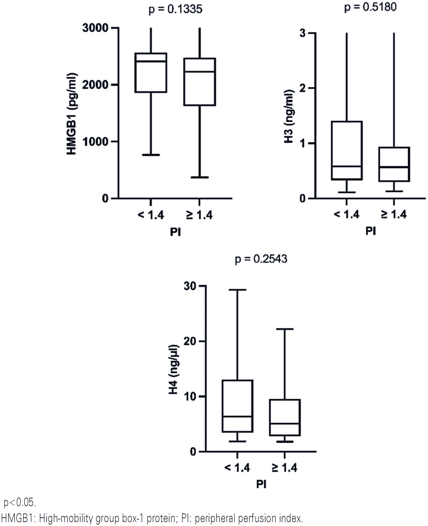
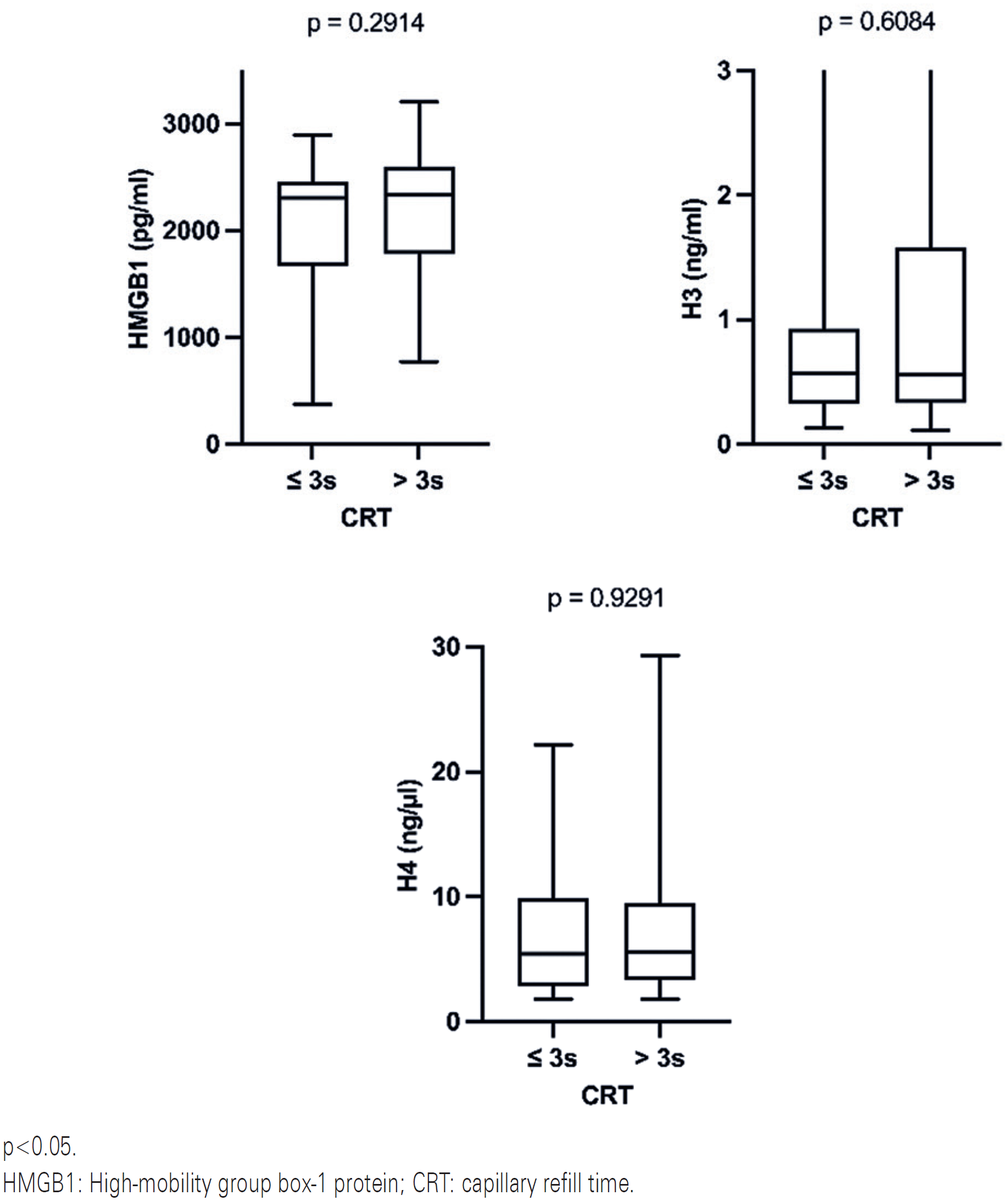
Results: Eighty patients were included in the study. Approximately half of the patients showed persistent peripheral hypoperfusion as determined by at least one of the techniques used. Surviving patients had better capillary refill time [2 (2-4) versus 4 (2-5), p=0.01] and perfusion index [1.9 (0.98-4.95) versus 0.54 (0.32-0.87) p<0.0001] compared with non-survivors. None of the evaluated damage-associated molecular patterns distinguished between survivors and non-survivors. In addition, damage-associated molecular patterns were not associated with perfusion index (versus HMGB1, p=0.13; versus H3, p=0.51 and versus H4, p=0.25) or capillary refill time (versus HMGB1, p=0.29; versus H3, p=0.60 and versus H4, p=0.92).
Conclusion: In septic patients evaluated in this retrospective cohort study, no statistically significant association was found between the levels of histones H3, H4, and HMGB1 protein, tissue hypoperfusion, and mortality.
Keywords:
Sepsis; Alarmins; HMGB1 protein; Histones; Perfusion index; Microcirculation
Highlights
-
■
This study aimed to evaluate the association between damage-associated molecular patterns and peripheral hypoperfusion in sepsis.
-
■
Surviving patients showed better peripheral perfusion than non-survivors.
-
■
No significant association was observed between mortality and serum levels of the evaluated damage-associated molecular patterns.
-
■
Damage-associated molecular patterns levels did not differ between patients with reduced or normal peripheral perfusion.

In Brief
Damage-associated molecular patterns mediate the immuneinflammatory response in sepsis, a process closely related to multiple organ dysfunction. Peripheral hypoperfusion has also been linked to increased mortality in septic patients. Therefore, this retrospective cohort study evaluated the relation between histone H3, histone H4, and high-mobility group box 1 protein with peripheral perfusion in sepsis.
INTRODUCTION
Sepsis remains the leading cause of death among critically ill patients.(1,2) For this reason, understanding the pathophysiological mechanisms behind the syndrome is of major importance to translate this knowledge into concrete results at the bedside.(3,4)
A condition related to its prognosis is the incoherence between macro- and microcirculation, which occurs when microcirculatory abnormalities persist after macrohemodynamic parameters have returned to normal, leading to tissue hypoperfusion and impaired oxygen delivery to cells.(5,6) Several events related to microcirculatory failure have been proposed, such as changes in vascular reactivity, endothelial dysfunction, glycocalyx barrier degradation, and microthrombosis.(6-8) However, evidence of these mechanisms in human studies are rare in the literature.(9-11)
When there is a loss of hemodynamic coherence, lactate can be used to assess systemic organ perfusion,(12,13) but hyperlactatemia may also be explained by other factors not related to a decrease in oxygen delivery.(7) Therefore, peripheral perfusion can be easily monitored at the bedside using capillary refill time (CRT)(14,15) or the peripheral perfusion index (PI).(16,17) Prior research have demonstrated a strong association between CRT and PI and outcome in sepsis.(14,18) Furthermore in the presence of persistent hyperlactatemia,(13) CRT indicates the subgroup with the highest mortality,(15) as does microvascular reactivity assessed by PI.(19)
Additionally, the role of mediators that interfere with immune activity has been discussed, particularly regarding the pathways leading to dysregulated response to infection.(3,4,20) Some mediators, known as damage-associated molecular patterns (DAMPs or alarmins), are host endogenous molecules released by the immune cells due to necrosis, tissue damage or cellular stress secondary to the inflammatory response.(21,22) In order to activate the innate immune system and eliminate the aggressor, DAMPs bind to cell receptors, triggering a complex signaling cascade that may also lead to immune and inflammatory dysfunction. These processes are frequently linked to multiple organ dysfunction and can worsen prognosis.(3,21,22)
Therefore, considering that a) persistent microcirculatory alterations are associated with high mortality in sepsis, particularly when macro- and microcirculation become uncoupled; b) the underlying mechanisms of these alterations are not well evidenced in human studies, and c) DAMPs are associated with immune dysregulation and may theoretically be associated with microcirculatory dysfunction in sepsis, this study aimed to verify the potential association between serum levels of DAMPs and hypoperfusion in septic patients.
OBJECTIVE
The purpose of this study was to determine whether HMGB1, histone H3, and histone H4 are associated with peripheral hypoperfusion in septic patients.
METHODS
A multicenter retrospective cohort study was conducted using a database and stored blood samples from patients enrolled in a previous major study protocol,(19) which is registered in the Brazilian Registry of Clinical Trials under the code RBR-35tv9ft. The study was approved by the Human Research Ethics Committee of the Hospital de Clínicas of the Universidade Federal do Paraná, (CAAE: 28824120.3.0000.0096; #3.913.982). This manuscript also adheres to The Strengthening the Reporting of Observational Studies in Epidemiology (STROBE) Statement.
The study included patients aged 18 years or older who were admitted to three intensive care units at a public tertiary hospital and at a private hospital in southern Brazil, between 2021 and 2022. Patients had sepsis or septic shock within the first 24 hours of diagnosis and received initial hemodynamic resuscitation in accordance with international guidelines.(1,20)
The exclusion criteria were defined at the implementation of the previous study protocol and included severe cirrhosis (Child Pugh class C); severe coagulopathy (platelets <20.000/mm3, activated partial thromboplastin time >70 seconds or International Normalized Ratio >2); severe active bleeding (such as hemorrhagic stroke and upper gastrointestinal bleeding); endocarditis; severe peripheral arterial insufficiency, scleroderma and pregnancy.
All patients or their legal surrogates provided written informed consent.
Epidemiological, clinical, and laboratory data were collected throughout hospitalization. Blood samples for the assessment of serum DAMPs and peripheral perfusion measurements were obtained simultaneously at a single time point: within the first 24 hours after a diagnosis of sepsis or septic shock and after hemodynamic stabilization and adequate fluid resuscitation.
Plasma measurements of three DAMPs - histone H3, histone H4 and high-mobility group box-1 protein (HMGB1) - were performed on the available samples using specific enzyme-linked immunosorbent assay (ELISA) kits according to the manufacturer's instructions: histone H3 (Cayman Chemical, Ann Arbor, MI, USA), histone H4 (EpigenTEK, Farmingdale, NY, USA) and HMGB1 protein (Elabscience, Houston, TX, USA).
Peripheral hypoperfusion was evaluated using PI and CRT values. A pulse oximeter (Masimo Radical®, Masimo-Corp, USA, and Mindray, China) was used to measure PI. The cut-off value defined as hypoperfusion was less than 1.4 for PI(18) and greater than 3 seconds for CRT.(14)
The association between HMGB1, histone H3, histone H4, and peripheral perfusion markers (CRT and PI) was the primary outcome. The correlation between DAMPs and mortality as well as the relation between peripheral perfusion variables and mortality were defined as secondary outcomes. We also examined the patients into shock and non-shock subgroups in a post-hoc analysis to search for an association between peripheral perfusion and DAMPs.
Sample size calculation was not feasible because the study was based on the database of a previous study protocol. Statistical analyses were performed using GraphPad Prism 10 software. Means and standard deviations were used to characterize quantitative variables, whereas absolute and relative frequencies were used to characterize categorical variables. The Shapiro-Wilk test was used to evaluate the normality of the variables. Since all the independent variables did not have a normal distribution, non-parametric data were described as median and interquartile range, and the Mann-Whitney U-test was the statistical test applied for all the analyses, with a 95% confidence interval and a 5% threshold of statistical significance.
RESULTS
Eighty patients were included in the study, with a mean age of 61 years, and half of them were male. Most patients had some type of comorbidity. The clinical variables are summarized in table 1.
When perfusion parameters were assessed, more than half of the patients had persistent hyperlactatemia despite adequate fluid resuscitation, 45% had prolonged CRT, and approximately 50% had hypoperfusion as determined by PI (Table 2). Perfusion index <1.4, extended CRT, and hyperlactatemia were all indicators of hypoperfusion in 25% of the patients.
There was a lack of statistical association between the evaluated DAMPs and PI (Figure 1) or CRT (Figure 2). Additionally, when the patients were separated into shock and non-shock subgroups, the results indicated no difference between DAMPs and either PI or CRT in these subgroups (p>0.05 in all data analyzed).


Moreover, there were no statistically significant associations between mortality and serum levels of histones H3, H4, or HMGB1 (Table 3). Nonetheless, there was a statistically significant relation between all the perfusion variables and mortality, as indicated in table 4.
DISCUSSION
Persistent tissue hypoperfusion is clearly related to increased mortality(23) and its mechanisms are still unclear. On the other hand, DAMPs are currently recognized as important factors in the immune-inflammatory response in sepsis and are closely associated with tissue damage and multiple organ dysfunction.(4,22) In addition, previous experimental studies have demonstrated that modulating DAMPs could improve the prognosis in sepsis.(24,25) Therefore, this study was the first to assess whether serum levels of DAMPs are related to hypoperfusion during the hemodynamic uncoupling phase in septic patients. Nevertheless, we found no association between tissue hypoperfusion and plasma levels of HMGB1, histone H3, and histone H4.
During sepsis, organ hypoperfusion is a dynamic event. In the early phase of resuscitation, hypoperfusion is more dependent on macrohemodynamics (flow-dependent), whereas local metabolic variables predominate in the later stages.(7-9,19) To highlight the microcirculatory mechanisms, which can be observed by the stability of the sample's macrohemodynamic parameters after fluid resuscitation — our study focused on evaluating this later phase. Additionally, peripheral perfusion is the first to be impaired and the last to recover due to the redistribution of blood flow in sepsis,(18,19) so, peripheral hypoperfusion could be a marker of this redistribution. Although our study did not directly measure the cardiac output, the stability of the macrohemodynamic variables makes this explanation less likely for the results.
Several mechanisms have been described to explain these persistent microcirculatory alterations in sepsis,(7-11,23) leading to a heterogeneity of microvascular blood flow, endothelial cell injury, and impaired vascular reactivity to local vasoconstriction-dilation mediators.(9) Previous studies have shown that the excessive release of histones and HMGB1 can damage the structure and function of the endothelium,(11,25) and, at least theoretically, DAMPs could be a linking factor between innate immune activation and the development of microcirculatory failure. However, the lack of difference in DAMPs levels between patients with reduced and normal peripheral perfusion may suggest that DAMPs release could be the consequence rather than a cause of microvascular alterations, representing an "epiphenomenon", since microcirculation failure is an independent predictor of outcome, well described in the literature.(5,6,15,18,19) Additional studies are needed to confirm these conclusions.
These assumptions may also be supported by the fact that an increasing amount of evidence correlates DAMPs with critical illnesses directly related to cell death triggered by the inflammatory process, such as different types of necrosis (pyroptosis, necroptosis and ferroptosis), rather than directly with tissue hypoperfusion.(26-28) Thus, DAMPs could act as a marker of the initial innate immune-inflammatory insult that contributes to a cascade of organ damages, the latter responsible for mortality.(22) This hypothesis is reinforced by evidence regarding another biomarker associated with the innate immune response, C-reactive protein, which is related to the development of sepsis but not to its prognosis.(29)
Another assumption deserves exploration. Since the proportion of perfused capillaries, a strong predictor of sepsis prognosis, cannot distinguish between survivors and nonsurvivors during the first 24 hours of septic shock, even though the value that differentiates these patients rises with time,(5,6) there is another hypothesis associated with DAMPs measurement at a single time point. The DAMPs examined in our investigation were unable to distinguish between septic patients with hypoperfusion and those with normoperfusion, which may be explained by the fact that HMGB1, histone H3, and histone H4 serum levels were tested within the first 24 hours of illness. The association between HMGB1 and sepsis prognosis(30-34) is another finding that supports this theory. In contrast to early measurement, studies that analyzed HMGB1 serially consistently indicated that late HMGB1 measurement was associated with mortality, unlike the initial measurement.(31,35,36)
Regarding the histones H3 and H4, the results also contrasted with the literature.(33,34) However, a recent review demonstrated that it is not possible to clearly confirm this association, given the lack of a gold standard technique and the variety of methodologies used to quantify histones in studies(37) Furthermore, efforts to reduce publication bias have attracted interest in the literature, as it affects the magnitude of evidence, reducing the precision of clinical management.(38) Thus, our negative findings may contribute to a subsequent meta-analysis focusing on the real prognostic accuracy of histones.
In addition, the concept of "enrichment" in precision medicine regarding sepsis is another interesting aspect related to our results.(39) Predictive enrichment prioritizes subgroups based on the same pathophysiological process (immunosuppression, for example), whereas prognostic enrichment focuses on a higher likelihood of a hard endpoint (such as mortality).(39) Therefore, our findings suggest that enrichment for any treatment guided by DAMPs measurements in sepsis should be predictive rather than prognostic, as it occurs with perfusion parameters.
Our study has some important limitations. First, we were unable to determine the sample size necessary to ensure the statistical power of the study because we had limited resources for processing serum DAMPs, although our findings suggest that the effect size would likely have minimal clinical significance if an association were found in a larger group. Second, the fact that a single serum measurement of DAMPs was performed during the early stage of sepsis does not exclude the possibility that DAMPs and peripheral hypoperfusion could be associated in an eventual serial analysis, as discussed above. Third, although the dose of noradrenaline in the study was similar in both groups, the use of noradrenaline as a vasopressor may affect the microcirculation, causing a stopped flow in capillaries as a result of excessive vasoconstriction, directly impairing tissue perfusion.(6) Additionally, only one-third of patients met the criteria for septic shock after initial hemodynamic stabilization,(20) which may indicate a subgroup with a poor prognosis. However, our statistical analysis suggests that this was independent of DAMPs levels. Finally, the patients included in our study were from hospitals in a middle-income country, which is known to affect sepsis prognosis.(40) Consequently, it is not feasible to generalize these findings to countries with more or fewer resources.
CONCLUSION
In conclusion, our findings demonstrated that among septic patients, there was no statistically significant association between the levels of histone H3, histone H4, and HMGB1 protein and hypoperfusion or mortality.
DATA AVAILABILITY
Data are available to reviewers upon request.
ACKNOWLEDGEMENT
The Conselho Nacional de Desenvolvimento Científico e Tecnológico (CNPq - Brazil) provided funding for this research under grant number 18/2021-Universal from CNPq/MCTI/FNDCT.
REFERENCES
- 1 Evans L, Rhodes A, Alhazzani W, Antonelli M, Coopersmith CM, French C, et al. Surviving Sepsis Campaign: International Guidelines for Management of Sepsis and Septic Shock 2021. Crit Care Med. 2021;49(11):e1063-143.
- 2 Fleischmann C, Scherag A, Adhikari NK, Hartog CS, Tsaganos T, Schlattmann P, Angus DC, Reinhart K; International Forum of Acute Care Trialists. Assessment of Global Incidence and Mortality of Hospital-treated Sepsis. Current Estimates and Limitations. Am J Respir Crit Care Med. 2016;193(3):259-72.
- 3 Murao A, Brenner M, Aziz M, Wang P. Exosomes in Sepsis. Front Immunol. 2020;11:2140.
- 4 Cheng Z, Abrams ST, Toh J, Wang SS, Wang Z, Yu Q, et al. The Critical Roles and Mechanisms of Immune Cell Death in Sepsis. Front Immunol. 2020;11:1918.
- 5 Sakr Y, Dubois MJ, De Backer D, Creteur J, Vincent JL. Persistent microcirculatory alterations are associated with organ failure and death in patients with septic shock. Crit Care Med. 2004;32(9):1825-31.
- 6 De Backer D, Donadello K, Sakr Y, Ospina-Tascon G, Salgado D, Scolletta S, et al. Microcirculatory alterations in patients with severe sepsis: impact of time of assessment and relationship with outcome. Crit Care Med. 2013;41(3):791-9.
- 7 He HW, Long Y, Liu DW, Ince C. Resuscitation incoherence and dynamic circulation-perfusion coupling in circulatory shock. Chin Med J (Engl). 2019;132(10):1218-27.
- 8 Mok G, Hendin A, Reardon P, Hickey M, Gray S, Yadav K. Macrocirculatory and Microcirculatory Endpoints in Sepsis Resuscitation. J Intensive Care Med. 2021;36(12):1385-91.
- 9 De Backer D, Orbegozo Cortes D, Donadello K, Vincent JL. Pathophysiology of microcirculatory dysfunction and the pathogenesis of septic shock. Virulence. 2014;5(1):73-9.
- 10 Guven G, Hilty MP, Ince C. Microcirculation: Physiology, Pathophysiology, and Clinical Application. Blood Purif. 2020;49(1-2):143-50.
- 11 Tang AL, Shen MJ, Zhang GQ. Intestinal microcirculation dysfunction in sepsis: pathophysiology, clinical monitoring, and therapeutic interventions. World J Emerg Med. 2022;13(5):343-8.
- 12 Bakker J, Gris P, Coffernils M, Kahn RJ, Vincent JL. Serial blood lactate levels can predict the development of multiple organ failure following septic shock. Am J Surg. 1996;171(2):221-6.
- 13 Nguyen HB, Rivers EP, Knoblich BP, Jacobsen G, Muzzin A, Ressler JA, et al. Early lactate clearance is associated with improved outcome in severe sepsis and septic shock. Crit Care Med. 2004;32(8):1637-42.
- 14 Hernández G, Ospina-Tascón GA, Damiani LP, Estenssoro E, Dubin A, Hurtado J, Friedman G, Castro R, Alegría L, Teboul JL, Cecconi M, Ferri G, Jibaja M, Pairumani R, Fernández P, Barahona D, Granda-Luna V, Cavalcanti AB, Bakker J; The ANDROMEDA SHOCK Investigators and the Latin America Intensive Care Network (LIVEN); Hernández G, Ospina-Tascón G, Petri Damiani L, Estenssoro E, Dubin A, Hurtado J, Friedman G, Castro R, Alegría L, Teboul JL, Cecconi M, Cecconi M, Ferri G, Jibaja M, Pairumani R, Fernández P, Barahona D, Cavalcanti AB, Bakker J, Hernández G, Alegría L, Ferri G, Rodriguez N, Holger P, Soto N, Pozo M, Bakker J, Cook D, Vincent JL, Rhodes A, Kavanagh BP, Dellinger P, Rietdijk W, Carpio D, Pavéz N, Henriquez E, Bravo S, Valenzuela ED, Vera M, Dreyse J, Oviedo V, Cid MA, Larroulet M, Petruska E, Sarabia C, Gallardo D, Sanchez JE, González H, Arancibia JM, Muñoz A, Ramirez G, Aravena F, Aquevedo A, Zambrano F, Bozinovic M, Valle F, Ramirez M, Rossel V, Muñoz P, Ceballos C, Esveile C, Carmona C, Candia E, Mendoza D, Sanchez A, Ponce D, Ponce D, Lastra J, Nahuelpán B, Fasce F, Luengo C, Medel N, Cortés C, Campassi L, Rubatto P, Horna N, Furche M, Pendino JC, Bettini L, Lovesio C, González MC, Rodruguez J, Canales H, Caminos F, Galletti C, Minoldo E, Aramburu MJ, Olmos D, Nin N, Tenzi J, Quiroga C, Lacuesta P, Gaudín A, Pais R, Silvestre A, Olivera G, Rieppi G, Berrutti D, Ochoa M, Cobos P, Vintimilla F, Ramirez V, Tobar M, García F, Picoita F, Remache N, Granda V, Paredes F, Barzallo E, Garcés P, Guerrero F, Salazar S, Torres G, Tana C, Calahorrano J, Solis F, Torres P, Herrera L, Ornes A, Peréz V, Delgado G, López A, Espinosa E, Moreira J, Salcedo B, Villacres I, Suing J, Lopez M, Gomez L, Toctaquiza G, Cadena Zapata M, Orazabal MA, Pardo Espejo R, Jimenez J, Calderón A, Paredes G, Barberán JL, Moya T, Atehortua H, Sabogal R, Ortiz G, Lara A, Sanchez F, Hernán Portilla A, Dávila H, Mora JA, Calderón LE, Alvarez I, Escobar E, Bejarano A, Bustamante LA, Aldana JL. Effect of a Resuscitation Strategy Targeting Peripheral Perfusion Status vs Serum Lactate Levels on 28-Day Mortality Among Patients With Septic Shock: The ANDROMEDA-SHOCK Randomized Clinical Trial. JAMA. 2019;321(7):654-664..
- 15 Lara B, Enberg L, Ortega M, Leon P, Kripper C, Aguilera P, et al. Capillary refill time during fluid resuscitation in patients with sepsis-related hyperlactatemia at the emergency department is related to mortality. PLoS One. 2017;12(11):e0188548.
- 16 Coutrot M, Dudoignon E, Joachim J, Gayat E, Vallée F, Dépret F. Perfusion index: physical principles, physiological meanings and clinical implications in anaesthesia and critical care. Anaesth Crit Care Pain Med. 2021;40(6):100964.
- 17 Elshal MM, Hasanin AM, Mostafa M, Gamal RM. Plethysmographic Peripheral Perfusion Index: Could It Be a New Vital Sign? Front Med (Lausanne). 2021;8:651909.
- 18 Mongkolpun W, Orbegozo D, Cordeiro CP, Franco CJ, Vincent JL, Creteur J. Alterations in Skin Blood Flow at the Fingertip Are Related to Mortality in Patients With Circulatory Shock. Crit Care Med. 2020;48(4):443-50.
- 19 de Miranda AC, De Stefani FD, Dal Vesco BC, Junior HC, Morello LG, Assreuy J, et al. Peripheral ischemic reserve in sepsis and septic shock as a new bedside prognostic enrichment tool: A Brazilian cohort study. PLoS One. 2023;18(7):e0288249.
- 20 Singer M, Deutschman CS, Seymour CW, Shankar-Hari M, Annane D, Bauer M, et al. The Third International Consensus Definitions for Sepsis and Septic Shock (Sepsis-3). JAMA. 2016;315(8):801-10.
- 21 Bianchi ME. DAMPs, PAMPs and alarmins: all we need to know about danger. J Leukoc Biol. 2007;81(1):1-5.
- 22 Denning NL, Aziz M, Gurien SD, Wang P. DAMPs and NETs in Sepsis. Front Immunol. 2019;10:2536.
- 23 Ince C. Hemodynamic coherence and the rationale for monitoring the microcirculation. Crit Care. 2015;19(Suppl 3):S8.
- 24 Stevens NE, Chapman MJ, Fraser CK, Kuchel TR, Hayball JD, Diener KR. Therapeutic targeting of HMGB1 during experimental sepsis modulates the inflammatory cytokine profile to one associated with improved clinical outcomes. Sci Rep. 2017;7(1):5850.
- 25 Li L, Lu YQ. The Regulatory Role of High-Mobility Group Protein 1 in Sepsis-Related Immunity. Front Immunol. 2021;11:601815.
- 26 Shah M, He Z, Rauf A, Beikoghli Kalkhoran S, Heiestad CM, Stensløkken KO, et al. Extracellular histones are a target in myocardial ischaemia-reperfusion injury. Cardiovasc Res. 2022;118(4):1115-25.
- 27 Pantalone D, Bergamini C, Martellucci J, Alemanno G, Bruscino A, Maltinti G, et al. The Role of DAMPS in Burns and Hemorrhagic Shock Immune Response: Pathophysiology and Clinical Issues. Review. Int J Mol Sci. 2021;22(13):7020.
- 28 Ludes PO, de Roquetaillade C, Chousterman BG, Pottecher J, Mebazaa A. Role of Damage-Associated Molecular Patterns in Septic Acute Kidney Injury, From Injury to Recovery. Front Immunol. 2021;12:606622.
- 29 Molano-Franco D, Arevalo-Rodriguez I, Muriel A, Del Campo-Albendea L, Fernández-García S, Alvarez-Méndez A, Simancas-Racines D, Viteri A, Sanchez G, Fernandez-Felix B, Lopez-Alcalde J, Solà I, Osorio D, Khan KS, Nuvials X, Ferrer R, Zamora J; SEPSIS-BIOMARKERS Collaborators. Basal procalcitonin, C-reactive protein, interleukin-6, and presepsin for prediction of mortality in critically ill septic patients: a systematic review and meta-analysis. Diagn Progn Res. 2023;7(1):15. Review.
- 30 Tian Y, Russo RM, Li Y, Karmakar M, Liu B, Puskarich MA, et al. Serum citrullinated histone H3 concentrations differentiate patients with septic verses non-septic shock and correlate with disease severity. Infection. 2021;49(1):83-93.
- 31 Gibot S, Massin F, Cravoisy A, Barraud D, Nace L, Levy B, et al. High-mobility group box 1 protein plasma concentrations during septic shock. Intensive Care Med. 2007;33(8):1347-53.
- 32 Rhodes A, Wort SJ, Thomas H, Collinson P, Bennett ED. Plasma DNA concentration as a predictor of mortality and sepsis in critically ill patients. Crit Care. 2006;10(2):R60.
- 33 Ekaney ML, Otto GP, Sossdorf M, Sponholz C, Boehringer M, Loesche W, et al. Impact of plasma histones in human sepsis and their contribution to cellular injury and inflammation. Crit Care. 2014;18(5):543.
- 34 Eichhorn T, Linsberger I, Lauková L, Tripisciano C, Fendl B, Weiss R, et al. Analysis of Inflammatory Mediator Profiles in Sepsis Patients Reveals That Extracellular Histones Are Strongly Elevated in Nonsurvivors. Mediators Inflamm. 2021;2021(1):8395048.
- 35 Sundén-Cullberg J, Norrby-Teglund A, Rouhiainen A, Rauvala H, Herman G, Tracey KJ, et al. Persistent elevation of high mobility group box-1 protein (HMGB1) in patients with severe sepsis and septic shock. Crit Care Med. 2005;33(3):564-73.
- 36 Matsuura R, Komaru Y, Miyamoto Y, Yoshida T, Yoshimoto K, Hamasaki Y, et al. Different Biomarker Kinetics in Critically Ill Patients with High Lactate Levels. Diagnostics (Basel). 2020;10(7):454.
- 37 Li Y, Wan D, Luo X, Song T, Wang Y, Yu Q, et al. Circulating Histones in Sepsis: Potential Outcome Predictors and Therapeutic Targets. Front Immunol. 2021;12:650184.
- 38 Murad MH, Chu H, Lin L, Wang Z. The effect of publication bias magnitude and direction on the certainty in evidence. BMJ Evid Based Med. 2018;23(3):84-6.
- 39 Stanski NL, Wong HR. Prognostic and predictive enrichment in sepsis. Nat Rev Nephrol. 2020;16(1):20-31.
- 40 Stephen AH, Montoya RL, Aluisio AR. Sepsis and Septic Shock in Low- and Middle-Income Countries. Surg Infect (Larchmt). 2020;21(7):571-8.
Edited by
-
Associate Editor:
Kenneth Gollob Hospital Israelita Albert Einstein, São Paulo, SP, Brazil https://orcid.org/0000-0003-4184-3867