 Transcranial Magnetic Stimulation on spasticity in Multiple Sclerosis – protocol for a feasibility study for a randomized, crossover clinical trial
ABSTRACT
Spasticity is a common sequela of multiple sclerosis (MS) that can cause pain, deformities, and impair movement. Transcranial magnetic stimulation (TMS) has been investigated for the treatment of spasticity; however, further investigation is needed. This study aimed to compare the effect of high and low frequency TMS on quadriceps spasticity in MS. Adults diagnosed with MS and who present bilateral quadriceps spasticity will undergo two treatment sessions with TMS. The first session will be randomized to receive high (≥5Hz) or low frequency (≤1Hz) TMS over the left motor cortex; after one week they will receive the second session, which will be the opposite. Spasticity will be assessed bilaterally, before and after each intervention, using the Ashworth scale, the latency of TMS to the quadriceps muscles, the amplitude of the motor evoked potential, the central motor conduction time, the latency time of the patellar reflex, and the amplitude of the quadriceps pendulum test. Statistical analyses will be carried out using the SPSS Statistic program, version 26, with a significance level of p<0.05. The Shapiro-Wilk test will be used to analyze the normality of the variables. Parametric data will be represented as mean and standard deviation and non-parametric data will be represented as median and interquartile range, and frequency and percentage for categorical variables. For the primary outcome, the two-way analysis of variance will be used for parametric data, and the Friedman’s test for non-parametric data.
Keywords
Multiple Sclerosis; Muscle Spasticity; Transcranial Magnetic Stimulation
RESUMO
A espasticidade é uma sequela comum na esclerose múltipla (EM) a qual pode causar dor, deformidades e interferir em movimentos. A estimulação magnética transcraniana (EMT) tem sido investigada para tratamento da espasticidade; no entanto, carece de mais investigações. O objetivo é comparar o efeito da EMT de alta e baixa frequência sobre a espasticidade do quadríceps na EM. Indivíduos adultos com diagnóstico de EM e que apresentem espasticidade em quadríceps bilateral serão submetidos a duas sessões de tratamento com EMT. A primeira sessão é randomizada para receber EMT de alta (≥ 5Hz) ou baixa frequência (≤1Hz) sobre o córtex motor esquerdo; após uma semana, receberá a segunda sessão, oposta à primeira. A espasticidade será avaliada bilateralmente, antes e após cada intervenção, pela escala de Ashworth, pela latência da EMT ao músculo quadríceps, pela amplitude do potencial evocado motor, pelo tempo de condução motora central, pelo tempo de latência do reflexo patelar e pela amplitude do teste do pêndulo do quadríceps. As análises estatísticas serão realizadas pelo programa SPSS Statistic versão 26, com nível de significância de p<0,05. Será utilizado o teste de Shapiro-Wilk para a análise de normalidade das variáveis. Os dados paramétricos são representados em média e desvio-padrão e os não paramétricos em mediana e intervalo interquartílico, e frequência e porcentagem para as variáveis categóricas. Para o desfecho primário, será utilizado o teste de análise de variância (Anova) de duas vias para os dados paramétricos, e o teste de Friedman para os dados não paramétricos.
Descritores
Esclerose Múltipla; Espasticidade Muscular; Estimulação Magnética Transcraniana
RESUMEN
La espasticidad es una secuela común en la esclerosis múltiple (EM) y puede provocar dolor, deformidades e interferir en los movimientos. Se ha investigado la estimulación magnética transcraneal (EMT) para el tratamiento de la espasticidad; sin embargo, requiere más investigaciones. El objetivo es comparar el efecto de la EMT de alta y baja frecuencia sobre la espasticidad del cuádriceps en la EM. Adultos diagnosticados con EM y que presenten espasticidad en cuádriceps bilateral se someterán a dos sesiones de tratamiento con EMT. La primera sesión es aleatorizada para recibir EMT de alta (≥5Hz) o baja frecuencia (≤1Hz) sobre la corteza motora izquierda; tras una semana, recibirá la segunda sesión, contraria a la primera. Se evaluará la espasticidad bilateralmente, antes y después de cada intervención, mediante la escala de Ashworth, la latencia de la EMT al músculo cuádriceps, la amplitud del potencial evocado motor, el tiempo de conducción motora central, el tiempo de latencia del reflejo rotuliano y la amplitud de la prueba del péndulo del cuádriceps. Los análisis estadísticos se realizarán a través del programa SPSS Statistic versión 26, con nivel de significación de p<0,05. Se utilizará la prueba de Shapiro-Wilk para el análisis de normalidad de las variables. Los datos paramétricos se representan en media y desviación estándar; los datos no paramétricos, en mediana y rango intercuartílico; y las variables categóricas, en frecuencia y porcentaje. Para el resultado primario, se utilizará la prueba de análisis de varianza (Anova) bidireccional para los datos paramétricos, y la prueba de Friedman para los datos no paramétricos.
Palabras clave
Esclerosis Múltiple; Espasticidad Muscular; Estimulación Magnética Transcraneal
INTRODUCTION
Multiple sclerosis (MS) is an autoimmune inflammatory disease of unknown etiology that affects the central nervous system (CNS). It destroys the myelin sheath with varying degrees of axonal loss. Clinical signs and symptoms depend on the affected site, which may be the pyramidal tract, the cerebellum, and the spinal cord, with spasticity being one of the prevalent symptoms in approximately 80% to 90% of affected patients 1 - 5 .
Spasticity is defined as a speed-dependent increase of tonic stretch reflexes with exaggerated movements due to hyperexcitability of stretch reflexes 6 . This characteristic results from an alteration in the descending inhibitory pathways of the CNS 7 . If untreated, spasticity may reduce joint mobility, which can lead to difficult-to-correct contractures, deformities, pain, difficulties in hygiene and sexual intercourse, and loss of quality of life 2 , 6 , 7 . In approximately one-third of MS patients, spasticity leads to moderate, severe, or total limitation in physical ability, which may be associated with pain, fatigue, gait disturbances, impaired sleep, and bladder dysfunction 5 .
There are a variety of treatments available to decrease spasticity. Non-medical treatments, including physical therapies, occupational therapies, and alternative medicine are effective adjuncts to major oral agents because, although oral agents are cheaper and easier to use in the short term, in the long term, they can present unwanted systemic effects such as sedation, weakness, and cognitive problems 8 , 9 , 10 . In MS, in some cases, the side effect of a drug to alleviate one symptom may exacerbate another symptom. For example, many treatments used for spasticity, fatigue, pain, and depression can cause erectile dysfunction, and decreased libido 11 .
Recently, transcranial magnetic stimulation (TMS) became one of the non-pharmacological methods investigated for treating spasticity 12 - 14 . This is a non-invasive brain stimulation based on electromagnetic induction. By a coil positioned on the surface of the head, a magnetic field is generated, inducing a potential difference across the neuronal membrane and, consequently, its depolarization 5 , 15 . When using a frequency equal to or less than 1Hz, it causes cortical inhibition, while a frequency equal to or greater than 5Hz increases cortical excitability 16 .
TMS has been shown to be a safe therapy, with positive effects for reducing spasticity in MS 2 , 13 , 14 . In healthy individuals, several electrophysiological studies have shown that stimulation of the primary motor cortex (M1) impacts the excitability of the contralateral homologous region 17 , 18 . Watanabe et al. 19 reported that excitatory repetitive TMS (rTMS) on M1 induced a decrease in resting state stimulated M1 inter-hemispheric functional connectivity—associated with an increase in motor evoked potentials (MEPs) of this region—while inhibitory rTMS induced an increase in functional connectivity, in healthy subjects. Therefore, the relative decrease in M1 connectivity with other brain areas could stimulate corticospinal descending activity, improving spasticity.
This mechanism may explain the correlation found between changes in inter-hemispheric balance and improvement in spasticity. Electrophysiological studies that showed how theta-burst TMS stimulation has an opposite effect on the stimulated and contralateral primary motor cortices were performed in healthy subjects 17 , 18 , 20 . More studies are needed to evaluate these effects on MS.
One of the few studies with MS 2 tested the effect of intermittent theta-burst stimulation (iTBS) applied on M1. Results have shown that stimulation combined with therapy improved spasticity, suggesting that the effect is associated with transient functional reorganization of bilateral homologous primary motor cortices. However, the authors highlight a negative point: spasticity was assessed only by the visual analog scale and not by the Modified Ashworth Scale.
Therefore, more studies are needed to understand the effects of TMS on MS spasticity. Such need is reinforced by Centonze et al. 12 . These authors reported that the bilateral evaluation of the limbs affected by spasticity is important because, due to inter-hemispheric communication, it is possible that the homolateral limb is also benefited by the therapy, and this deserves to be investigated. A recent systematic review reinforces the importance of more randomized clinical trials with TMS for spasticity in this population 21 .
Thus, this study aims to contribute to the current literature, investigating the effects of high and low frequency TMS on the bilateral spasticity of the quadriceps muscles of adults with multiple sclerosis, and will investigate which protocol (high or low frequency) would be the most suitable for this population.
METHODOLOGY
Study design
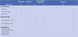
This is a randomized, crossover, blinded (participant) clinical study in which adults diagnosed with multiple sclerosis and quadriceps femoris spasticity will receive two intervention protocols with single sessions for each intervention ( Figure 1 )
Participants
Adult participants diagnosed with multiple sclerosis will be recruited “RETIRADO” in the same location where the survey will also be conducted.
Eligibility criteria
Participants must be aged over 18 years, have a confirmed diagnosis of MS and a minimum grade 3 spasticity bilaterally in the quadriceps muscles, measured with the Ashworth Scale, in addition to being right-handed (so that it is a more homogeneous population). Individuals whose motor thresholds are not obtained via transcranial magnetic stimulation, individuals who have a contraindication for TMS (metallic implant in the skull, cardiac pacemaker, history of epilepsy) 22 , who take oral medication for spasticity or who have undertook Botox or phenol application in the quadriceps in the last six months will not be included.
Intervention
Participants eligible for the study will receive all information about the procedures and risks, giving written informed consent to participate in the research. Then, data will be collected from the participants, such as gender, age, time of diagnosis, MS subtype, if they have any associated diseases, if they use medication and which ones, if they do any interventionist therapy and which type, if they feel pain, what they feel when they have spasticity. The evaluation of outcomes will also be performed, such as the clinical evaluation of quadriceps spasticity (using the Ashworth scale), evaluation of spinal cord excitability by the response of the patellar reflex and the pendulum test, evaluation of cortical excitability by the parameters of the motor evoked potential (latency and amplitude).
At the end of the first (baseline) assessment of the outcomes, the participant will receive the first intervention session, which may be I1 (high-frequency magnetic stimulation) or I2 (low-frequency magnetic stimulation) according to randomization. Then, the outcome evaluations will be repeated (post-intervention)
Participants will receive a single session of each protocol with an interval of one week between them. Assessments and interventions will always take place at the same time of day to not interfere with spasticity.
Transcranial Magnetic Stimulation – TMS
Before starting the intervention, it will be necessary to identify the hotspot (location on the scalp where TMS consistently produces the highest amplitude motor evoked potentials) 23 of the dominant (right) quadriceps of each participant.
For this, a Magpro R20 TMS equipment (Magventure – Denmark), a satellite coil model MMC-140-II, an electromyography device (EMG System) that will be coupled to the TMS and a computer (Lenovo 300e 2nd Gen), disposable electrodes and Ag-AgCl electromyography stickers will be used.
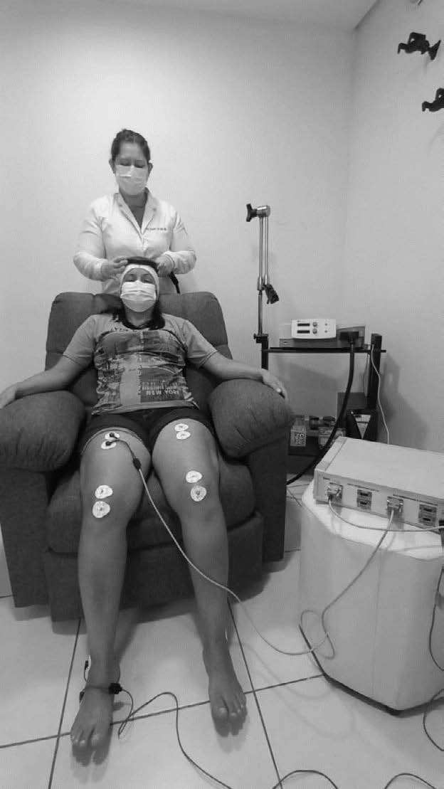
The participant will be seated in a chair, with the upper and lower limbs relaxed. A white fabric cap will be placed on their head, in which the anatomical position corresponding to the motor area (M1) and the CZ point will be marked according to the references of the International Electroencephalogram System (EEG) 10-20 24 . The electromyography electrodes will be positioned in the rectus femoris muscle of both legs, following the guidelines of the surface EMG for a non-invasive assessment of muscles (SENIAM) project 25 , which recommends the use of a measuring tape to measure the distance between the upper iliac crest and the line of the upper edge of the patella; thus, the electrodes will be positioned over the rectus femoris at 50% of the distance between the patella and the anterosuperior iliac spine with a ground electrode positioned over the iliac crest.
Then, to find the hotspot, the coil will be moved slowly over the left motor cortex during rest, and the place where a larger contraction of the target muscle (right quadriceps) appears, after a single pulse series of TMS, will be identified as the motor hotspot. Once identified, it will be marked on the cap so that all TMS stimuli (evaluation and treatment) are applied on this exact point. Then, the motor threshold of each participant will be identified, which will be defined as the lowest intensity (μV) to elicit a contraction in the muscle with peak amplitude of motor evoked potential (MEP) above 50μV, in at least five of 10 unique pulses applied over the hotspot. Treatment will commence after obtaining this data.
High frequency protocol - Excitability
The participant should be seated in an armchair with a backrest, arms and legs relaxed, and will be instructed to remain relaxed, not to speak and not to move during the stimulation. The therapist will position the coil of the TMS equipment in the left hemisphere, over the quadriceps hotspot. The parameters that will be used for this stimulation are frequencies of 5Hz, 50 pulses, 18 waves, with an interval of 40 seconds between waves and the intensity of the stimulus at 90% of the motor threshold at rest 12 .
Low Frequency Protocol
For the low-frequency TMS treatment protocol, the positioning and orientations will be the same as for the high-frequency protocol. However, the parameters will be 1Hz, 100 pulses per wave, nine waves, with an interval of one second and stimulus intensity at 90% of the motor threshold at rest 12 .
At the end of the stimulation of each protocol, which should take approximately 30 minutes, an adverse effects questionnaire will be applied 22 .
Outcomes
Assessments will be made before and after each session.
Primary outcome – Ashworth scale
The clinical assessment of bilateral quadriceps spasticity will be performed using the Ashworth scale 26 , by a physical therapist trained to use it.
The participant should be lying in lateral decubitus with the hip in a neutral position, while the physical therapist performs the knee flexion. The results of muscle resistance to passive stretching can be classified as: 1 – normal tone; 2 – slight increase in muscle tone when the joint is moved in flexion or extension; 3 – more pronounced increase in tone, but the limb is easily flexed; 4 – considerable increase in muscle tone and passive movement is performed with difficulty; 5 – rigid affected joint in flexion or extension.
Spasticity between right (contralateral to TMS stimulus) and left (homolateral to TMS stimulus) quadriceps will be compared pre- and post-intervention.
Secondary outcomes
Assessment of Spasticity by varying the knee joint angle
For this assessment, the pendulum test developed by Badj and Vodovnik 27 will be used. The test offers a simple approach in which gravity induces the stretch reflex, making it possible to observe its oscillatory movement along the passive movement. The greater the oscillation during the fall, the greater the degree of spasticity.
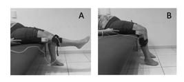
To perform it, the participant must be lying in the supine position, the examiner asks the participant to keep their knees out of the stretcher. The participant is asked to leave the leg relaxed, the examiner should extend the knee joint to the horizontal position and release it ( Figure 2 ).
The measurement of the knee angle will be performed with an electrogoniometer positioned on the lateral condyle of the knee joint and connected to a surface electromyography (EMG) equipment (EMG System), used to capture the activity of the quadriceps during the pendulum test. The EMG equipment will be used with a sampling rate of 4,000Hz and a capture duration of 10 seconds. A 23Hz high-pass filter and a 500Hz low-pass filter will be used; no 60% filter will be used during the capture of the noise filtering signals from the power grid, as the device has an internal battery and will not be connected directly to the outlet.
The goniometric data for each pendulum test will be analyzed according to the average of sinusoidal oscillations captured by the EMG and produced by the oscillating limb after the release of the heel of the evaluated limb. The criterion for each oscillation will be a displacement of the knee joint by at least three angles in the extension direction.
Assessment of spinal cord excitability
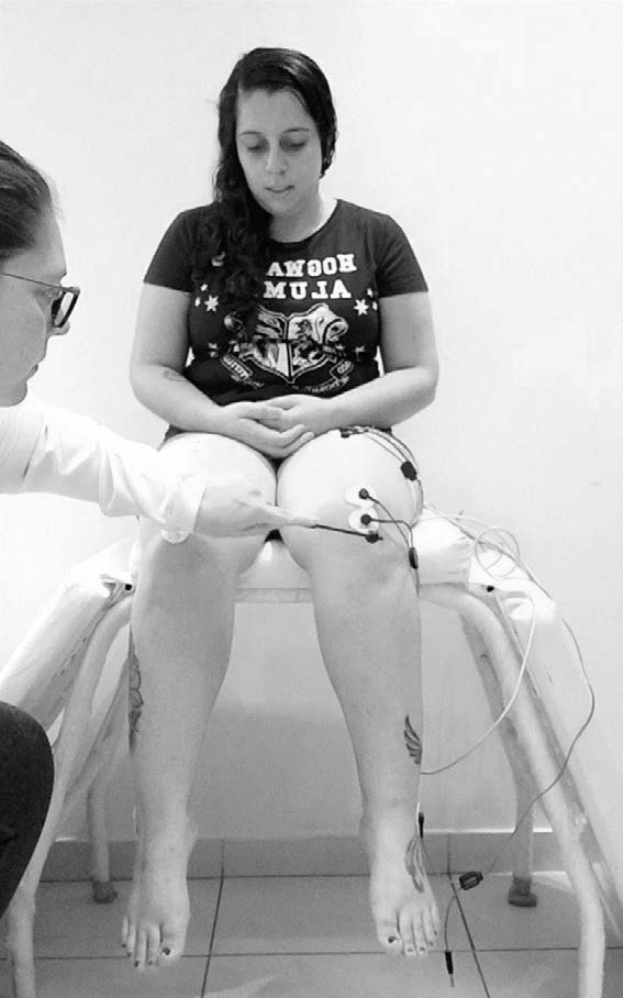
To assess spinal cord excitability, the patellar tendon stretch reflex will be performed. The participant will be seated with the knee flexed at 90°, forming a pendulum. Using a reflex hammer, the researcher will strike the participant’s patellar tendon ( Figure 3 ).
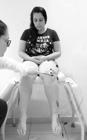
The reflex latency (ms) and the amplitude of the electromyographic signal (ms) of the muscle will be measured, which will be recorded by the EMG System electromyography device, with a sampling rate of 4,000Hz and 10 seconds of capture duration. A 23Hz high-pass filter and a 500Hz low-pass filter will be used.
The electromyographic signal will be captured with two bipolar, round, adhesive and disposable electrodes, one of them positioned on the rectus femoris at 50% of the distance between the patella and the patient’s anterior-superior iliac spine, as described by SENIAM, and the other just below, with a distance of 20mm between them.
For the analysis of spasticity, the latency time between tendon percussion and muscle response will be evaluated. The higher the latency response, the higher the spasticity. The quadriceps reflex of individuals with spasticity was defined in three factors: velocity-dependent excitation; length-dependent inhibition and fatigability; and the greater the stretch velocity, the greater the magnitude of the patellar reflex, thus the greater the latency of this reflex 28 .
Cortical Excitability
The cortical excitability of the quadriceps motor area will be measured by the latency time (ms), central motor conduction time of the motor evoked potential (ms), and amplitude and duration of the MEP peak versus the EMG sampling rate time. For this evaluation, the MagPro R20 (Magventure, Denmark) TMS equipment will be used with the MMC-140-II parabolic coil model on its concave face ( Figure 4 ), using the average of five simple pulses of 120% and 140% of the motor threshold.
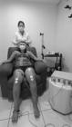
The coil will be positioned over the quadriceps hotspot. The MEP will be recorded by the EMG system, with an input rate of 4.00Hz and 10 seconds of capture duration, a 500Hz high pass filter and a 500Hz low pass filter will be used.
The electromyographic signal will be obtained by means of two round, adhesive and disposable bipolar electrodes, positioned in the rectus femoris with a distance of 20mm between them, at 50% of the distance between the patella and the superior iliac crest, following the SENIAM marking. The electrode site will be previously sanitized with alcohol and cotton, and excess hair will be removed.
Assessments for subjects’ characterization
Spasms frequency assessment
Spasms frequency will be assessed by the Penn Spasm Frequency Scale 29 prior to each intervention protocol. The Penn Spasm Frequency Scale is a scale characterized as a patient outcome report (PRO), in which the individual classifies their number of spasms in a one-hour period; the scale is scored from 0 to 4, being: 0 – no spasm per hour; 1 – mild spasms; 2 – complete spasms occurring less than one per hour; 3 – complete spasms occurring more than once per hour; and 4 – more than 10 spontaneous spasms per hour. The scale was developed to measure the individual’s report of their experience with spasticity.
Patient’s perception of their spasticity
For this evaluation, the Numerical Rating Scale for Spasticity (NRS-S) 30 will be used, which is a variation of the Visual Analog Scale and the Numeric Ranting Scale for Pain. The NRS-S asks the participant to rate the severity of their symptom on a scale of 0 to 10, with 0 for no spasticity and 10 for the worst possible spasticity in a 24-hour period. The evaluation will be applied before each intervention protocol.
Confounding factors
Before starting each session, the Pittsburgh Sleep Quality Index (PSQI) 31 will be applied, as lack of sleep can interfere with fatigue and voluntary limb control, increasing the number of spasms and spasticity throughout the day. The PSQI contains 19 self-assessment questions and five questions assessed by the bed partner or roommate (if one is available). Only self-assessment questions are included in the score.
The 19 self-assessment items are combined to form seven “component” scores, each with a range of zero to three points. In all cases, a score of “0” indicates no difficulty, while a score of “3” indicates severe difficulty. The scores of the seven components are summed to produce an “overall” score, with a range of zero to 21 points, “0” indicates no difficulty, and “21” indicates severe difficulties in all areas.
TMS Adverse Effects Questionnaire
An adverse effects questionnaire will be applied after each treatment session. The questionnaire consists of an intensity scale from 1 to 4 (1 – absent; 2 – mild; 3 – moderate; 4 – severe) and a scale of how much the participant thinks that symptom is from TMS from 1 to 5 (1 – no chance; 2 – remote; 3 – possible; 4 – probable; 5 – definitive.) 22
The symptoms of the scale are headache, neck pain, muscle tension, scalp pain, scalp burning, nausea, dizziness, hearing difficulty, difficulty concentrating, mental confusion, positive mood, negative mood, and seizure 22 .
Randomization
The order of interventions I1 (high-frequency TMS) and I2 (low-frequency TMS) will be chosen through a random ordering performed by a researcher not involved in the evaluation and intervention, who will use the website: www.randomizer.org.
Sample size
For this feasibility study, 10 individuals will be recruited, based on a study by Centonze et al. 12 .
Data analysis
Statistical analyzes will be performed with the aid of the SPSS Statistic program, version 26, with a significance level established for the analyses of p<0.05. The Shapiro-Wilk test will be used to analyze the normality of the variables. Parametric data will be represented as mean and standard deviation; for non-parametric variables median and interquartile range, and frequency and percentage for categorical variables. For the outcome, the two-way analysis of variance test will be used for parametric data, and the Friedman test for non-parametric data.
ACKNOWLEDGMENTS
To Universidade Nove de Julho, for the support during the research. To the Coordination for the Improvement of Higher Education Personnel (CAPES), for the funding with scholarship for the first author.
REFERENCES
-
1. Perez CA, Cuascut FX, Hutton GJ. Immunopathogenesis, diagnosis, and treatment of multiple sclerosis: a clinical update. Neurol Clin. 2023;41(1):87-106. doi: 10.1016/j.ncl.2022.05.004
» https://doi.org/10.1016/j.ncl.2022.05.004 -
2. Boutiére C, Rey C, Zaaraoui W, Le Troter A, Rico A, Crespy L, et al. Improvement of spasticity following intermittent theta burst stimulation in multiple sclerosis is associated with modulation of resting-state functional connectivity of the primary motor cortices. Mult Scler. 2017;23(6):855-63. doi: 10.1177/1352458516661640
» https://doi.org/10.1177/1352458516661640 -
3. Oliveira EML, Souza NA. Esclerose Múltipla. Rev Neuro. 1998;6(3):114-8. doi: 10.34024/rnc.1998.v6.1032
» https://doi.org/10.34024/rnc.1998.v6.1032 -
4. Khan F, Amatya B, Bensmail B, Yelnik A. Non-pharmacological interventions for spasticity in adults: An overview of systematic reviews. Ann Phys Rehabil Med. 2019;62(4):265-73. doi: 10.1016/j.rehab.2017.10.001
» https://doi.org/10.1016/j.rehab.2017.10.001 -
5. Rizzo MA, Hadjimichael OC, Preiningerova J, Vollmer TL. Prevalence and treatment of spasticity reported by multiple sclerosis patients. Mult Scler. 2004;10(5):589-95.doi: 10.1191/1352458504ms1085oa
» https://doi.org/10.1191/1352458504ms1085oa -
6. Lance JW. What is spasticity? The Lancet. 1990;335(8689):606. doi: 10.1016/0140-6736(90)90389-m
» https://doi.org/10.1016/0140-6736(90)90389-m -
7. Kheder A, Nair KPS. Spasticity: pathophysiology, evaluation and management. Pract Neurol. 2012;12(5):289-98. doi: 10.1136/practneurol-2011-000155
» https://doi.org/10.1136/practneurol-2011-000155 -
8. Teixeira LF, Olney SJ, Brouwer B. Mecanismos e medidas de espasticidade. Fisioter Pesqui. 2011;5(1):4-19. doi: 10.1590/fpusp.v5i1.76781
» https://doi.org/10.1590/fpusp.v5i1.76781 -
9. Abbruzzese G. The medical management of spasticity. Eur J Neurol. 2002;9(Suppl 1):30-4. doi: 10.1046/j.1468-1331.2002.0090s1030.x
» https://doi.org/10.1046/j.1468-1331.2002.0090s1030.x -
10. Rekand T, Grønning M. Treatment of spasticity related to multiple sclerosis with intrathecal baclofen: a long-term follow-up. J Rehabil Med. 2011;43(6):511-4. doi: 10.2340/16501977-0811
» https://doi.org/10.2340/16501977-0811 -
11. Fletcher SG, Castro-Borrero W, Remington G, Treadaway K, Lemack GE, Frohman EM. Sexual dysfunction in patients with multiple sclerosis: a multidisciplinary approach to evaluation and management. Nat Clin Pract Urol. 2009;6(2):96-107. doi: 10.1038/ncpuro1298
» https://doi.org/10.1038/ncpuro1298 -
12. Centonze D, Koch G, Versace V, Mori F, Rossi S, Brusa L, et al. Repetitive transcranial magnetic stimulation of the motor cortex ameliorates spasticity in multiple sclerosis. Neurology. 2007;68(13):1045-50. doi: 10.1212/01.wnl.0000257818.16952.62
» https://doi.org/10.1212/01.wnl.0000257818.16952.62 -
13. Mori F, Ljoka C, Magni E, Codecà C, Kusayanagi H, Monteleone F, et al. Transcranial magnetic stimulation primes the effects of exercise therapy in multiple sclerosis. J Neurol. 2011;258(7):1281-7. doi: 10.1007/s00415-011-5924-1
» https://doi.org/10.1007/s00415-011-5924-1 -
14. Mori F, Codecà C, Kusayanagi H, Monteleone F, Boffa L, Rimano A, et al. Effects of intermittent theta burst stimulation on spasticity in patients with multiple sclerosis. Eur J Neurol. 2010;17(2):295-300. doi: 10.1111/j.1468-1331.2009.02806.x
» https://doi.org/10.1111/j.1468-1331.2009.02806.x -
15. Hugos CL, Cameron MH. Assessment and measurement of spasticity in MS: State of the evidence. Curr Neurol Neurosci Rep. 2019;19(10):79. doi: 10.1007/s11910-019-0991-2
» https://doi.org/10.1007/s11910-019-0991-2 -
16. Şan AU, Yılmaz B, Kesikburun S. The Effect of Repetitive Transcranial Magnetic Stimulation on Spasticity in Patients with Multiple Sclerosis. J Clin Neurol. 2019;15(4):461-7. doi: 10.3988/jcn.2019.15.4.461.
» https://doi.org/10.3988/jcn.2019.15.4.461. -
17. Di Lazzaro V, Profice P, Pilato F, Dileone M, Oliviero A, Ziemann U. The effects of motor cortex rTMS on corticospinal descending activity. Clin Neurophysiol. 2010;121(4):464-73. doi: 10.1016/j.clinph.2009.11.007
» https://doi.org/10.1016/j.clinph.2009.11.007 -
18. Suppa A, Ortu E, Zafar N, Deriu F, Paulus W, Berardelli A, et al. Theta burst stimulation induces after-effects on contralateral primary motor cortex excitability in humans. J Physiol. 2008;586(18):4489-500. doi: 10.1113/jphysiol.2008.156596
» https://doi.org/10.1113/jphysiol.2008.156596 -
19. Watanabe T, Hanajima R, Shirota Y, Ohminami S, Tsutsumi R, Terao Y, et al. Bidirectional effects on interhemispheric resting-state functional connectivity induced by excitatory and inhibitory repetitive transcranial magnetic stimulation. Hum Brain Mapp. 2014;35(5):1896-905. doi: 10.1002/hbm.22300
» https://doi.org/10.1002/hbm.22300 -
20. Di Lazzaro V, Pilato F, Dileone M, Profice P, Oliviero A, Mazzone P, et al. The physiological basis of the effects of intermittent theta burst stimulation of the human motor cortex. J Physiol. 2008;586(16):3871-9. doi: 10.1113/jphysiol.2008.152736
» https://doi.org/10.1113/jphysiol.2008.152736 -
21. Su D, Wang A, Zhu M, Yang F, Li W, Ma B, et al. Repetitive transcranial magnetic stimulation for treatment of limb spasticity following multiple sclerosis: a systematic review and meta-analysis. Braz J Med Biol Res. 2003;56:e12708. doi: 10.1590/1414-431X2023e12708
» https://doi.org/10.1590/1414-431X2023e12708 -
22. Rossi S, Antal A, Bestmann S, Bikson M, Brewer C, Brockmöller J, et al.. Safety and recommendations for TMS use in healthy subjects and patient populations, with updates on training, ethical and regulatory issues: Expert Guidelines. Clin Neurophysiol. 2021;132(1):269-306. doi: 10.1016/j.clinph.2020.10.003
» https://doi.org/10.1016/j.clinph.2020.10.003 -
23. Ahdab R, Ayache SS, Brugières P, Farhat WH, Lefaucheur JP. The hand motor hotspot is not always located in the hand knob: a neuronavigated transcranial magnetic stimulation study. Brain Topogr. 2016;29(4):590-7. doi: 10.1007/s10548-016-0486-2
» https://doi.org/10.1007/s10548-016-0486-2 - 24. Klem GH, Lüders HO, Jasper HH, Elger C. The ten-twenty electrode system of the International Federation. International Federation of Clinical Neurophysiology. Electroencephalogr Clin Neurophysiol Suppl. 1999;52:3-6.
-
25. Hermens HJ, Freriks B, Disselhorst-Klug C, Rau G. Development of recommendations for SEMG sensors and sensor placement procedures. J Electromyogr Kinesiol. 2000;10(5):361-74. doi: 10.1016/s1050-6411(00)00027-4
» https://doi.org/10.1016/s1050-6411(00)00027-4 - 26. Ashworth B. Preliminary Trial of Carisoprodol in Multiple Sclerosis. Practitioner. 1964;192:540-2.
-
27. Bajd T, Vodovnik L. Pendulum testing of spasticity. J Biomed Eng. 1984;6(1):9-16. doi: 10.1016/0141-5425(84)90003-7
» https://doi.org/10.1016/0141-5425(84)90003-7 -
28. Fagius J, Wallin BG, Sympathetic reflex latencies and conduction velocities in normal man. J Neuro Sci. 1980;47(3):433-48. doi: 10.1016/0022-510X(80)90098-2
» https://doi.org/10.1016/0022-510X(80)90098-2 -
29. Penn RD, Savoy SM, Corcos D, Latash M, Gottlieb G, Parke B, et al. Intrathecal baclofen for severe spinal spasticity. N Engl J Med. 1989;320(23):1517-21. doi: 10.1056/NEJM198906083202303.
» https://doi.org/10.1056/NEJM198906083202303. -
30. Farrar JT, Troxel AB, Stott C, Duncombe P, Jensen MP. Validity, reliability, and clinical importance of change in a 0-10 numeric rating scale measure of spasticity: a post hoc analysis of a randomized, double-blind, placebo-controlled trial. Clin Ther. 2008;30(5):974-85. doi: 10.1016/j.clinthera.2008.05.011
» https://doi.org/10.1016/j.clinthera.2008.05.011 -
31. Buysse DJ, Reynolds Third CF, Monk TH, Berman SR, Kupfer DJ. The Pittsburgh Sleep Quality Index: a new instrument for psychiatric practice and research. Psychiatry Res. 1989;28(2):193-213. doi: 10.1016/0165-1781(89)90047-4
» https://doi.org/10.1016/0165-1781(89)90047-4
-
Financing source:
Universidade Nove de Julho (Uninove), Coordination for the Improvement of Higher Education Personnel (CAPES), Process Number: 88887.818169/2023-00
-
this study was approved by local Research Ethics Committee No. 5,803,048. – Brazilian Registry of Clinical Trials: RBR-2crspbc.

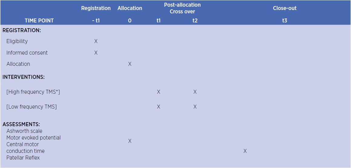 *TMS: Transcranial magnetic stimulation
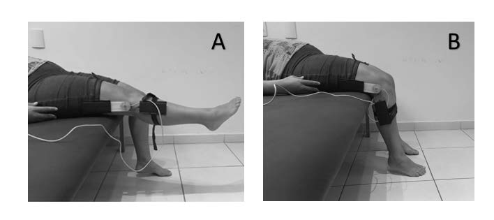 (A) initial position; (B) final position.