 Hereditary thrombocytopenias: the challenge of increasing frequency and differential diagnosis
Abstract
Hereditary thrombocytopenias are often difficult to diagnose. Since most of them are rare diseases, their characteristics are less known by physicians who deal with adults. While pediatricians are accustomed to considering genetic diseases in the differential diagnosis of diseases affecting their young patients, clinicians who treat adults often overlook this possibility. Making a definitive diagnosis usually requires complex laboratory techniques. In addition, because this is a very dynamic field half of the patients have forms that have not yet been identified with new disease definitions being made every month with next-generation sequencing (NGS). These patients are often mistakenly diagnosed with immune thrombocytopenia, and, accordingly, they are at risk of receiving unnecessary immunosuppressive therapy. Misdiagnosis is widespread in patients whose low platelet count is discovered in adulthood because, in these cases, the hereditary origin of thrombocytopenia can be overlooked. The age of manifestation and the duration/chronicity of symptoms are crucial clinical features for identifying hereditary thrombocytopenia disorders. It is important to establish the correct diagnosis because it has recently been shown that some hereditary forms of thrombocytopenia predispose to other diseases such as leukemia, renal failure, and bone marrow failure; therefore, affected individuals should be kept under close surveillance and, when necessary, treated for concomitant diseases. This review aims to determine when to suspect and how to diagnose and manage inherited thrombocytopenias. It also intends to detail the less common kinds of isolated thrombocytopenias highlighting that not all isolated thrombocytopenias that emerge in adults are immune thrombocytopenia.
Keywords
Hereditary thrombocytopenia; Acquired thrombocytopenia; Immune thrombocytopenic purpura
Introduction
An acquired condition rather than an inherited genetic mutation is more likely to be the underlying cause of newly diagnosed thrombocytopenia in a person of any age. Autoimmune disorders, elevated platelet consumption, splenomegaly, bone marrow suppression (infectious or drug-mediated), and bone marrow failure are among the different reasons for acquired thrombocytopenia [1]. The most frequent causes of acquired thrombocytopenia will differ according to the patient's age at onset, reflecting the prevalent illnesses in each age group. Acute viral infections and ımmune thrombocytopenia (ITP) are more prevalent in children. Because chronic diseases and cancer are more common and diverse in adults, the differential diagnosis is rather extensive [1].
Hereditary thrombocytopenias (HTs) are a rare, heterogeneous group of disorders that result in early-onset thrombocytopenia with marked variability in bleeding. The age at manifestation and the duration/chronicity of symptoms are crucial clinical features for identifying HT disorders. The prenatal period or the time the child starts to crawl and walk are often when acute thrombocytopenia or profound platelet dysfunction is identified. Throughout life, milder abnormalities are observed at periods of hemostatic stress (beginning of menses, surgery/trauma, delivery). However, a significant proportion of individuals with HT may not have clinically significant bleeding and are diagnosed with mild to moderate thrombocytopenia (>20×109/L) during routine blood tests [1]. Because alterations in platelet shape can identify HTs, all patients with recently diagnosed thrombocytopenia should have their peripheral blood smear carefully examined. The diagnostic options are reduced when big platelets or microthrombocytes are identified. Furthermore, due to the fixed particle size, automatic cell counts may underreport very big and very tiny platelets.
In 1948, a patient with Bernard-Soulier syndrome (BSS), a congenital bleeding condition, was described, marking the beginning of the history of HTs [2]. Nearly 70 years later, 33 distinct types of HT caused by genetic abnormalities affecting at least 32 genes were defined as a result of developments in clinical and scientific research [2]. The advent of whole-exome sequencing (WES) and whole-genome sequencing (WGS) significantly advanced our understanding of HTs. This is a very dynamic field with new disease definitions being made every month with next-generation sequencing (NGS). Numerous national and international consortiums devoted to identifying the genes causing HT in patients who did not receive a molecular diagnosis after examining one or more candidate genes have embraced them. These high-throughput sequencing techniques have identified genes that cause illness, such as PRKACG, GFI1b, STIM1, FYB, SLFN14, ETV6, DIAPH1, and SRC [3-11]. New genes will continue to be identified in the future. This review aims to determine when to suspect and how to diagnose inherited thrombocytopenias. It also intends to detail the less common kinds of isolated thrombocytopenias highlighting the fact that not all isolated thrombocytopenias that emerge in adults are ITP.
When to suspect and how to diagnose hereditary thrombocytopenias
Inappropriate therapies and incorrect prognostic criteria might arise from misdiagnosing hereditary illnesses as acquired conditions. Furthermore, misdiagnosis hinders genetic counseling, which might impact subsequent generations. The differential diagnosis between ITP and HT can be challenging due to the absence of specific tests, particularly in patients with mild bleeding symptoms. Recently, the feasibility of using parameters of the complete blood count (CBC) to support this differential diagnosis was illustrated by a series of studies, which demonstrated and validated that the mean platelet volume (MPV) can help the segregation of patients with ITP and HT [12,13]. In recent years, new parameters have been incorporated into the CBC, including the immature platelet fraction (IPF), which represents a population of newly formed platelets containing a greater amount of residual RNA [14]. Initially, the IPF was measured by flow cytometry, and described as reticulated platelets [15]. According to Ferreira et al., IPF helps distinguish hereditary macrothrombocytopenia from ITP; it is elevated in the former when compared to both ITP and other thrombocytopenias [16]. It is still unclear what processes underlie the rise in IPF in hereditary macrothrombocytopenia. One consistent feature linked to the biological causes of thrombocytopenia in these patients may be the higher RNA content of these platelets [16]. Two characteristics of the HTs that are more commonly found in adulthood are that they do not cause spontaneous bleeding and are not syndromic at birth [17]. The abnormalities linked to thrombocytopenia are apparent from the first few months of life in the majority of syndromic types, which makes accurate diagnosis easier [18-20]. However, in some syndromic types, thrombocytopenia-related problems show up later in life, making it easy to overlook their genetic roots [21,22].
The best diagnostic approach to HTs is still debated. Mutation screening is always necessary for a conclusive diagnosis. Instead of sequencing one or a small number of genes selected based on patient features, a single-step diagnostic process that can assess all potential mutations causing HTs is now feasible. Massive sequencing, however, often identifies some genetic alterations, and it is frequently difficult to discern between pathogenetic and non-pathogenetic variations. With the use of these techniques, new HTs continue to be identified. Because NGS-targeted systems enable the simultaneous investigation of a predetermined set of genes, molecular diagnosis of hereditary illnesses may be completed more quickly and at a lower cost. Ninety percent of patients who had not previously undergone molecular-level investigation were able to have the disease-causing gene detected thanks to a targeted sequencing platform that covered 63 genes associated with thrombotic and bleeding disorders. The platform demonstrated 100 % sensitivity in identifying causal variants previously found by Sanger sequencing [2,23]. An appropriate clinical approach combined with NGS techniques may provide the best chance for diagnostic success. Since NGS is widely used because of its high throughput and high efficiency, additional gene variants linked to HT have been found. It will grow to be a significant addition to first-line diagnostic techniques like the patient’s history, physical examination, and morphological evaluations. Laboratory and clinical features suggestive of HT are shown in Table 1.
Classification of hereditary thrombocytopenias: clinical, biological, and cytogenetic features
From minor disorders that may go undiagnosed even in adulthood to severe bleeding diatheses that are identified in the first few weeks of birth, HT has a wide variety of clinical manifestations. To prevent needless and sometimes hazardous therapies, it is crucial to differentiate between HT and acquired thrombocytopenia, particularly ITP, for the category of disorders. An effective diagnosis of HT cannot ignore the meticulous collection of personal and family history, careful physical examination, and analysis of peripheral blood smears. The HT categories that work best for diagnostic reasons are those based on the existence of other abnormalities in addition to thrombocytopenia and platelet size, which vary significantly across the different types. Even if NGS is becoming more widely available, a logical and economical method of diagnosing HTs is still needed. Molecular analysis is necessary to confirm the diagnosis and offer patients individualized treatment, counseling, and follow-up. At the very least, a definite diagnosis must always be sought for persons in whom a predisposing form is suspected. The large number of sporadic instances caused by de novo mutations in some conditions and the inadequate penetrance of the same mutations, frequently making it difficult to discern the inheritance pattern, limits the usefulness of the inheritance-based categorization. In this article, HTs are classified into three groups based on more recent perspectives and research on the condition as depicted in Figure 1: forms that only have the platelet defect; disorders in which the platelet phenotype is associated with other congenital defects (syndromic forms); and forms that have an elevated risk of developing other diseases in life [2,3,11,20,24-46]. As a result, this categorization has both diagnostic and prognostic value.
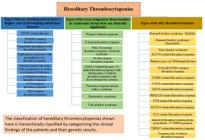
Classification of Hereditary Thrombocytopenias based on more recent perspectives and research.
Types that are predisposed or have a higher risk of developing additional diseases
The distribution of hereditary thrombocytopenia types associated with an elevated risk of developing secondary comorbidities or related clinical disorders is delineated in Table 2. A non-syndromic type of macrothrombocytopenia with a tendency to bleed generally correlated with the severity of platelet shortage is the initial manifestation of myosin-9-related disease (MYH9-RD) [21]. This diagnosis is supported by the finding of Döhle-like structures in the neutrophil cytoplasm [47]. Monoallelic mutations in DIAPH1, a gene implicated in proplatelet expansion and cytoskeleton assembly, cause abnormally big platelets in DIAPH1-related thrombocytopenia [10]. Congenital megakaryocytic thrombocytopenia (CAMT) typically manifests as isolated, severe, and symptomatic thrombocytopenia from birth. If a hematopoietic stem cell transplant (HSCT) is unsuccessful, this condition almost always progresses into bone marrow aplasia during childhood and results in death before adulthood [44]. In addition to additional skeletal abnormalities and/or sensorineural deafness, radio-ulnar synostosis with amegakaryocytic thrombocytopenia disease is typified by congenital bilateral or unilateral proximal synostosis of the radius and ulna, which results in restricted forearm supination [45]. ANKRD26-related thrombocytopenia (ANKRD26-RT), ETV6-related thrombocytopenia (ETV6-RT), and familial platelet disorder predisposing to acute myeloid leukemia (FPD/AML) are three HT linked to specific predispositions to hematological malignancies [13,48]. Platelets in SRC-related thrombocytopenia are dysmorphic, exhibit a wide range of sizes, and have few granules [11].
Hereditary thrombocytopenia types that are predisposed or have a higher risk of developing additional diseases.
Types with extra congenital abnormalities or syndromic forms that are clinically significant
Clinically significant hereditary thrombocytopenia types with extra congenital abnormalities or syndromic forms are listed in Table 3. Almost primarily affecting men, Wiskott-Aldrich syndrome (WAS) is characterized by moderate to severe thrombocytopenia, immunosuppression, and eczema. Microthrombocytes, with a steady platelet count of 5.0-50.0×10⁹/L and a markedly decreased mean platelet volume, are a defining feature of various associated illnesses [49]. In Jacobsen syndrome (and Paris-Trousseau thrombocytopenia, a major and consistent feature of Jacobsen syndrome), probably, the psychomotor retardation and facial and cardiac defects characteristic of Jacobsen syndrome are caused by the loss of a contiguous gene [50,51]. Periventricular nodular heterotopia (PNH) and the otopalatodigital syndrome spectrum diseases, which include congenital deformities, mental retardation, and skeletal abnormalities, are the two primary phenotypes associated with FLNA mutations [52]. From birth, patients with GATA-related diseases are anemic and have symptoms of severe thrombocytopenia. The bone marrow includes many tiny, dysplastic megakaryocytes in addition to dyserythropoiesis and dysplastic platelet alterations [53]. Bilateral radial aplasia is the defining feature of thrombocytopenia with missing radius syndrome, however, most patients may also have other anatomical abnormalities [54]. The endoplasmic reticulum protein that STIM1 encodes controls the entry of Ca2+ into cells via the calcium release-activated channels of the plasma membrane in a variety of cell types. In Stormorken syndrome and York platelet syndrome, platelets exhibit many in vitro abnormalities related to aggregation, activation, and granule secretion [6,20].
Hereditary thrombocytopenia types with extra congenital abnormalities or syndromic forms that are clinically significant.
Forms with only thrombocytopenia
Bernard-Soulier syndrome, the most prevalent isolated form of thrombocytopenia, is characterized by recurrent and spontaneous hemorrhages and is frequently fatal since it is linked to severe platelet function abnormalities and thrombocytopenia [28].
Patients with HT vary widely in the presence and intensity of their bleeding tendency. While some patients may only experience hemorrhages during hemostatic challenges, others may present with no bleeding at all. A small percentage of individuals experience spontaneous bleeding at first, and some may even experience potentially fatal bleeding episodes. Petechiae, ecchymoses, epistaxis, menorrhagia, and gastrointestinal hemorrhages are all examples of mucocutaneous bleeding. In those HTs that are not linked to severe platelet dysfunction, the degree of bleeding tendency correlates with the platelet count [24]. On the other hand, in forms linked to altered platelet function, the tendency to hemorrhage is typically more severe than anticipated based on platelet count [28]. For the vast majority of HTs, the severity of thrombocytopenia does not change throughout the patients’ lives. A classification of hereditary thrombocytopenias presenting with isolated thrombocytopenia is provided in Table 4.
Treatment and follow-up of individuals with hereditary Thrombocytopenias
The treatment and follow-up of these patients is a very important issue in itself and should be discussed in detail in another article. Avoiding the use of medications that might reveal the platelet abnormality is a crucial first step [55]. Thus, it is important to carefully weigh the advantages and disadvantages of various therapies. Preventing the need for medical procedures that increase the risk of bleeding is another crucial step. New therapy avenues in this area were made possible by the recent discovery that eltrombopag, an oral nonpeptide agonist of the thrombopoietin receptor, might raise the platelet count in a small case series of individuals with MYH9-RD [56]. Patients with HTs who are predisposed or have a higher risk of developing additional diseases need close follow-up, whereas individuals with HTs who are not at risk of developing new disorders need medical care for hemostatic problems or bleeding episodes [57].
Conclusions and future perspectives for the hereditary thrombocytopenias
Once thought to be rare, HT syndromes are now more widely recognized as a group of clinical abnormalities ranging from mild disorders discovered incidentally in adults to severe diseases in neonates. In recent years, our knowledge of HTs has shown that these syndromes will not only present with thrombocytopenia and bleeding disorders, but also with complex diseases and even malignancies, bone marrow failure, and renal, hearing, and vision disorders. A multidisciplinary approach to patients with HT is needed, taking into account additional disorders that have already developed or are at risk of developing soon. The classification of HTs should be evaluated together with the clinical findings of the patient and categorized together with the genetic results. Studying the clinical features of patients and identifying new genes and their association will greatly contribute to the understanding of the pathophysiology of HT syndromes and the development of new treatments.
Data availability statement
The data that support the findings of this study are available from the corresponding author upon reasonable request.
-
Funding information
No funding was received for this article.
References
- 1 Drachman JG. Inherited thrombocytopenia: when a low platelet count does not mean ITP. Blood. 2004;103(2):390-8.
- 2 Noris P, Pecci A. Hereditary thrombocytopenias: a growing list of disorders. Hematol 2014 Am Soc Hematol Educ Program Book. 2017;2017(1):385-99.
- 3 Kahr WH, Hinckley J, Li L, Schwertz H, Christensen H, Rowley JW, et al. Mutations in NBEAL2, encoding a BEACH protein, cause gray platelet syndrome. Nat Genet. 2011;43(8):738-40.
- 4 Manchev VT, Hilpert M, Berrou E, Elaib Z, Aouba A, Boukour S, et al. A new form of macrothrombocytopenia induced by a germ-line mutation in the PRKACG gene. Blood J Am Soc Hematol. 2014;124(16):2554-63.
- 5 Monteferrario D, Bolar NA, Marneth AE, Hebeda KM, Bergevoet SM, Veenstra H, et al. A dominant-negative GFI1B mutation in the gray platelet syndrome. N Engl J Med. 2014;370(3):245-53.
- 6 Nesin V, Wiley G, Kousi M, Ong E-C, Lehmann T, Nicholl DJ, et al. Activating mutations in STIM1 and ORAI1 cause overlapping syndromes of tubular myopathy and congenital miosis. Proc Natl Acad Sci. 2014;111(11):4197-202.
- 7 Levin C, Koren A, Pretorius E, Rosenberg N, Shenkman B, Hauschner H, et al. Deleterious mutation in the FYB gene is associated with congenital autosomal recessive small-platelet thrombocytopenia. J Thromb Haemost. 2015;13(7):1285-92.
- 8 Fletcher SJ, Johnson B, Lowe GC, Bem D, Drake S, Lordkipanidzé M, et al. SLFN14 mutations underlie thrombocytopenia with excessive bleeding and platelet secretion defects. J Clin Invest. 2015;125(9):3600-5.
- 9 Noetzli L, Lo RW, Lee-Sherick AB, Callaghan M, Noris P, Savoia A, et al. Germline mutations in ETV6 are associated with thrombocytopenia, red cell macrocytosis and predisposition to lymphoblastic leukemia. Nat Genet. 2015;47(5):535-8.
- 10 Stritt S, Nurden P, Turro E, Greene D, Jansen SB, Westbury SK, et al. A gain-of-function variant in DIAPH1 causes dominant macrothrombocytopenia and hearing loss. Blood J Am Soc Hematol 2016;127(23):2903-14.
- 11 Turro E, Greene D, Wijgaerts A, Thys C, Lentaigne C, Bariana TK, et al. A dominant gain-of-function mutation in universal tyrosine kinase SRC causes thrombocytopenia, myelofibrosis, bleeding, and bone pathologies. Sci Transl Med. 2016;8(328):328ra30-ra30.
- 12 Noris P, Klersy C, Zecca M, Arcaini L, Pecci A, Melazzini F, et al. Platelet size distinguishes between inherited macrothrombocytopenias and immune thrombocytopenia. J Thromb Haemost. 2009;7(12):2131-6.
- 13 Noris P, Biino G, Pecci A, Civaschi E, Savoia A, Seri M, et al. Platelet diameters in inherited thrombocytopenias: analysis of 376 patients with all known disorders. Blood J Am Soc Hematol. 2014;124(6):e4-e10.
- 14 Buttarello M, Plebani M. Automated blood cell counts: state of the art. Am J Clin Pathol. 2008;130(1):104-16.
- 15 Rinder H, Munz U, Ault K, Bonan J, Smith B. Reticulated platelets in the evaluation of thrombopoietic disorders. Arch Pathol Lab Med. 1993;117(6):606-10.
- 16 Ferreira FLB, Colella MP, Medina SS, Costa-Lima C, Fiusa MML, Costa LNG, et al. Evaluation of the immature platelet fraction contribute to the differential diagnosis of hereditary, immune and other acquired thrombocytopenias. Sci Rep. 2017;7(1):3355.
- 17 Balduini C, Savoia A, Seri M. Inherited thrombocytopenias frequently diagnosed in adults. J Thromb Haemost. 2013;11(6):1006-19.
- 18 Notarangelo LD, Miao CH, Ochs HD. Wiskott-aldrich syndrome. Curr Opin Hematol. 2008;15(1):30-6.
- 19 Mattina T, Perrotta CS, Grossfeld P. Jacobsen syndrome. Orphanet J Rare Dis. 2009;4:1-10.
- 20 Albers CA, Paul DS, Schulze H, Freson K, Stephens JC, Smethurst PA, et al. Compound inheritance of a low-frequency regulatory SNP and a rare null mutation in exon-junction complex subunit RBM8A causes TAR syndrome. Nat Genet. 2012;44(4):ng. 1083.
- 21 Balduini CL, Pecci A, Savoia A. Recent advances in the understanding and management of MYH9-related inherited thrombocytopenias. Br J Haematol. 2011;154(2):161-74.
- 22 Rees DC, Iolascon A, Carella M, O'marcaigh AS, Kendra JR, Jowitt SN, et al. Stomatocytic haemolysis and macrothrombocytopenia (Mediterranean stomatocytosis/macrothrombocytopenia) is the haematological presentation of phytosterolaemia. Br J Haematol. 2005;130(2):297-309.
- 23 Simeoni I, Stephens JC, Hu F, Deevi SV, Megy K, Bariana TK, et al. A high-throughput sequencing test for diagnosing inherited bleeding, thrombotic, and platelet disorders. Blood J Am Soc Hematol 2016;127(23):2791-803.
- 24 Pecci A, Klersy C, Gresele P, Lee KJ, De Rocco D, Bozzi V, et al. MYH 9-related disease: A novel prognostic model to predict the clinical evolution of the disease based on genotype-Phenotype correlations. Hum Mutat. 2014;35(2):236-47.
- 25 Noris P, Perrotta S, Bottega R, Pecci A, Melazzini F, Civaschi E, et al. Clinical and laboratory features of 103 patients from 42 Italian families with inherited thrombocytopenia derived from the monoallelic Ala156Val mutation of GPIbα (Bolzano mutation). haematologica. 2012;97(1):82.
- 26 Noris P, Perrotta S, Seri M, Pecci A, Gnan C, Loffredo G, et al. Mutations in ANKRD26 are responsible for a frequent form of inherited thrombocytopenia: analysis of 78 patients from 21 families. Blood J Am Soc Hematol. 2011;117(24):6673-80.
- 27 Pleines I, Woods J, Chappaz S, Kew V, Foad N, Ballester-Beltrán J, et al. Mutations in tropomyosin 4 underlie a rare form of human macrothrombocytopenia. J Clin Invest. 2017;127(3):814-29.
- 28 Savoia A, Kunishima S, De Rocco D, Zieger B, Rand ML, Pujol-Moix N, et al. Spectrum of the mutations in B ernard-S oulier Syndrome. Hum Mutat. 2014;35(9):1033-45.
- 29 Kunishima S, Okuno Y, Yoshida K, Shiraishi Y, Sanada M, Muramatsu H, et al. ACTN1 mutations cause congenital macrothrombocytopenia. Am J Hum Genet. 2013;92(3):431-8.
- 30 Guerrero JA, Kyei M, Russell S, Liu J, Gartner TK, Storrie B, et al. Visualizing the von Willebrand factor/glycoprotein ib-IX axis with a platelet-type von Willebrand disease mutation. Blood J Am Soc Hematol. 2009;114(27):5541-6.
- 31 Nurden AT, Pillois X, Fiore M, Heilig R, Nurden P, editors. Glanzmann thrombasthenia-like syndromes associated with macrothrombocytopenias and mutations in the genes encoding the αIIbβ3 integrin. Seminars in thrombosis and hemostasis, © Thieme Medical Publishers; 2011.
- 32 Kunishima S, Kobayashi R, Itoh TJ, Hamaguchi M, Saito H. Mutation of the β1-tubulin gene associated with congenital macrothrombocytopenia affecting microtubule assembly. Blood J Am Soc Hematol 2009;113(2):458-61.
- 33 Morison IM, Cramer Bordé EM, Cheesman EJ, Cheong PL, Holyoake AJ, Fichelson S, et al. A mutation of human cytochrome c enhances the intrinsic apoptotic pathway but causes only thrombocytopenia. Nat Genet. 2008;40(4):387-9.
- 34 Stevenson WS, Rabbolini DJ, Beutler L, Chen Q, Gabrielli S, Mackay JP, et al. Paris-Trousseau thrombocytopenia is phenocopied by the autosomal recessive inheritance of a DNA-binding domain mutation in FLI1. Blood J Am Soc Hematol. 2015;126(17):2027-30.
- 35 Noris P, Marconi C, De Rocco D, Melazzini F, Pippucci T, Loffredo G, et al. A new form of inherited thrombocytopenia due to monoallelic loss of function mutation in the thrombopoietin gene. Br J Haematol. 2018;181(5).
- 36 Stritt S, Nurden P, Favier R, Favier M, Ferioli S, Gotru SK, et al. Defects in TRPM7 channel function deregulate thrombopoiesis through altered cellular Mg2+ homeostasis and cytoskeletal architecture. Nat Commun. 2016;7(1):11097.
- 37 Massaad MJ, Ramesh N, Geha RS. Wiskott-Aldrich syndrome: a comprehensive review. Ann N Y Acad Sci. 2013;1285(1):26-43.
- 38 Albert MH, Bittner TC, Nonoyama S, Notarangelo LD, Burns S, Imai K, et al. X-linked thrombocytopenia (XLT) due to WAS mutations: clinical characteristics, long-term outcome, and treatment options. Blood J Am Soc Hematol 2010;115(16):3231-8.
- 39 Favier R, Akshoomoff N, Mattson S, Grossfeld P, editors. Jacobsen syndrome: advances in our knowledge of phenotype and genotype. American Journal of Medical Genetics Part C: Seminars in Medical Genetics. Wiley Online Library; 2015.
- 40 Nurden P, Debili N, Coupry I, Bryckaert M, Youlyouz-Marfak I, Solé G, et al. Thrombocytopenia resulting from mutations in filamin A can be expressed as an isolated syndrome. Blood J Am Soc Hematol. 2011;118(22):5928-37.
- 41 Millikan PD, Balamohan SM, Raskind WH, Kacena MA, editors. Inherited thrombocytopenia due to GATA-1 mutations. Seminars in thrombosis and hemostasis, © Thieme Medical Publishers; 2011.
- 42 Toriello HV, ed. Thrombocytopenia-absent radius syndrome. Seminars in thrombosis and hemostasis, © Thieme Medical Publishers; 2011.
- 43 Markello T, Chen D, Kwan JY, Horkayne-Szakaly I, Morrison A, Simakova O, et al. York platelet syndrome is a CRAC channelopathy due to gain-of-function mutations in STIM1. Mol Genet Metab. 2015;114(3):474-82.
- 44 Ballmaier M, Germeshausen M, editors. Congenital amegakaryocytic thrombocytopenia: clinical presentation, diagnosis, and treatment. Seminars in thrombosis and hemostasis, © Thieme Medical Publishers; 2011.
- 45 Niihori T, Ouchi-Uchiyama M, Sasahara Y, Kaneko T, Hashii Y, Irie M, et al. Mutations in MECOM, encoding oncoprotein EVI1, cause radioulnar synostosis with amegakaryocytic thrombocytopenia. Am J Hum Genet. 2015;97(6):848-54.
- 46 Liew E, Owen C. Familial myelodysplastic syndromes: a review of the literature. Haematologica. 2011;96(10):1536.
- 47 Pecci A, Panza E, Pujol-Moix N, Klersy C, Di Bari F, Bozzi V, et al. Position of nonmuscle myosin heavy chain IIA (NMMHC-IIA) mutations predicts the natural history of MYH9-related disease. Hum Mutat. 2008;29(3):409-17.
- 48 Hirata S, Takayama N, Jono-Ohnishi R, Endo H, Nakamura S, Dohda T, et al. Congenital amegakaryocytic thrombocytopenia iPS cells exhibit defective MPL-mediated signaling. J Clin Invest. 2013;123(9):3802-14.
- 49 Ochs HD. The Wiskott-Aldrich syndrome. Clin Rev Allergy Immunol. 2001;20:61-86.
- 50 Breton-Gorius J, Favier R, Guichard J, Cherif D, Berger R, Debili N, et al. A new congenital dysmegakaryopoietic thrombocytopenia (Paris-Trousseau) associated with giant platelet alpha-granules and chromosome 11 deletion at 11q23 [see comments]. 1995.
- 51 Krishnamurti L, Neglia J, Nagarajan R, Berry S, Lohr J, Hirsch B, et al. Paris-Trousseau syndrome platelets in a child with Jacobsen's syndrome. Am J Hematol. 2001;66(4):295-9.
- 52 Parrini E, Ramazzotti A, Dobyns W, Mei D, Moro F, Veggiotti P, et al. Periventricular heterotopia: phenotypic heterogeneity and correlation with Filamin A mutations. Brain. 2006;129(7):1892-906.
- 53 Nichols KE, Crispino JD, Poncz M, White JG, Orkin SH, Maris JM, et al. Familial dyserythropoietic anaemia and thrombocytopenia due to an inherited mutation in GATA1. Nat Genet. 2000;24(3):266-70.
- 54 Khincha PP, Savage SA, editors. Genomic characterization of the inherited bone marrow failure syndromes. Seminars in hematology. Elsevier; 2013.
- 55 George JN, Shattil SJ. The clinical importance of acquired abnormalities of platelet function. N Engl J Med. 1991;324(1):27-39.
- 56 Pecci A, Gresele P, Klersy C, Savoia A, Noris P, Fierro T, et al. Eltrombopag for the treatment of the inherited thrombocytopenia deriving from MYH9 mutations. Blood J Am Soc Hematol 2010;116(26):5832-7.
- 57 Melazzini F, Zaninetti C, Balduini C. Bleeding is not the main clinical issue in many patients with inherited thrombocytopaenias. Haemophilia. 2017;23(5):673-81.
Edited by
-
Editor name
Eduardo Rego