 Modification of restorative glass ionomer cement with zinc oxide nanoparticles and calcium glycerophosphate microparticles: in vitro assessment of mechanical properties and antimicrobial activity
Abstract
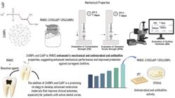
The incorporation of bioactive agents into resin-modified glass ionomer cement (RMGIC) is a promising strategy to improve its mechanical strength and biofilm control, especially for patients with active dental caries.
Objective This study aimed to evaluate the effects of incorporating ZnONPs and CaGP into RMGIC on its mechanical and microbiological properties.
Design Six groups were tested: 1) RMGIC (without CaGP/ZnONPs); 2) RMGIC-1.0%ZnONPs; 3) RMGIC-2.0%ZnONPs; 4) RMGIC-3.0%CaGP; 5) RMGIC-3.0%CaGP-1.0%ZnONPs; and 6) RMGIC-3.0%CaGP-2.0%ZnONPs. The compressive strength (CS), diametral tensile strength (DTS), and surface hardness (SH) were evaluated after 24 hours and 7 days. Antimicrobial and antibiofilm activity were evaluated using agar diffusion and biofilm metabolic activity (XTT) assays.
Results After 24 hours, all the groups showed similar DTS values (p>0.05), except for RMGIC-3.0%CaGP-1.0%ZnONPs, which showed the highest DTS value (p<0.05). Comparing 24 hours and 7 days, the DTS values of RMGIC-3.0%CaGP-2.0%ZnONPs, RMGIC-3.0%CaGP, and RMGIC-3.0%CaGP-2.0%ZnONPs were similar (p=0.360). After 24 hours, the RMGIC group showed the CS highest value, followed by RMGIC-2.0%ZnONPs (p < 0.05). After 7 days, the RMGIC-3.0%CaGP-1.0%ZnONPs group exhibited the highest CS value, approximately 15% higher than RMGIC (p<0.05). The RMGIC-1.0%ZnONPs group exhibited significantly higher SH at 24 hours (p=0.621). At 7 days, the highest SH value was observed for the RMGIC-3.0%CaGP-1.0%ZnONPs group (p<0.05). Regarding antimicrobial and antibiofilm activity, including results from biofilm metabolism assays, the RMGIC-3.0%CaGP-1.0%ZnONPs group demonstrated the most effective antimicrobial and inhibitory effects (p<0.05).
Conclusion This study demonstrated that adding ZnONPs and CaGP to RMGIC enhanced its mechanical and antimicrobial and antibiofilm properties, suggesting enhanced mechanical performance and improved protection against cariogenic biofilms—critical factors for successful restorative treatments. Therefore, the addition of ZnONPs and CaGP is a promising strategy to develop advanced restorative materials that improve clinical outcomes, especially for patients with active dental caries.
Keywords
Resin-modified glass ionomer cement; Phosphate; Zinc oxide nanoparticles; Biofilm; Mechanical properties
Introduction
Dental caries is one of the most prevalent human diseases, resulting from dental demineralization caused by acid production from bacterial plaque. It is characterized as a dynamic, multifactorial, and non-transmissible disease, leading to the loss of minerals from hard dental tissues. Its progression is mediated by biofilm, modulated by diet, and influenced by biological, behavioral, psychosocial, and environmental factors.1,2 Prevention is the primary method to control carious lesions, emphasizing regular use of fluoridated toothpaste, maintenance of a balanced diet with reduced consumption of fermentable carbohydrates, and mechanical control of biofilm.3 Secondary treatment is based on minimally invasive approaches and maximal preservation of mineralized dental tissue.4
Clinical procedures that modify the oral environment, such as temporary and definitive sealing of open cavities and removal of biofilm-retentive factors, can reduce caries risk and activity.5,6 According to Machiulskiene, et al.1 (2020), the main goal of Minimal Intervention Dentistry (MID) is to preserve dental tissues. This involves early detection of caries, non-surgical treatment, minimally invasive restorative procedures, and selective removal of infected tissue7. In this context, materials with biomineralizing capacity have been studied to enhance chemical reactions on the tooth surface, promoting tissue mineralization. Glass ionomer cements (GICs) are considered bioactive materials because they exhibit biomineralizing potential,8 biocompatibility, and bioactivity. However, they exhibit limited antibacterial performance and insufficient load-bearing capacity. To address these limitations, various strategies have been proposed to improve their mechanical and antimicrobial properties, aiming to expand their clinical applications. Nevertheless, it remains unclear which bioactive agent should be incorporated into restorative materials to adequately meet this requirement, making the selection of such agents highly relevant for the oral environment.
One way to enhance the biomineralizing effect and properties of GICs is the addition of Ca and P, as remineralization depends on the presence of these ions in the medium. Among these, Calcium Glycerophosphate (CaGP) is an organic phosphate known for its anticariogenic action, pH buffering effect on plaque, capacity to reduce plaque levels and increase calcium and phosphate concentrations, and direct effects on dental tissues.9-11 Santos, et al.12(2019) evaluated the effect of incorporating CaGP into resin-modified GICs (RMGIC) on physical-mechanical properties, ion release, and enamel demineralization. The authors concluded that adding 3% CaGP to resin-modified GIC increased F, P, and Ca release, reduced enamel demineralization, and maintained physical-mechanical properties within acceptable parameters for this material, proving it to be a promising strategy.
In recent years, nanotechnology has significantly impacted numerous applications across various fields, including Dentistry.13,14 Structures at the nanoscale exhibit unique functional properties not found at the macroscale,15,16 such as their small size, large surface area, and high chemical reactivity.17 Studies show that it is possible to enhance the effectiveness of GICs by incorporating nanoparticles to improve their anticaries effect, as well as their physical-mechanical, microbiological, and cytotoxic properties.13,14 Zinc oxide nanoparticles (ZnONPs) are highlighted in this context, as they exhibit intrinsic catalytic bactericidal activity, antimicrobial effect, chemical stability, and biocompatibility.18,19 According to Huang, et al.20 (2008), ZnONPs are believed to be effective in controlling various microorganisms by altering components of the bacterial cell membrane, leading to the loss of intracellular components and, consequently, cell death. Incorporation of ZnONPs into GIC has emerged as a promising approach to improve its mechanical strength and antibacterial performance. ZnO is known for its antimicrobial activity and bone-regenerative potential, making it a valuable additive in restorative dental materials. In a recent study, Azimi, Shahgholi, and Khandan21 (2021) reported that GICs modified with 4% wt% ZnONPs exhibited a notable increase in compressive strength while maintaining cytocompatibility. Additionally, antibacterial efficacy was improved, as demonstrated by well diffusion, Minimum Inhibitory Concentration (MIC), and Minimum Bactericidal Concentration (MBC) assays. These results highlight the potential of ZnONPs in GIC formulations, although further studies are needed to understand the long-term impact of Zn2⁺ release in the oral environment.
The results available in the literature so far warrant further studies on these compounds, as well as on their mechanisms of action within the formulations in which they are employed, given the lack of studies combining active agents in GIC. Also emphasized is the importance of the dose-response relationship between active agent concentration and the resulting physical-mechanical properties. This relationship appears closely linked to the impact of additives on the acid-base reaction fundamental to GIC setting. Therefore, this study aimed to evaluate the effects of incorporating ZnONPs and CaGP into RMGIC on its mechanical and microbiological properties. The null hypothesis tested was that the addition of ZnONPs and CaGP to RMGIC would not result in improvements in physical-mechanical and microbiological properties compared to unmodified RMGIC.
Methodology
Synthesis and characterization of ZnONPs
For the synthesis of ZnONPs, the sol-gel reaction method was used,22,23 obtaining NPs with an average size of ⁓ 50 nm. For the synthesis, zinc chloride (ZnCl2) and sodium hydroxide (NaOH) 98% (Sigma-Aldrich) were used. Two solutions of each of the reagents were prepared separately. In the first solution, 8 g of NaOH was dissolved in 200 mL of Milli-Q water to obtain a solution with a concentration of 1 M. Then, this solution was heated, under constant stirring, until it reached a temperature of 90°C. The second solution (precursor) was obtained by dissolving 13.6 g of ZnCl2 in 200 mL of Milli-Q water, resulting in a solution with a concentration of 0.5 M. The precursor solution (containing Zn2(aq) ions) was added slowly (rate of 16 mL/min) to the solution containing 1 M NaOH at 90°C. This procedure was carried out under constant temperature and agitation. Slow addition of ZnCl2 solution (2) into an alkaline aqueous solution (1) resulted in the immediate precipitation of ZnONPs, with a color change from transparent to white. After complete addition, the solution remained under stirring for another two hours at 90°C. The resulting sample was washed several times with Milli-Q water and dried in an oven at 80°C for 12 hours.
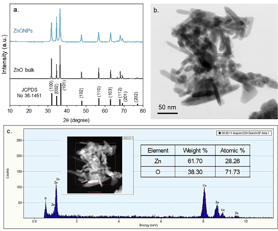
The crystalline structure of the ZnONPs samples was obtained by X-ray diffraction (XRD) measurements, using a Rigaku diffractometer coupled to a 40 kV and 20 mA Cu Kα source. The scanning range was between 20 and 80° (2θ), with a step of 0.02° and a speed of 1 degree per min. Morphological and structural characterization was performed using transmission electron microscopy (TEM, JEOL JEM-2100, 200 kV) to assess particle shape, size distribution, and structural features. Energy-Dispersive X-ray Spectroscopy (EDS), coupled with TEM, was employed to analyze the elemental composition of the nanoparticles.
Sample size calculation
The sample size was determined using SigmaPlot software (version 12.0), based on power analysis with the following parameters: for the mechanical tests, a statistical power of 80%, a minimum detectable difference of 18.0, and a standard deviation (SD) of 5.2 obtained from a pilot study; for the antimicrobial and antibiofilm assays, the analysis considered a power of 80%, an expected difference of 14 between groups, and a standard deviation of 3.9, also derived from preliminary data. To account for possible specimen loss or variability, additional samples were included. As a result, the final sample sizes were n=5 for mechanical tests, n=6 for the Streptococcus mutans adherence assay, and n=6 for biofilm viability and metabolic activity assessments.
Preparation of the experimental RMGICs
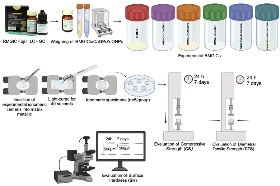
The RMGIC used in this study was Fuji II LC (GC Corporation, Tokyo, Japan). Initially, ZnONPs were added to the RMGIC powder at concentrations of 1.0% and 2.0% (% m/m) (concentrations based on the study by Garcia, et al.24 (2017). For microparticulate CaGP, the concentration was 3.0% (% m/m), based on the study by Santos, et al.12 (2019). To prepare the RMGIC/ZnONPs/CaGP mixtures, it was assumed that 100 g of the final product would contain either 1.0 or 2.0 g of ZnONPs and/or 3.0 g of CaGP. The RMGIC and each mixture were homogenized in an agate ball mill (Planetary Micro Mill PULVERISETTE 7 classic line, Fritsch GmbH, Idar-Oberstein, Germany) with a frequency of 100 rpm in five repetition cycles (normal/reverse) lasting 1 minute each.14 For the manipulation of the experimental ionomer cements, the powder to liquid ratio recommended by the manufacturer was maintained (3.2 g of powder to 1.0 g of liquid) (Figure 1).

Experimental design: Preparation of experimental RMGICs; Evaluation of Diametral Tensile Strength (DTS), Compressive Strength (CS), and Surface Hardness (SH).
Evaluation of the physical and mechanical properties of RMGICs
Evaluation of compressive strength (CS)
Specimens (n=5) of each experimental ionomeric cements were prepared using a split steel mold with internal dimensions of 6 mm height and 4 mm diameter,25,26 following the manufacturer’s specifications (powder: liquid ratio), at room temperature (23±1°C) and relative humidity of 50% (± 5%). Polyester strips were placed on both sides of the mold, and the material was compressed using two steel plates and a screw clamp, then light-cured on both sides according to the manufacturer’s instructions. The set was stored at 37 °C and relative humidity of approximately 90% for 1 hour. Afterwards, excess cement was removed by polishing both sides of the mold with 600-grit silicon carbide paper under continuous water irrigation. The experimental ionomer cement samples were carefully removed from the molds. Subsequently, they were stored in distilled water at 37°C, completing the total remaining time of 24 hours and 7 days. Then, they were submitted to a Compression Resistance test (Instron universal test equipment - DL3000, Instron Co., Canton, MA, USA) at a speed of 0.75 mm/min, in a vertical position, until fracture occurred. The values obtained in the compression tests were calculated (kgf/cm2) by dividing the force (F) by the area and converted to MPa, according to ISO 9917−1:2017.25 (Figure 1)
Evaluation of diametral tensile strength (DTS)
Specimens (n=5) of each experimental ionomeric cement were prepared using a split steel mold with internal dimensions of 3 mm height × 6 mm diameter,26,27 following manufacturer’s specifications, at room temperature (23±1 °C) and relative humidity of 50% (± 5%). Cement manipulation, mold insertion and clamping, storage, and excess removal were performed as previously described for CS. After storage the same period, the samples were submitted to a DTS test at a speed of 0.5 mm/min. Each specimen was covered with a sheet of damp filter paper on both the top and bottom sides that were in contact with the machine platens, applying stress on the specimen’s diameter until fracture occurred. For this purpose, an Instron universal testing machine (DL3000, Instron Co., Canton, MA, USA) was used. DTS values (kgf/cm2) were estimated using the equation: DTS=2F/3.14DT, where F is the load, D is the diameter, and T is the height of the sample (Figure 1).
Evaluation of Surface Hardness (SH)
Specimens (n=5) (3 mm height × 6 mm diameter)14,26,27 were manipulated and inserted into a mold, as previously described for CS and DTS. Afterwards, excess cement was removed by polishing both sides of the specimens with 600-grit silicon carbide paper under continuous water irrigation. The experimental ionomer cement samples were carefully removed from the molds. Then, they were stored in distilled water at 37°C, completing the total remaining time of 24 hours and 7 days. The hardness test was determined using the Shimadzu Micro Hardness Tester HMV-2.000 microhardness meter (Shimadzu Corporation, Kyoto, Japan), under a static load (Knoop) of 100 grams and a time of 5 seconds. Five impressions were made on the surface of the material 500 μm apart. Then, the specimens were stored at 37°C (in relative humidity).14 After 7 days, test was repeated, with 5 impressions taken at a distance of 250 µm from those made after the initial hardness (Figure 1).
Determination of the antimicrobial properties of experimental RMGICs
Streptococcus mutans adherence assay
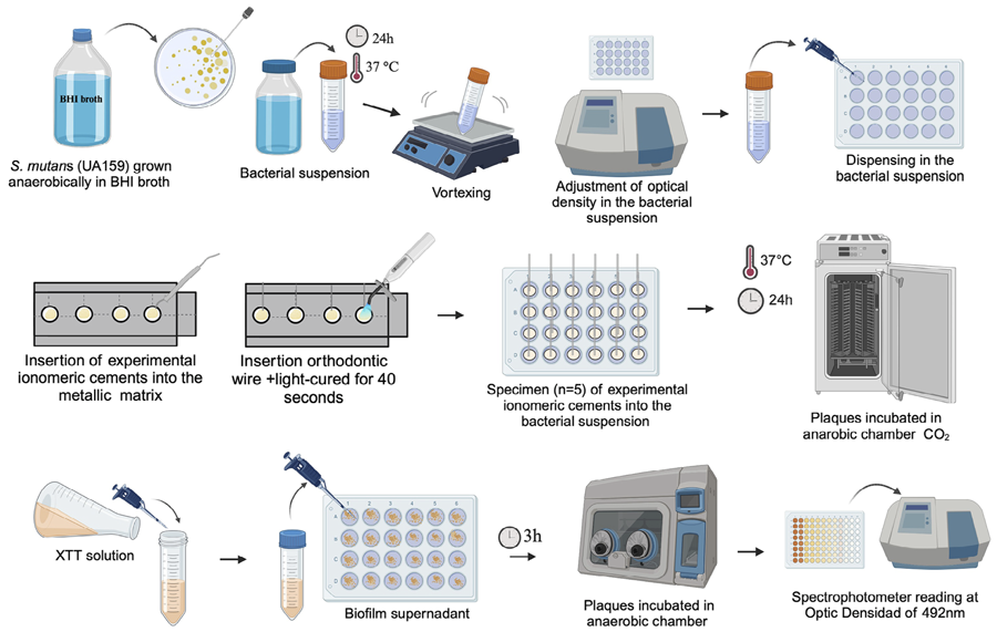
The S. mutans species (UA159) reactivation plates were incubated at 37°C for 48 h. Each specimens (n=6) was exposed under static conditions to 25µL of inoculum and then adjusted to an Optical Density (OD) of 0.6 at 550 nm (approximately 8 × 10¹¹ CFU/mL). After 2 hours at room temperature, non-adherent bacteria were removed by washing twice with 0.9% NaCl solution (saline). Each disk was then inserted into 3 mL of saline containing three glass beads and vortexed for 1 min. The suspension was diluted in decimal series from 10-1 to 10-4 in saline and inoculated in triplicate on BHI agar. These plates were incubated at 37°C for 48 hours in an environment supplemented with 10% CO2. Colonies were counted and the number of viable bacteria was determined as CFU/mL, corresponding to the cells that adhered to the specimens28 (Figure 2).
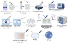
Experimental design: Determination of the antimicrobial/antibiofilm properties of experimental RMGICs - Streptococcus mutans adherence assay.
Biofilm assay
A new biofilm assay proposal was presented based on the methodologies proposed by Cassanho, et al.29 (2005), Kubota, et al.30 (2008), and Bastos, et al.16(2021), with modifications. The aim was to evaluate cell viability in biofilms formed on the surface of the specimens and the influence of the components released from the specimens on biofilm formation at the bottom of the wells of the polystyrene plates. Streptococcus mutans (UA159) were cultured in BHI broth at 37°C for 18 to 24 hours. The bacterial suspension obtained was measured in a spectrophotometer (PowerWave 340, Biotek) and adjusted to an optical density of 0.3 at 600 nm (approximately 3 × 10⁸ CFU/mL). Five disk-shaped specimens (3 × 2 × 1mm) were made in metal matrices for each group tested, in independent experiments. After 1 h in the environment at 100% humidity, the specimens were washed with distilled water and disinfected in a laminar flow chamber under UV light for 15 min. Specimens were inserted into individual wells of 24-well plates (Corning Inc., Corning, NY, USA) and maintained in suspension within a BHI broth medium. To achieve this, when manipulating the RMGICs, an orthodontic wire was inserted into the middle of the specimens and, after light-curing, this wire was fixed with utility wax on the cover of the polystyrene plate and maintained during the period of the biofilm tests. A solution of 1 mL of BHI broth supplemented with 1% sucrose and 5 uL of bacterial suspension was inoculated into each well. Biofilms grown at the bottom of 24-well plates after adding bacterial suspension at a distance from the specimens were evaluated under the action of components released from groups of RMGICs. After incubating the plates for 24 hours at 37°C under anaerobic conditions, the specimens were removed, washed with 1 mL of saline solution to remove non-adherent cells, and inserted individually into microtubes containing 1 mL of saline solution. Soon after, the specimens were sonicated for 30 s at 30W (Misonix Ultrasonic, USA) and vortexed for 1 min. The solutions were serially diluted and inoculated into BHI agar plates. After 48 h, the number of Colony Forming Units (CFU/mL) was determined. The wells containing the formed biofilm were gently washed with saline solution, then stained with XTT solution (2,3-Bis-(2-Methoxy-4-Nitro-5-Sulfophenyl - Sigma-Aldrich) for 3 hours. Then, 200 μL of the solubilized suspension was transferred to a new 96-well plate and analyzed in a spectrophotometer, considering an optical density of 492 nm (Figure 3).
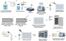
Experimental design: Determination of the antimicrobial/antibiofilm properties of experimental RMGICs - Biofilm viability and metabolism assessment.
Data analysis
For statistical analysis, the Sigmaplot© statistical software program for Windows version 12.0 was used, with significance at the 5% level. All data showed normal (Shapiro-Wilk) and homogeneous (Bartlett) distribution. The data of CS, DTS and SH were submitted to ANOVA (two-way), followed by the Student-Newman-Keuls test (p<0.05). Microbiological data (CFU/mL) were analyzed by ANOVA (one-way), followed by the Student-Newman-Keuls test (p<0.05).
Results
Figure 4a shows the X-ray diffraction (XRD) pattern of the synthesized ZnONPs compared with bulk ZnO. The reference pattern for the wurtzite hexagonal ZnO structure (JCPDS 36-1451) is also included. The ZnONPs showed a highly crystalline structure consistent with the reference pattern. The milling process effectively reduced the particle size of the ZnONPs powder without compromising its crystallinity. Transmission electron microscopy (TEM) image (Figure 4b) shows nanoparticles with an average size of approximately 50 nm. The particles exhibit shapes ranging from spherical to more elongated structures. Compositional analysis (Energy Dispersive X-ray Spectroscopy, EDS; Figure 4c) confirmed a high percentage of zinc (Zn) and oxygen (O) with weight percentages of 61.7% and 38.3%, respectively.
Table 1 presents the DTS values (MPa). After 24 hours, all the groups showed similar values (p>0.05), except for RMGIC-3.0%CaGP-1.0%ZnONPs, which exhibited the highest DTS at both periods (p<0.05). When comparing the two time intervals (24 hours and 7 days), all groups showed significant differences, except for RMGIC-3.0%CaGP and RMGIC-3.0%CaGP-2.0%ZnONPs, which maintained similar values over time (p=0.36). Regarding the CS values (MPa), after 24 hours, the RMGIC group demonstrated the highest value, followed by RMGIC-2.0%ZnONPs (p<0.05). After 7 days, the RMGIC-3.0%CaGP-1.0%ZnONPs group exhibited the highest value, which was 15% greater than that of the RMGIC group (p<0.05). When comparing the two time periods, all groups showed significant differences, except for RMGIC-3.0%CaGP, which remained stable over time. Regarding SH (KHN), after 24 hours, the RMGIC-1.0%ZnONPs group showed the highest value (p=0.621). The RMGIC, RMGIC-3.0%CaGP, and RMGIC-3.0%CaGP-1.0%ZnONPs groups presented similar values (p=0.071). After 7 days, the RMGIC-3.0%CaGP-1.0%ZnONPs group showed the highest value (p<0.05). Between the two times, all groups showed significantly increased values, except for RMGIC-1.0%ZnONPs, which remained unchanged over time (p<0.05).
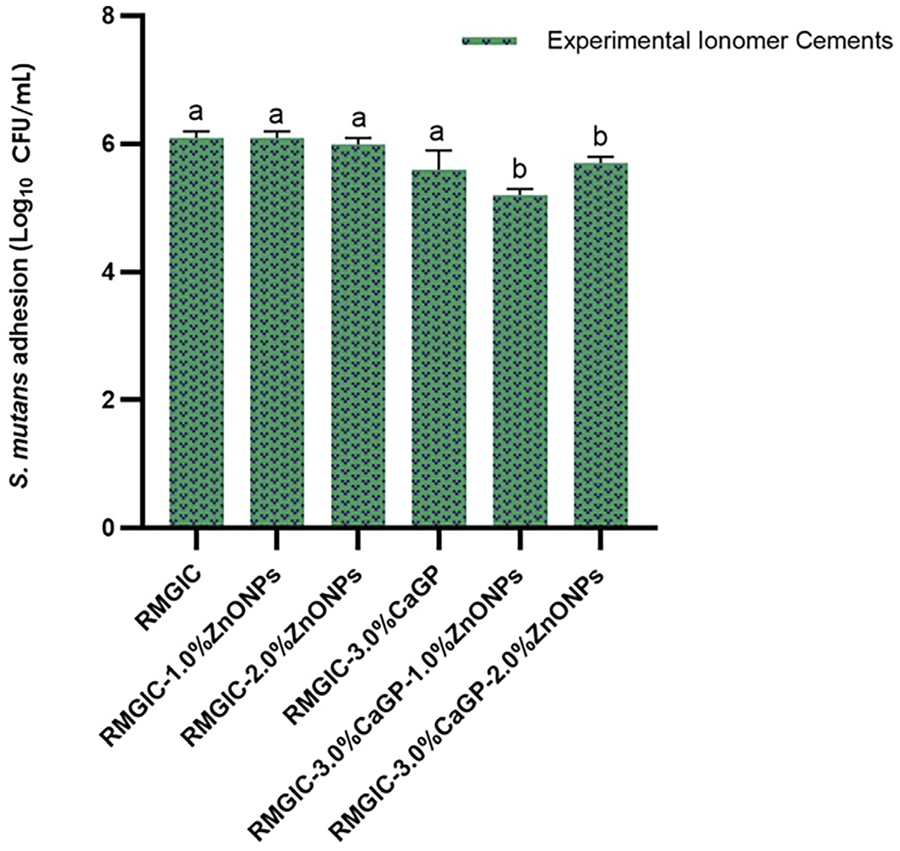
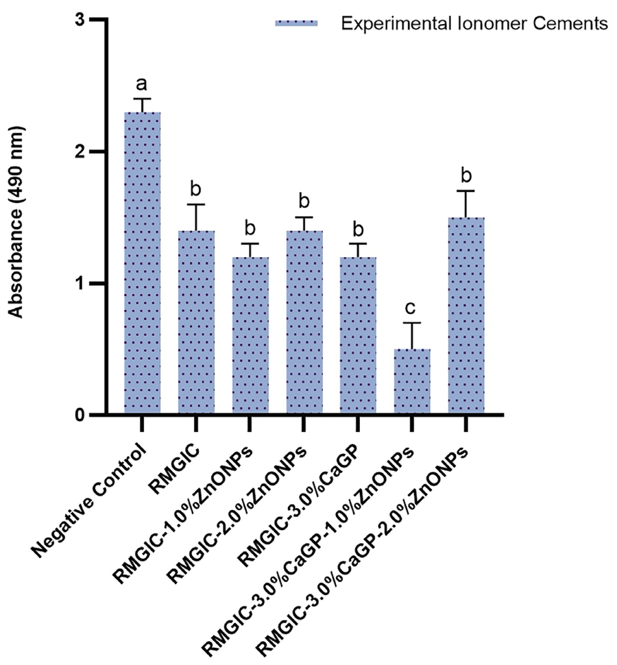
Figure 5 presents the antimicrobial effect results obtained by the adhesion assay with S. mutans on the experimental cements (Log10 CFU/mL). All tested groups showed statistically similar values (p=0.881), except for RMGIC-3.0%CaGP-1.0%ZnONPs and RMGIC-3.0%CaGP-2.0%ZnONPs, which demonstrated a ~17% reduction in S. mutans adhesion compared to the RMGIC group (p<0.05). Figure 6 shows the biofilm formation results (Log10 CFU/mL) for the experimental groups. The results indicate an overall antibacterial effect against S. mutans across all tested groups, with no significant differences between them (p>0.05), except for the RMGIC-3.0%CaGP-1.0%ZnONPs group, which showed the greatest antimicrobial/inhibitory effect (p<0.05). When compared to RMGIC, it showed a higher effect of ~22% (p<0.05). Figure 7 shows the results of S. mutans biofilm reduction (by the XTT method). The incorporation of active agents (ZnONPs and CaGP) into the experimental ionomeric cement significantly reduced the metabolic activity of the S. mutans biofilm compared to the Negative Control and the RMGIC group (p<0.05). All groups showed similar values (p=0.908), except for the Negative Control and RMGIC-3.0%CaGP-1.0%ZnONPs groups (p<0.05). Moreover, the addition of 1.0%ZnONPs and 3.0%CaGP into RMGIC resulted in reductions of approximately 64% and 58%, respectively, when compared to RMGIC and RMGIC-3.0%CaGP/RMGIC-1.0%ZnONPs (p=0.0051).
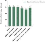
Mean values (±SD, n=4) of S. mutans adhesion (Log10 CFU/mL) according to the Experimental Ionomer Cements. Lowercase letters indicate statistical differences between groups (one-way ANOVA – Experimental Ionomer Cements; Student-Newman-Keuls, p<0.05).
Mean values (±SD, n=6) of biofilm growth (S. mutans, Log10 CFU/mL) according to the Experimental Ionomer Cements. Lowercase letters indicate statistical differences between groups (one-way ANOVA – Experimental Ionomer Cements; Student-Newman-Keuls, p<0.05).
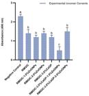
Mean values (±SD, n=6) of cell viability (S. mutans biofilm, Absorbance) according to the Experimental Ionomer cements. Lowercase letters indicate statistical differences between groups (one-way ANOVA – Experimental Ionomer Cements; Student-Newman-Keuls, p<0.05).
Discussion
Dental caries is considered a multifactorial disease, characterized by an imbalance between the demineralization and remineralization of tooth structure due to biofilm metabolism, cariogenic diet, and other factors. Initially, the management of cavitated lesions focused on the complete removal of carious tissue to halt lesion progression. However, in recent years, there has been a significant paradigm shift towards minimally invasive intervention, emphasizing the selective removal of infected tissue. This concept is based on the practice of biologically derived methods for caries management using atraumatic techniques. The goal is to conserve as much tooth structure as possible, in contrast to conventional, more traumatic treatments.31 The aim of this study was to evaluate the effect of incorporating ZnONPs and CaGP into RMGIC on its physical-mechanical and microbiological properties. The study’s null hypothesis was that the addition of these active agents into RMGIC would not improve physical-mechanical and microbiological properties compared to unmodified RMGIC. Based on the results obtained, the null hypothesis was rejected. The incorporation of nanoparticles into dental materials, including cements and sealants, has emerged as a promising strategy to enhance their functional properties. GICs are particularly important in restorative dentistry due to their broad clinical applications. However, their well-documented limitations highlight the need for further optimization. ZnONPs have been extensively investigated owing to their safety, favorable mechanical properties, biocompatibility, high stability, low cost, antibacterial action, and low toxicity.32 In our study, the incorporation of ZnONPs into RMGIC demonstrated positive effects, with 1% and 2% concentrations leading to improvements in physical-mechanical properties.
ZnONPs may influence both the organic (light-curable resin) and the inorganic (acid-base reaction) phases of RMGIC, thereby directly affecting its physicochemical characteristics. Within the resin matrix, ZnONPs can act as reinforcing agents due to their high specific surface area and their ability to form physical or chemical bonds with the monomer chains. This interaction contributes to improved polymer network compaction and increased mechanical strength.33,34 Additionally, studies suggest that ZnONPs may improve photopolymerization efficiency, either by acting as secondary photosensitizers or by enhancing light dispersion within the resin matrix.35 In the inorganic phase, ZnONPs can participate in the acid-base reaction through the gradual release of Zn2⁺. These ions may compete with other cations (such as Ca2⁺ or Sr2⁺) in forming the metallic polysalt network that constitutes the ionic matrix of the cement. This interaction can affect the degree of ionic crosslinking, setting time, and potentially the fluoride release, either by altering porosity or the ionic diffusion channels. Although XRD, TEM, and EDS analyses confirmed the successful synthesis of highly crystalline and compositionally pure ZnONPs, a direct assessment of their spatial distribution within the RMGIC matrix was not performed in the present study.
However, it is important to highlight that the sol-gel method, combined with a controlled mixing protocol, was specifically employed to minimize nanoparticle agglomeration and promote uniform dispersion in the cement. Beyond the synthesis method, several physicochemical factors support the likely uniform distribution of ZnONPs within the RMGIC matrix: a) dry phase incorporation: Incorporating ZnONPs into the powder phase, followed by homogenization, allows for even distribution prior to the acid-base and photopolymerization reactions, limiting opportunities for nanoparticle aggregation during cement manipulation; b) kinetic stability of the system: RMGICs are kinetically controlled systems in which phase separation mechanisms such as sedimentation or Ostwald ripening are unlikely. Given the small particle size and absence of excess liquid, the system inherently favors stable dispersion and c) surface properties of the ZnONPs: The nanoparticles exhibited high crystallinity (confirmed by XRD) and large surface area (observed by TEM), which favor stable interactions with the ionic and resinous phases of the cement. Future characterization studies should be conducted to assess and validate the homogeneity of nanoparticle distribution within the ionomeric matrix. Regarding CS (Table 1), although RMGIC showed the highest value among the groups at 24 hours, these values were at the limit established by ISO 9917-1:2017.25 Notably, the combination of 3.0%CaGP-1.0%ZnONPs in RMGIC resulted in the highest CS at both times. According to Malekhoseini, et al.33 (2021), this improvement may be attributed to the increased Zn2 content in the glass powder, which enhances polymer network connectivity. Therefore, additives should be incorporated at optimal concentrations to improve the material’s properties, as demonstrated by the RMGIC-3.0%CaGP-1.0%ZnONPs group. Our findings are consistent with those of Agarwal, et al.36 (2018), who reported that the incorporation of ZnONPs into conventional GIC improved its physical and mechanical properties. The SH results in our study also align with the observations of Panahandeh, et al.37 (2018), who found that higher concentrations of ZnONPs increased the risk of agglomeration, thereby leading to a reduction in the mechanical properties of the cement.
Regarding CaGP, its incorporation into RMGIC represents a promising strategy for the gradual and controlled release of calcium ions (Ca2⁺), as already observed in the studies by Santos, et al.12 (2019) and de Azevedo Santana, et al.38 (2025). These ions not only contribute to the material’s bioactive and remineralizing properties but may also interact directly with the ionic matrix of the RMGIC, influencing its internal structure. The Ca2⁺ released from CaGP can compete with other divalent cations (such as Sr2⁺ or Zn2⁺) during the formation of the metallic polysalt network, altering the degree of crosslinking and, consequently, affecting properties such as mechanical strength and dimensional stability.12,37 Moreover, CaGP contains an organic component that may influence ion release dynamics and the microstructure of the matrix, promoting the formation of apatite nuclei in fluid-rich environments, such as at the tooth/material interface. The formation of these mineralized deposits within the matrix can reduce porosity and alter fluoride ion diffusion, which may lead to an initially increased release (due to competitive displacement of F⁻ ions) and modulate prolonged fluoride release over time.12 Therefore, future studies should focus on evaluating the fluoride release and remineralizing effects of these new ionomeric cements.
In addition to active agents that enhance the mechanical properties of the cements, it is important that they also have the potential to reduce and/or inhibit biofilm formation and, consequently, bacterial metabolism.39 In this study, the colony-forming unit counts (Figure 5) were similar across the experimental groups, which can be explained by the cements’ initial capacity to provide adhesion for microorganisms such as S. mutans species; however, this colonization is superficial. It is important to note that the presence of a substrate or medium that provides suitable conditions for microorganisms to perform their functions over time can evolve into biofilm formation. In the analysis of biofilm growth and bacterial viability (Figures 6 and 7), the incorporation of 3.0%CaGP and 1.0%ZnONPs into the RMGIC powder resulted in a more significant antimicrobial effect against S. mutans and a greater reduction in bacterial viability (Figure 7). These results can be attributed to the mechanism of action of ZnONPs, which directly interact with the microorganisms, leading to the disruption of the bacterial cell wall, making it more permeable and facilitating the entry of nanoparticles into the bacterial cytoplasm.40-42 This interaction promotes the photocatalytic generation of reactive oxygen species, causing oxidative stress and irreversible damage to bacterial cells.42 In addition, the production of Zn2 by the nanoparticles contributes to the inhibition of active transport and sugar metabolism, as well as interrupting the enzyme systems of the dental biofilm (leading to the displacement of magnesium ions, which are essential for biofilm activity). Zn2 can also reduce the production of acids by the S. mutans biofilm and inhibit the activity of the enzyme glycosyltransferase, thus preventing tooth demineralization. According to the study by Malekhoseini, et al.33 (2021), the addition of ZnONPs to RMGIC caused a significant increase in its antibacterial properties against S. mutans, similar to our findings. In addition, an in vivo split-mouth clinical trial demonstrated that adding 2 wt% ZnONPs into GIC significantly reduced subgingival colony counts of S. mutans and lactobacilli around orthodontic bands—underscoring real-world antimicrobial efficacy.44 On the other hand, CaGP can increase the pH of the biofilm and raise calcium and phosphate levels in the environment, creating less favorable conditions for microbial proliferation and metabolism. Recent studies have indicated that CaGP may also contribute to reducing S. mutans biofilm formation.45,46 In dual-species biofilm models (with S. mutans and Candida albicans), even CaGP without fluoride significantly reduced bacterial viability, and its combination with fluoride led to a notable decrease in extracellular matrix components such as carbohydrates and nucleic acids.46
An important point to mention is that, in the case of bioactive materials such as RMGIC containing ZnONPs and CaGP, ion release is not only expected but also considered a desirable therapeutic feature. Literature reports have shown that the release of Zn2 contributes to sustained antimicrobial activity by disrupting bacterial metabolism and inhibiting biofilm formation.33,37,45,46,47 The study by Azimi, et al.21 (2021) also highlighted a significant improvement in the antibacterial activity of GIC containing 4 wt% ZnONPs, as confirmed through microbiological analyses. Similarly, the release of calcium and phosphate ions plays a fundamental role in the remineralization of demineralized dental tissues, supporting the prevention and control of caries progression.10,45,48 In the present study, the incorporation of ZnONPs and CaGP improved both the antimicrobial properties and mechanical performance of RMGIC, suggesting that the addition of these agents does not compromise material stability. On the contrary, the controlled release of ions may enhance the clinical functionality of the cement, particularly in patients at high risk of caries. Although some degree of material dissolution may occur, this process is considered clinically beneficial in the context of bioactive restorative strategies.
Future investigations should focus on evaluating the long-term ion release profiles and structural stability of these modified ionomeric materials under simulated oral conditions to confirm their clinical durability and effectiveness. Additionally, to deepen the understanding of their behavior and therapeutic potential, future studies are recommended to encompass a comprehensive analysis of their chemical composition, biocompatibility, fluoride release capacity, and performance on relevant dental substrates such as enamel and dentin. The inclusion of well-designed clinical trials is also essential to provide more accurate and clinically relevant data regarding the behavior and efficacy of these innovative restorative materials in real-world scenarios.
Conclusion
This study demonstrated that adding ZnONPs and CaGP to RMGIC enhanced its mechanical and antimicrobial/antibiofilm properties, suggesting enhanced mechanical performance and better protection against cariogenic biofilms—critical factors for the success of restorative treatments. Therefore, the addition of ZnONPs and CaGP is a promising strategy to develop advanced restorative materials that improve clinical outcomes, especially for patients with active dental caries.
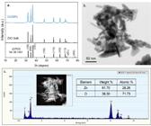
ZnONPs characterization: a) X-ray diffraction (XRD) pattern of the synthesized ZnO nanoparticles (ZnONPs) compared with bulk ZnO. The reference pattern of the wurtzite hexagonal ZnO structure (JCPDS 36-1451) is also shown. The ZnONPs exhibit a highly crystalline structure matching the reference pattern. b) Transmission electron microscopy (TEM) image of ZnONPs, showing nanoparticles with an average size of around 50 nm. c) Energy-dispersive X-ray spectroscopy (EDX) spectrum and image of the ZnONPs. The elemental composition reveals the presence of zinc (Zn) and oxygen (O).
Acknowledgments
The authors are grateful to São Paulo Research Foundation (FAPESP) (#2021/06408-8) for the concession of a scholarship to the first author and FAPESP (#2013/07793-6) (Cepid).
References
-
1 - Machiulskiene V, Campus G, Carvalho JC, Dige I, Ekstrand KR, Jablonski-Momeni A, et al. Terminology of dental caries and dental caries management: consensus report of a workshop organized by ORCA and Cariology Research Group of IADR. Caries Res 2020;54:7-14. doi: 10.1159/000506797.
» https://doi.org/10.1159/000506797 -
2 - Xu Y, You Y, Yi L, Wu X, Zhao Y, Yu J, et al. Dental plaque-inspired versatile nanosystem for caries prevention and tooth restoration. Bioact Mater 2022;20:418-33. doi: 10.1016/j.bioactmat.2022.02.007.
» https://doi.org/10.1016/j.bioactmat.2022.02.007 -
3 - Van Loveren C. Sugar restriction for caries prevention: amount and frequency. Which is more important? Caries Res. 2019;53:168-75. doi: 10.1159/000495471
» https://doi.org/10.1159/000495471 -
4 - Leal SC, Barros BV, Cabral RN, Ferrari JC, Menezes Abreu DM, Ribeiro AP. Dental caries lesions in primary teeth without obvious cavitation: treatment decision-making process. Int J Paediatr Dent. 2019;29:422-8. doi: 10.1111/ipd.12483
» https://doi.org/10.1111/ipd.12483 -
5 - Dorri M, Dunne SM, Walsh T, Schwendicke F. Micro-invasive interventions for managing proximal dental decay in primary and permanent teeth. Cochrane Database Syst Rev 2015;2015(11):CD010431. doi: 10.1002/14651858.CD010431.pub2
» https://doi.org/10.1002/14651858.CD010431.pub2 -
6 - Urquhart O, Tampi MP, Pilcher L, Slayton RL, Araujo MW, Fontana M, et al. Nonrestorative treatments for caries: systematic review and network meta-analysis. J Dent Res. 2019;98(1):14-26. doi: 10.1177/0022034518800014
» https://doi.org/10.1177/0022034518800014 -
7 - Moradi S, Sabbagh S, Timms L, Ravaghi V. Teaching minimally invasive interventions in paediatric dentistry: A cross-sectional survey of dental schools in Iran. BMC Oral Health. 2021;21(1):368. doi: 10.1186/s12903-021-01735-5
» https://doi.org/10.1186/s12903-021-01735-5 -
8 - Zandi Karimi A, Rezabeigi E, Drew RA. Glass ionomer cements with enhanced mechanical and remineralizing properties containing 45S5 bioglass-ceramic particles. J Mech Behav Biomed Mater. 2019;97:396-405. doi: 10.1016/j.jmbbm.2019.05.033
» https://doi.org/10.1016/j.jmbbm.2019.05.033 -
9 - Zaze AC, Dias AP, Sassaki KT, Delbem AC. The effects of low-fluoride toothpaste supplemented with calcium glycerophosphate on enamel demineralization. Clin Oral Investig. 2014;18(6):1619-24. doi: 10.1007/s00784-013-1140-y
» https://doi.org/10.1007/s00784-013-1140-y -
10 - Emerenciano NG, Delbem AC, Gonçalves FM, Quinteiro JP, Camargo ER, Silva-Sousa YTC, et al. Effect of the association of microparticles and nano-sized ß-calcium glycerophosphate in conventional toothpaste on enamel remineralization: in situ study. J Dent. 2023;138:104719. doi: 10.1016/j.jdent.2023.104719
» https://doi.org/10.1016/j.jdent.2023.104719 -
11 - Nunes GP, Delbem AC, Gonçalves FM, Rischka K, Camargo ER, Sousa YT, et al. Biomineralization and remineralizing potential of toothpastes containing nanosized ß-calcium glycerophosphate: an in vitro study. Odontology. 2024;112:1186-96. doi: 10.1007/s10266-024-00927-z
» https://doi.org/10.1007/s10266-024-00927-z -
12 - Santos SS, Delbem AC, Moraes JC, Souza JA, Oliveira LQ, Pedrini D. Resin-modified glass ionomer containing calcium glycerophosphate: physico-mechanical properties and enamel demineralization. J Appl Oral Sci. 2019;27:e20180188. doi: 10.1590/1678-7757-2018-0188.
» https://doi.org/10.1590/1678-7757-2018-0188 -
13 - Kheur M, Kantharia N, Lakha T, Kheur S, Al-Haj Husain N, Özcan M. Evaluation of mechanical and adhesion properties of glass ionomer cement incorporating nano-sized hydroxyapatite particles. Odontology. 2020;108:66-73. doi: 10.1007/s10266-019-00447-2
» https://doi.org/10.1007/s10266-019-00447-2 -
14 - Fernandes GL, Vanim MM, Delbem AC, Martorano AS, Raucci LM, Oliveira PT, et al. Antibacterial, cytotoxic and mechanical properties of an orthodontic cement with phosphate nano-sized and phosphorylated chitosan: an in vitro study. J Dent 2024;146:105073. doi: 10.1016/j.jdent.2024.105073
» https://doi.org/10.1016/j.jdent.2024.105073 -
15 - Chau CF, Wu SH, Yen GC. The development of regulations for food nanotechnology. Trends Food Sci Technol. 2007;18:269-80. doi: 10.1016/j.tifs.2007.01.007
» https://doi.org/10.1016/j.tifs.2007.01.007 -
16 - Bastos NA, Bitencourt SB, Martins EA, Souza GM. Review of nano-technology applications in resin-based restorative materials. J Esthet Restor Dent. 2021;33:567-82. doi: 10.1111/jerd.12699
» https://doi.org/10.1111/jerd.12699 -
17 - Oberdörster G, Oberdörster E, Oberdörster J. Nanotoxicology: an emerging discipline evolving from studies of ultrafine particles. Environ Health Perspect. 2005;113:823-39. doi: 10.1289/ehp.7688
» https://doi.org/10.1289/ehp.7688 - 18 - Pourhajibagher M, Salehi Vaziri A, Takzaree N, Ghorbanzadeh R. Physico-mechanical and antimicrobial properties of an orthodontic adhesive containing cationic curcumin doped zinc oxide nanoparticles subjected to photodynamic therapy. Photodiagnosis Photodyn Ther 2019;25:239-46. doi: 10.1016/j.pdpdt.2019.01.002
-
19 - Jowkar Z, Fattah Z, Ghanbarian S, Shafiei F. The effects of silver, zinc oxide, and titanium dioxide nanoparticles used as dentin pretreatments on the microshear bond strength of a conventional glass ionomer cement to dentin. Int J Nanomedicine. 2020;15:4755-62. doi: 10.2147/IJN.S272098.
» https://doi.org/10.2147/IJN.S272098 - 20 - Huang Z, Zheng X, Yan D, Yin G, Liao X, Kang Y, et al. Toxicological effect of ZnO nanoparticles based on bacteria. Langmuir. 2008;24:4140-4. doi: 10.1021/la703550z
-
21 - Azimi R, Shahgholi M, Khandan A. Fabrication and characterization of reinforced glass ionomer cement by zinc oxide and hydroxyapatite nanoparticles. Heliyon. 2024;10:e39063. doi: 10.1016/j.heliyon.2024.e39063
» https://doi.org/10.1016/j.heliyon.2024.e39063 -
22 - Ismail SM, Ahmed SM, Abdulrahman AF, Almessiere MA. Characterization of green synthesized of ZnO nanoparticles by using pinus brutia leaves extracts. J Mol Struct. 2023;1280:135063. doi: 10.1016/j.molstruc.2023.135063
» https://doi.org/10.1016/j.molstruc.2023.135063 -
23 - Changle Wu, Xueliang Qiao, Jianguo Chen, Hongshui Wang, Fatang Tan, Shitao LiA novel chemical route to prepare ZnO nanoparticles Mater Lett 2006;60:1828-1832. https://doi.org/10.1016/j.matlet.2005.12.061
» https://doi.org/10.1016/j.matlet.2005.12.061 - 24 - Garcia PP, Cardia MF, Francisconi RS, Dovigo LN, Spolidório DM, Souza Rastelli AN, et al. Antibacterial activity of glass ionomer cement modified by zinc oxide nanoparticles. Microsc Res Tech. 2017;80:456-61. doi: 10.1002/jemt.22814
- 25 - International Organization for Standardization. ISO 9917-2:2017-Dentistry - Water based cements. Part 2: Resin-modified cements. Geneva: ISO; 2017.
-
26 - Menezes-Silva R, Oliveira BM, Magalhães AP, Bueno LS, Borges AF, Baesso ML, et al. Correlation between mechanical properties and stabilization time of chemical bonds in glass-ionomer cements. Braz Oral Res. 2020;34:e053. doi: 10.1590/1807-3107bor-2020.vol34.0053
» https://doi.org/10.1590/1807-3107bor-2020.vol34.0053 - 27 - Anusavice KJ. Phillips' Science of Dental Materials. Philadelphia: WB Saunders; 1994.
-
28 - Fúcio SB, Paula AB, Sardi JC, Duque C, Correr-Sobrinho L, Puppin-Rontani RM. Streptococcus mutans biofilm influences on the antimicrobial properties of glass ionomer cements. Braz Dent J. 2016;17:681-7. doi: 10.1590/0103-6440201600655
» https://doi.org/10.1590/0103-6440201600655 -
29 - Cassanho AC, Fernandes AM, Oliveira LD, Carvalho CA, Jorge AO, Koga-Ito CY. In vitro activity of zinc oxide-eugenol and glass ionomer cements on Candida albicans. Braz Oral Res. 2005;19:134-8. doi: 10.1590/s1806-83242005000200011
» https://doi.org/10.1590/s1806-83242005000200011 -
30 - Kubota H, Senda S, Nomura N, Tokuda H, Uchiyama H. Biofilm formation by lactic acid bacteria and resistance to environmental stress. J Biosci Bioeng. 2008;106:381-6. doi: 10.1263/jbb.106.381
» https://doi.org/10.1263/jbb.106.381 -
31 - Alam BF, Najmi MA, Qasim SB, Almulhim KS, Ali S. A bibliometric analysis of minimally invasive dentistry: a review of the literature from 1994 to 2021. J Prosthet Dent. 2023;130:179-86. doi: 10.1016/j.prosdent.2021.09.023
» https://doi.org/10.1016/j.prosdent.2021.09.023 -
32 - Moradpoor H, Safaei M, Mozaffari HR, Sharifi R, Imani MM, Golshah A, et al. An overview of recent progress in dental applications of zinc oxide nanoparticles. RSC Adv. 2021;11:21189-21206. doi: 10.1039/D1RA02792B
» https://doi.org/10.1039/D1RA02792B -
33 - Malekhoseini Z, Rezvani MB, Niakan M, Atai M, Bassir MM, Alizade HS, et al. Effect of zinc oxide nanoparticles on physical and antimicrobial properties of resin-modified glass ionomer cement. Dent Res J (Isfahan). 2021;18:73. doi: 10.4103/1735-3327.326646
» https://doi.org/10.4103/1735-3327.326646 -
34 - Shojaei S, Shahgholi M, Karimipour A. The effects of atomic percentage and size of Zinc nanoparticles, and atomic porosity on thermal and mechanical properties of reinforced calcium phosphate cement by molecular dynamics simulation. J Mech Behav Biomed Mater. 2023;141:105785. doi: 10.1016/j.jmbbm.2023.105785
» https://doi.org/10.1016/j.jmbbm.2023.105785 -
35 - Burunkova JA, Denisyuk IY, Semina AS. Self-Organization of ZnO Nanoparticles on UV-curable acrylate nanocomposites. J Nanotechnology. 2011:951036. doi: 10.1155/2011/951036
» https://doi.org/10.1155/2011/951036 -
36 - Agarwal P, Nayak R, Upadhya NP, Ginjupalli K. Evaluation of properties of glass ionomer cement reinforced with zinc oxide nanoparticles - an in vitro study. Materials Today: Proceedings. 2018;5:16065-72. doi: 10.1016/j.matpr.2018.05.088
» https://doi.org/10.1016/j.matpr.2018.05.088 -
37 - Panahandeh N, Torabzadeh H, Aghaee M, Hasani E, Safa S. Effect of incorporation of zinc oxide nanoparticles on mechanical properties of conventional glass ionomer cements. J Conserv Dent. 2018;21(2):130-5. doi: 10.4103/JCD.JCD_170_17
» https://doi.org/10.4103/JCD.JCD_170_17 -
38 - Azevedo Santana JA, Navarro MF, Gomes LF, Brighenti FL, Oliveira AB, Fernandes GL, et al. Eur J Oral Sci 2025;133:e70001. doi: 10.1111/eos.70001
» https://doi.org/10.1111/eos.70001 -
39 - Benoit DS, Sims KR Jr, Fraser D. Nanoparticles for oral biofilm treatments. ACS Nano. 28;13(5):4869-4875. doi: 10.1021/acsnano.9b02816
» https://doi.org/10.1021/acsnano.9b02816 -
40 - Almutairi B, Albahser G, Almeer R, Alyami NM, Almukhlafi H, Yaseen KN, et al. Investigation of cytotoxicity, apoptotic and inflammatory responses of biosynthesized zinc oxide nanoparticles from Ocimum sanctum Linn in human skin keratinocyte (HaCaT) and human lung epithelial (A549) cells. Oxid Med Cell Longev. 2020;2020:1835475. doi: 10.1155/2020/1835475
» https://doi.org/10.1155/2020/1835475 -
41 - Czyzowska A, Barbasz A. A review: zinc oxide nanoparticles - friends or enemies? Int J Environ Health Res. 2022;32:885-901. doi: 10.1080/09603123.2020.1805415
» https://doi.org/10.1080/09603123.2020.1805415 -
42 - Hasanin MS, El Saied H, Morsy FA, Hassan Abdel Latif Rokbaa H. Green nanocoating-based polysaccharides decorated with ZnONPs doped Egyptian kaolinite for antimicrobial coating paper. Sci Rep. 2023;13:11461. doi: 10.1038/s41598-023-38467-1
» https://doi.org/10.1038/s41598-023-38467-1 -
43 - Valtsifer VA, Sivtseva AV, Kondrashova NB, Shamsutdinov AS, Averkina AS, Valtsifer IV, et al. Influence of synthesis conditions on the properties of zinc oxide obtained in the presence of nonionic structure-forming compounds. Nanomaterials (Basel). 2023;13:2537. doi: 10.3390/nano13162537
» https://doi.org/10.3390/nano13162537 - 44 - Shirazi M, Qazvini FF, Mohamadrezaie S. Antimicrobial properties of glass-ionomer cement incorporated with zinc oxide nanoparticles against mutans streptococci and lactobacilli under orthodontic bands: an in vivo split-mouth study. Dent Res J (Isfahan). 2023;20:45.
-
45 - Miranda ML, Danelon M, Delbem AC, Kopp W, Nunes GP, Brighenti FL. Enhanced anti-biofilm and anti-caries potential of arginine combined with calcium glycerophosphate and fluoride. J Dent. 2024;146:105039. doi: 10.1016/j.jdent.2024.105039
» https://doi.org/10.1016/j.jdent.2024.105039 -
46 - Cavazana TP, Hosida TY, Sampaio C, Morais LA, Monteiro DR, Pessan JP, et al. The activity of calcium glycerophosphate and fluoride against cariogenic biofilms of streptococcus mutans and candida albicans formed in vitro. Antibiotics (Basel). 2023;12:422. doi: 10.3390/antibiotics12020422
» https://doi.org/10.3390/antibiotics12020422 -
47 - Danelon M, Nunes GP, Sterzenbach T, Hannig C. Enhancing antimicrobial properties of glass ionomer cement through metallic agent reinforcement: a systematic review and meta-analysis. J Dent. 2025;160:105892. doi: 10.1016/j.jdent.2025.105892
» https://doi.org/10.1016/j.jdent.2025.105892 -
48 - Emerenciano NG, Delbem AC, Gonçalves FM, Camargo ER, Souza FN Neto, Gorup LF, et al. Effect of nanometric ß-calcium glycerophosphate supplementation in conventional toothpaste on enamel demineralization: an in vitro study. J Mech Behav Biomed Mater 2024;151:106354. doi: 10.1016/j.jmbbm.2023.106354
» https://doi.org/10.1016/j.jmbbm.2023.106354
-
Data availability:
All data generated or analyzed during this study are included in this published article
Edited by
-
Editor:
Linda Wang
-
Associate Editor:
Daniela Rios Honório
All data generated or analyzed during this study are included in this published article