 Design, Synthesis and Biological Evaluation of Lupeol-3-Thiadiazole Derivatives as Novel Glutaminase 1 Inhibitors
Abstract
Cancer cells rely on different metabolic pathways to maintain their unchecked cell proliferation and resistance to apoptosis. As an integral component in glutamine hydrolysis, glutaminase 1 (GLS1) has been proposed as a valuable target for cancer therapy. In this study, a series of novel lupeol derivatives were successfully synthesized using a structure-based drug design methodology. The anti-proliferative activity of these derivatives against four human cancer cell lines (A549: human non-small-cell lung cancer cells; Hep G2: human hepatocellular cancer cells; MCF-7: human breast cancer cells; MDA-MB-231: human triple-negative breast cancer cells) was assessed through the MTT (3-(4,5-dimethylthiazol-2-yl)-2,5-diphenyltetrazolium bromide) assay. Compound 9c exhibited the most potent anti-proliferative activity against A549 cells (half-maximal inhibitory concentration, IC50 = 4.12 μM), and had a selectivity index of 18.27 (IC50 MRC-5 (human embryonic lung fibroblasts)/IC50 A549). Bioassay optimization further identified compound 9c as a novel GLS1 inhibitor (IC50 = 6.71 µM). Compound 9c arrested the cell cycle in the S phase. By blocking the glutamine hydrolysis pathway, it reduced cellular glutamate levels, which in turn triggered the production of reactive oxygen species (ROS) and induced apoptosis. Molecular docking revealed that compound 9c interacts via the reaction site of the variable binding pocket of GLS1. Considering these results, compound 9c holds promise as a novel GLS1 inhibitor and could be further evaluated for non-small-cell lung cancer (NSCLC) therapy.
Keywords:
lupeol; GLS1 inhibitors; molecular docking; antitumor; NSCLC
Introduction
Cancer is characterized by a metabolic shift that leads to uncontrolled cell proliferation, increased energy production, and metabolite synthesis.1, 2 Glutamine, an amino acid abundant in blood and muscle, is the favored carbon source for pyrimidine ring biosynthesis in tumor cells under hypoxic conditions and plays a crucial role as a cellular intermediate metabolite.3, 4 Glutamine deficiency can cause intestinal mucosal necrosis and induces MYC-dependent apoptosis in human cells.5, 6, 7 Glutaminase converts glutamine to glutamate, which then enters the tricarboxylic acid cycle, where it is further processed by glutamate dehydrogenase (GLUD), or participates in the biosynthesis of non-essential amino acids via transaminase to provide nutrition for cells by transaminase.8, 9 Glutamine hydrolysis is essential for maintaining redox homeostasis, and many cancer cells exhibit high glutamine dependence, so further development of drugs targeting glutamine-related pathways is considered an attractive cancer treatment strategy.10
Human glutamine isoenzymes are divided into two isoforms: kidney-type glutaminase (GLS1) and liver-type glutaminase (GLS2). GLS1 is expressed in various tissues, whereas GLS2 is restricted to the brain, pancreas, and liver.11 Both isoforms have long and short splice variants, which play different roles in various biological processes and participate in the occurrence and progression of different types of cancer.12, 13, 14, 15 The short splice variant of GLS1 has been found to be highly expressed in triple negative breast cancer (TNBC), acute random leukemia, and non-small-cell lung cancer (NSCLC).16, 17 The inhibition of glutaminase has also emerged as a valuable target for cancer therapy.18 BPTES (bis-2-(5-phenylacetamido-1,3,4-thiadiazol-2-yl)ethyl sulfide) interferes with the process of glutamine synthesis in tumor cells by inhibiting GLS1, thereby inhibiting the growth and proliferation of tumor cells. An in vitro study showed its high efficiency and specificity in inhibiting cancer cell proliferation.19 However, poor bioavailability and water solubility hinder the use of BPTES. A large number of derivatives have been synthesized based on the BPTES backbone structures and have been tested in clinical trials.20, 21, 22, 23, 24, 25
Natural products are important sources for the discovery and development of novel anticancer drugs.26, 27, 28, 29 The structural modification of natural products to increase the drug potential is one of the current research hotspots.30, 31, 32, 33 Lupeol (lup-20(29)-en-3β-ol, 1, Figure 1), as a natural pentacyclic triterpene, is present in various common traditional Chinese medicines, such as Astragalus membranaceus and Euphorbia fischeriana Steud.34, 35, 36 A wealth of research37, 38, 39, 40, 41, 42 has consistently demonstrated that lupeol exerts inhibitory effects on a spectrum of malignant neoplasms, including liver cancer, lung cancer, colorectal cancer, prostate cancer, osteosarcoma, and melanoma. In our prior investigations, we discovered that lupeol exhibits moderate inhibitory activity against GLS1, with an half-maximal inhibitory concentration (IC50) value of 49.74 μM. Therefore, lupeol is an ideal lead compound for antitumor development. In the present study, according to the concept of molecular hybridization, significant groups or substituents from the structure of BPTES were incorporated into lupeol (Figure 2), leading to the development of a series of compounds that exhibit inhibitory effects on GLS1. Among them, the most potent inhibitor, compound 9c, exhibited a remarkable inhibitory effect on the proliferation of A549 (human non-small-cell lung cancer) cells, indicating its potential as a novel GLS1 inhibitor for the treatment of NSCLC and deserves further development.


Experimental
Chemistry
All reagents were sourced from reputable commercial suppliers (Xilong Scientific, Wuhan, China), and were deemed sufficiently pure for direct use in our experiments, obviating the requirement for further purification. For analytical monitoring of reaction progress, thin-layer chromatography (TLC) was conducted using silica gel GF254 as the stationary phase, provided by Thermo (Shanghai, China). The TLC was developed with mobile phases comprised of either dichloromethane (CH2Cl2) and methanol (CH3OH) mixtures. Subsequent purification of the intermediates and target compounds was achieved through column chromatography, employing 300-400 mesh silica gel procured from Haiyang Chemical (Qingdao, China). Spectroscopic analysis was performed using a spectrometer Avance DRX400 NMR (Aunion, Shanghai, China), with tetramethylsilane (TMS) serving as the reference for both 1H and 13C nuclear magnetic resonance (NMR). The chemical shifts are reported in delta (δ) notation, in parts per million (ppm). Additionally, high-resolution mass spectrometry (HRMS) analysis was conducted on a mass spectrometer Esquire 6000 (Bruker, Massachusetts, USA), providing further confirmation of the structure of the synthesized compounds.
Synthesis of intermediates 5, 6, 7
Starting from lupeol (1, 500 mg, 1.2 mmol) in 10 mL of dichloromethane, intermediates 2, 3, 4 (3.5 mmol, 3 eq) and the catalyst triethylamine (Et3N, 0.6 mmol, 0.5 eq) were respectively introduced, after which the mixture was heated in a water bath for 12 h. Once the reaction was confirmed to be complete by TLC, the reaction mixture was diluted with 50 mL of dichloromethane, extracted with water, and the water layer was saturated with sodium chloride solution to absorb the water. Finally, anhydrous sodium sulfate was used to remove moisture, ensuring the complete removal of water. The solvent was then removed under reduced pressure to yield the intermediates 5, 6, 7.
Lupeol-3-(4-oxobutanoic acid) (5)
White solid; 77% yield; mp 167.8-168.4 °C; 1H NMR (600 MHz, CDCl3) δ 4.69 (s, 1H, H-29), 4.57 (s, 1H, H-29), 4.51-4.48 (m, 1H, H-3), 2.69-2.66 (m, 2H, H-32), 2.63-2.60 (m, 2H, H-33), 2.37 (td, 1H, J 11.0, 5.8 Hz), 1.94-1.89 (m, 1H), 1.68 (s, 3H, CH3), 1.67 (s, 1H), 1.65 (d, 2H, J 3.5 Hz), 1.63-1.60 (m, 2H), 1.57 (dd, 1H, J 13.2, 3.8 Hz), 1.50-1.45 (m, 2H), 1.42-1.40 (m, 1H), 1.40-1.36 (m, 6H), 1.34-1.32 (m, 1H), 1.30 (d, 1H, J 2.5, Hz), 1.28 (d, 1H, J 2.8 Hz), 1.25 (s, 1H), 1.22 (dd, 1H, J 7.9, 4.8 Hz), 1.19 (d, 1H, J 11.3 Hz), 1.07 (dd, 1H, J 12.8, 4.6 Hz), 1.03 (s, 3H, CH3), 0.98 (dd, 1H, J 4.2, 2.5 Hz), 0.94 (s, 3H, CH3), 0.85 (s, 3H, CH3), 0.83 (d, 6H, J 3.1 Hz, CH3), 0.78 (s, 3H, CH3); 13C NMR (150 MHz, CDCl3) δ 176.81 (C-34), 170.82 (C-31), 149.96 (C-20), 108.35 (C-29), 80.60 (C-3), 54.37 (C-5), 49.30 (C-9), 47.26 (C-18), 46.98 (C-19), 41.97 (C-17), 41.81 (C-14), 39.82 (C-8), 38.97 (C-22), 37.32 (C-13), 37.01 (C-4), 36.82 (C-1), 36.05 (C-10), 34.54 (C-16), 33.17 (C-7), 28.80 (C-21), 28.30 (C-32), 27.99 (C-33), 26.86 (C-15), 26.41 (C-23), 24.05 (C-12), 22.60 (C-2), 19.92 (C-11), 18.26 (C-30), 17.16 (C-28), 16.98 (C-24), 15.48 (C-6), 15.14 (C-25), 14.95 (C-26), 13.50 (C-27).
Lupeol-3-(4-oxobut-2-enoic acid) (6)
Yellow solid; 70% yield; mp 165.6-166.2 °C; 1H NMR (600 MHz, CDCl3) δ 6.48 (d, 1H, J 12.9 Hz, H-32), 6.37 (d, 1H, J 12.9 Hz, H-33), 4.69 (s, 1H, H-29), 4.66 (dd, 1H, J 9.3, 7.0 Hz, H-29), 4.57 (s, 1H, H-3), 2.38 (td, 1H, J 11.1, 5.8 Hz), 1.92 (ddd, 1H, J 13.2, 7.1, 2.9 Hz), 1.74 (d, 1H, J 3.7 Hz), 1.72 (d, 2H, J 6.2 Hz), 1.70 (d, 1H, J 3.5 Hz), 1.68 (s, 3H, CH3), 1.65 (dd, 2H, J 12.3, 3.8 Hz), 1.52 (dd, 1H, J 6.4, 2.8 Hz), 1.48 (ddd, 2H, J 12.9, 4.6, 2.6 Hz), 1.42 (s, 1H), 1.42-1.40 (m, 4H), 1.39 (d, 2H, J 5.8 Hz), 1.37 (d, 1H, J 4.6 Hz), 1.34 (d, 1H, J 2.5 Hz), 1.33 (d, 1H, J 2.9 Hz), 1.30 (d, 1H, J 2.6 Hz), 1.24-1.22 (m, 1H), 1.04 (s, 3H, CH3), 1.02-0.99 (m, 2H), 0.95 (s, 3H, CH3), 0.88 (d, 9H, J 3.9 Hz, CH3), 0.79 (s, 3H, CH3); 13C NMR (150 MHz, CDCl3) δ 168.25 (C-34), 163.89 (C-31), 151.08 (C-20), 138.03 (C-32), 129.52 (C-33), 109.55 (C-29), 85.47 (C-3), 55.43 (C-5), 50.45 (C-9), 48.39 (C-18), 48.14 (C-19), 43.13 (C-17), 42.99 (C-14), 40.97 (C-8), 40.12 (C-22), 38.39 (C-13), 38.13 (C-4), 38.07 (C-1), 37.20 (C-10), 35.67 (C-16), 34.26 (C-7), 29.95 (C-21), 29.84 (C-15), 28.13 (C-23), 27.55 (C-12), 25.16 (C-2), 21.09 (C-11), 19.43 (C-30), 18.29 (C-28), 18.14 (C-24), 16.58 (C-6), 16.30 (C-25), 16.11 (C-26), 14.65 (C-27).
Lupeol-3-(5-oxopentanoic acid) (7)
White solid; 73% yield; mp 163.1-164.3 °C; 1H NMR (600 MHz, CDCl3) δ 4.62 (s, 1H, H-29), 4.50 (s, 1H, H-29), 4.42 (dd, 1H, J 10.6, 5.7 Hz, H-3), 2.37 (d, 2H, J 7.1 Hz, H-32), 2.33 (d, 2H, J 7.5 Hz, H-34), 2.29 (s, 1H), 1.90 (d, 2H, J 7.2 Hz, H-33), 1.87-1.82 (m, 1H), 1.61 (s, 3H, CH3), 1.59 (d, 2H, J 4.7 Hz), 1.56-1.53 (m, 2H), 1.43 (d, 2H, J 8.7 Hz), 1.40 (s, 1H), 1.35 (d, 2H, J 4.1 Hz), 1.33 (s, 2H), 1.31 (d, 2H, J 4.2 Hz), 1.29 (d, 2H, J 2.8 Hz), 1.28 (d, 2H, J 4.8 Hz), 1.25 (s, 2H), 1.24 (s, 1H), 1.21 (d, 2H, J 2.5 Hz), 1.18 (s, 2H), 0.96 (s, 3H, CH3), 0.87 (s, 3H, CH3), 0.84 (s, 3H, CH3), 0.77 (d, 6H, J 3.8 Hz, CH3), 0.72 (s, 3H, CH3);13C NMR (150 MHz, CDCl3) δ 171.66 (C-35), 166.78 (C-31), 149.97 (C-20), 108.34 (C-29), 80.12 (C-3), 67.14 (C-5), 54.33 (C-9), 49.30 (C-18), 47.25 (C-19), 46.98 (C-17), 41.97 (C-14), 41.80 (C-8), 39.81 (C-22), 38.97 (C-13), 37.69 (C-4), 37.00 (C-1), 36.05 (C-10), 34.54 (C-16), 33.16 (C-32), 32.63 (C-34), 29.33 (C-7), 28.68 (C-21), 27.90 (C-15), 26.97 (C-23), 22.71 (C-12), 21.96 (C-2), 19.91 (C-11), 19.00 (C-33), 16.97 (C-30), 15.54 (C-28), 15.14 (C-24), 14.94 (C-6), 13.49 (C-25), 13.03 (C-26), 9.93 (C-27).
Synthesis of target compounds 8a-8f, 9a-9f, 10a-10f
The intermediates 5, 6, or 7 (150 mg, 0.3 mmol) were dissolved in 15 mL of dichloromethane, and then the thiazol-2-yl amine substituents containing amino groups (0.3 mmol, 1.5 eq), Et3N (0.1 mmol, 0.5 eq), and benzotriazol-1-yl-oxytripyrrolidinophosphonium hexafluorophosphate (PyBOP, 0.2 mmol) were added respectively. The mixture was reacted at room temperature for 4 h. After the reaction was confirmed to be complete by TLC, the solvent was removed thorough reduced pressure. The product was purified by silica gel column chromatography (dichloromethane/methanol, 100:1, v/v) to yield the target compounds 8a-8f, 9a-9f, 10a-10f.
Lupeol-3-(4-((1,3,4-thiadiazol-2-yl) amino)-4-oxobutanoate) (8a)
White solid; 65% yield; mp 150.8-152.1 °C; 1H NMR (600 MHz, CDCl3) δ 8.78 (s, 1H, H-36), 4.68 (s, 1H, H-29), 4.56 (s, 1H, H-29), 4.49 (dd, 1H, J 11.6, 4.7 Hz, H-3), 3.10 (d, 2H, J 2.8 Hz, H-33), 2.83 (d, 2H, J 6.6 Hz, H-32), 2.39-2.34 (m, 1H), 1.94-1.87 (m, 1H), 1.69 (s, 1H), 1.67 (s, 3H, CH3), 1.66 (s, 1H), 1.65-1.63 (m, 2H), 1.62 (s, 1H), 1.60 (s, 1H), 1.55 (s, 1H), 1.47 (d, 2H, J 8.4 Hz), 1.39 (d, 2H, J 3.1 Hz), 1.37 (d, 2H, J 3.3 Hz), 1.35 (s, 2H), 1.33 (s, 1H), 1.30 (s, 1H), 1.27 (s, 1H), 1.26 (s, 1H), 1.21 (s, 1H), 1.19 (s, 1H), 1.08-1.03 (m, 1H), 1.01 (s, 3H, CH3), 0.99-0.95 (m, 1H), 0.92 (s, 3H, CH3), 0.90-0.85 (m, 1H), 0.81 (d, 6H, J 4.6 Hz, CH3), 0.77 (d, 6H, J 4.3 Hz, CH3); 13C NMR (150 MHz, CDCl3) δ 171.84 (C-34), 170.68 (C-31), 160.54 (C-35), 150.98 (C-20), 147.33 (C-36), 109.39 (C-29), 81.64 (C-3), 55.38 (C-5), 50.30 (C-9), 48.27 (C-18), 48.00 (C-19), 43.00 (C-17), 42.82 (C-14), 40.83 (C-8), 40.00 (C-22), 38.35 (C-13), 38.02 (C-4), 37.82 (C-1), 37.05 (C-10), 35.57 (C-16), 34.17 (C-7), 30.97 (C-32), 29.82 (C-33), 29.33 (C-21), 27.92 (C-15), 27.43 (C-23), 25.07 (C-12), 23.61 (C-2), 20.93 (C-11), 19.30 (C-30), 18.18 (C-28), 18.00 (C-24), 16.46 (C-6), 16.15 (C-25), 15.96 (C-26), 14.52 (C-27); HRMS (ESI) m/z, calcd. for C36H55N3O3S [M + Na]+: 632.3862, found: 632.3859.
Lupeol-3-(4-((5-methyl-1,3,4-thiadiazol-2-yl) amino)-4-oxobutanoate) (8b)
White solid; 67% yield; mp 154.2-155.1 °C; 1H NMR (600 MHz, CDCl3) δ 4.68 (s, 1H, H-29), 4.56 (s, 1H, H-29), 4.49 (dd, 1H, J 11.5, 4.7 Hz, H-3), 3.03 (d, 2H, J 5.6 Hz, H-33), 2.81 (d, 2H, J 6.6 Hz, H-32), 2.68 (s, 3H, CH3), 2.40-2.35 (m, 1H), 1.93-1.88 (m, 1H), 1.68 (s, 3H, CH3), 1.66 (s, 1H), 1.65 (d, 2H, J 2.6 Hz), 1.63 (s, 1H), 1.60 (s, 1H), 1.57 (s, 1H), 1.47 (d, 2H, J 8.2 Hz), 1.39 (d, 2H, J 3.4 Hz), 1.37 (s, 2H), 1.36 (s, 1H), 1.35 (s, 1H), 1.33 (s, 1H), 1.31 (s, 1H), 1.30 (s, 1H), 1.28 (d, 2H, J 3.6 Hz), 1.26 (d, 2H, J 3.0 Hz), 1.22 (s, 1H), 1.20 (s, 1H), 1.09-1.04 (m, 1H), 1.01 (s, 3H, CH3), 0.93 (s, 3H, CH3), 0.82 (d, 6H, J 4.6 Hz, CH3), 0.78 (d, 6H, J 2.8 Hz, CH3); 13C NMR (150 MHz, CDCl3) δ 171.92 (C-34), 170.42 (C-31), 160.56 (C-35), 159.77 (C-20), 150.99 (C-36), 109.38 (C-29), 81.56 (C-3), 55.38 (C-5), 50.31 (C-9), 48.28 (C-18), 48.01 (C-19), 43.00 (C-17), 42.82 (C-14), 40.83 (C-8), 40.00 (C-22), 38.36 (C-13), 38.02 (C-4), 37.83 (C-1), 37.05 (C-10), 35.57 (C-16), 34.17 (C-7), 31.61 (C-32), 30.93 (C-33), 29.42 (C-21), 27.94 (C-15), 27.43 (C-23), 25.07 (C-12), 23.62 (C-2), 22.67 (C-11), 20.93 (C-30), 18.00 (C-28), 16.48 (C-24), 16.15 (C-6), 15.97 (C-25), 15.33 (C-26), 14.52 (C-37), 14.14 (C-27); HRMS (ESI) m/z, calcd. for C37H57N3O3S [M + Na]+: 646.4018, found: 646.4016.
Lupeol-3-(4-((5-ethyl-1,3,4-thiadiazol-2-yl) amino)-4-oxobutanoate) (8c)
White solid; 70% yield; mp 167.6-169.1 °C; 1H NMR (600 MHz, CDCl3) δ 4.68 (s, 1H, H-29), 4.56 (s, 1H, H-29), 4.49 (dd, 1H, J 11.5, 4.7 Hz, H-3), 3.05 (d, 4H, J 7.4 Hz, H-33, H-32), 2.82 (d, 2H, J 6.7 Hz, H-37), 2.40-2.35 (m, 1H), 1.94-1.89 (m, 1H), 1.68 (s, 3H, CH3), 1.66 (s, 1H), 1.64 (d, 2H, J 3.7 Hz), 1.62 (s, 1H), 1.60 (s, 1H), 1.57 (s, 1H), 1.47 (d, 2H, J 8.2 Hz), 1.41 (s, 1H), 1.39 (s, 3H, CH3), 1.38 (s, 2H), 1.37 (s, 2H), 1.35 (d, 2H, J 3.1 Hz), 1.33 (s, 1H), 1.30 (s, 1H), 1.28 (s, 1H), 1.26 (d, 2H, J 2.6 Hz), 1.22 (s, 1H), 1.20 (s, 1H), 1.08-1.04 (m, 1H), 1.01 (s, 3H, CH3), 0.98-0.95 (m, 1H), 0.93 (s, 3H, CH3), 0.82 (d, 6H, J 5.1 Hz, CH3), 0.78 (d, 6H, J 4.6 Hz, CH3); 13C NMR (150 MHz, CDCl3) δ 171.92 (C-34), 170.49 (C-31), 166.39 (C-35), 160.22 (C-20), 150.98 (C-36), 109.38 (C-29), 81.51 (C-3), 55.37 (C-5), 50.31 (C-9), 48.28 (C-18), 48.01 (C-19), 43.00 (C-17), 42.82 (C-14), 40.83 (C-8), 40.00 (C-22), 38.36 (C-13), 38.02 (C-4), 37.82 (C-1), 37.05 (C-10), 35.57 (C-16), 34.17 (C-7), 31.60 (C-32), 30.94 (C-33), 29.82 (C-21), 29.46 (C-15), 27.94 (C-23), 27.43 (C-12), 25.07 (C-2), 23.62 (C-37), 23.53 (C-11), 20.93 (C-30), 18.18 (C-28), 18.00 (C-24), 16.48 (C-6), 16.14 (C-25), 15.97 (C-26), 14.52 (C-27), 14.13 (C-38); HRMS (ESI) m/z, calcd. for C38H59N3O3S [M + Na]+: 660.4175, found: 660.4175.
Lupeol-3-(4-((5-isopropyl-1,3,4-thiadiazol-2-yl) amino)-4-oxobutanoate) (8d)
Yellow solid; 66% yield; mp 170.4-171.5 °C; 1H NMR (600 MHz, CDCl3) δ 4.68 (s, 1H, H-29), 4.56 (s, 1H, H-29), 4.49 (dd, 1H, J 11.6, 4.7 Hz, H-3), 3.40-3.35 (m, 1H, H-37), 3.05 (d, 2H, J 7.6 Hz, H-33), 2.80 (d, 2H, J 6.7 Hz, H-32), 2.40-2.35 (m, 1H), 1.95-1.88 (m, 1H), 1.68 (s, 3H, CH3), 1.66 (s, 1H), 1.64 (d, 2H, J 3.8 Hz), 1.62 (s, 1H), 1.59 (s, 1H), 1.54 (s, 1H), 1.47 (d, 2H, J 10.9 Hz), 1.41 (d, 6H, J 7.0 Hz, CH3), 1.39 (s, 1H), 1.37 (d, 2H, J 2.6 Hz), 1.36 (d, 2H, J 4.3 Hz), 1.33 (s, 1H), 1.30 (s, 1H), 1.28 (s, 1H), 1.26 (d, 2H, J 2.0 Hz), 1.22 (s, 1H), 1.20 (s, 1H), 1.08-1.04 (m, 1H), 1.01 (s, 3H, CH3), 0.99-0.95 (m, 1H), 0.93 (s, 3H, CH3), 0.89 (d, 2H, J 6.5 Hz), 0.82 (d, 6H, J 6.1 Hz, CH3), 0.77 (d, 6H, J 6.7 Hz, CH3);13C NMR (150 MHz, CDCl3) δ 171.89 (C-34), 171.37 (C-31), 170.54 (C-35), 160.09 (C-20), 150.96 (C-36), 109.39 (C-29), 81.47 (C-3), 55.37 (C-5), 50.30 (C-9), 48.27 (C-18), 48.00 (C-19), 42.99 (C-17), 42.82 (C-14), 40.83 (C-8), 40.00 (C-22), 38.35 (C-13), 38.02 (C-4), 37.81 (C-1), 37.05 (C-10), 35.57 (C-16), 34.17 (C-7), 30.96 (C-37), 30.62 (C-32), 29.82 (C-33), 29.50 (C-21), 27.94 (C-15), 27.43 (C-23), 25.07 (C-38, C-39), 23.63 (C-12), 23.09 (C-2), 20.93 (C-11), 19.29 (C-30), 18.18 (C-28), 18.00 (C-24), 16.47 (C-6), 16.13 (C-25), 15.96 (C-26), 14.52 (C-27); HRMS (ESI) m/z, calcd. for C39H61N3O3S [M + Na]+: 652.4512, found: 652.4510.
Lupeol-3-(4-((5-bromo-1,3,4-thiadiazol-2-yl) amino)-4-oxobutanoate) (8e)
White solid; 65% yield; mp 164.8-165.6 °C; 1H NMR (600 MHz, CDCl3) δ 4.68 (s, 1H, H-29), 4.56 (s, 1H, H-29), 4.50 (dd, 1H, J 11.1, 5.3 Hz, H-3), 2.99 (d, 2H, J 6.6 Hz, H-33), 2.80 (d, 2H, J 5.9 Hz, H-32), 2.39-2.35 (m, 1H), 1.94-1.89 (m, 1H), 1.68 (s, 3H, CH3), 1.66 (s, 1H), 1.64 (d, 2H, J 4.0 Hz), 1.62 (s, 1H), 1.61 (s, 1H), 1.59 (s, 1H), 1.55 (s, 1H), 1.47 (d, 2H, J 9.2 Hz), 1.38 (s, 2H), 1.36 (d, 2H, J 4.7 Hz), 1.34 (d, 2H, J 3.2 Hz), 1.31 (s, 1H), 1.30 (s, 1H), 1.28 (s, 1H), 1.25 (s, 2H), 1.22 (d, 2H, J 4.1 Hz), 1.19 (d, 2H, J 11.1 Hz), 1.02 (s, 3H, CH3), 0.93 (s, 3H, CH3), 0.83 (s, 6H, CH3), 0.79 (d, 6H, J 7.0 Hz, CH3); 13C NMR (150 MHz, CDCl3) δ 171.82 (C-34, C-31), 170.77 (C-35), 162.56 (C-20), 151.00 (C-36), 109.38 (C-29), 81.85 (C-3), 55.38 (C-5), 50.31 (C-9), 48.28 (C-18), 48.01 (C-19), 43.01 (C-17), 42.83 (C-14), 40.84 (C-8), 40.00 (C-22), 38.35 (C-13), 38.03 (C-4), 37.85 (C-1), 37.06 (C-10), 35.57 (C-16), 34.17 (C-7), 30.81 (C-32), 29.83 (C-33), 29.72 (C-21), 29.22 (C-15), 27.97 (C-23), 27.43 (C-12), 25.07 (C-2), 23.65 (C-11), 20.94 (C-30), 18.19 (C-28), 18.01 (C-24), 16.52 (C-6), 16.16 (C-25), 15.98 (C-26), 14.53 (C-27); HRMS (ESI) m/z, calcd. for C36H54BrN3O3S [M + Na]+: 710.2967, found: 710.2961.
Lupeol-3-(4-((5-chloro-1,3,4-thiadiazol-2-yl) amino)-4-oxobutanoate) (8f)
Yellow solid; 77% yield; mp 174.6-175.2 °C; 1H NMR (600 MHz, CDCl3) δ 4.68 (s, 1H, H-29), 4.56 (s, 1H, H-29), 4.50 (dd, 1H, J 11.1, 5.3 Hz, H-3), 2.98 (d, 2H, J 6.7 Hz, H-33), 2.80 (d, 2H, J 5.8 Hz, H-32), 2.39-2.35 (m, 1H), 1.95-1.91 (m, 1H), 1.68 (s, 3H, CH3), 1.66 (s, 1H), 1.64 (d, 2H, J 3.9 Hz), 1.63 (s, 1H), 1.61 (s, 1H), 1.59 (s, 1H), 1.57 (s, 1H), 1.48 (d, 2H, J 10.6 Hz), 1.39 (s, 1H), 1.38 (d, 2H, J 2.9 Hz), 1.36 (d, 2H, J 4.7 Hz), 1.33 (s, 1H), 1.32 (s, 1H), 1.30 (s, 1H), 1.28 (s, 1H), 1.25 (s, 2H), 1.22 (d, 2H, J 4.2 Hz), 1.20 (s, 1H), 1.18 (s, 1H), 1.02 (s, 3H, CH3), 0.93 (s, 3H, CH3), 0.83 (s, 6H, CH3), 0.79 (d, 6H, J 8.0 Hz, CH3); 13C NMR (150 MHz, CDCl3) δ 171.81 (C-34, C-31), 170.72 (C-35), 151.00 (C-20, C-36), 109.38 (C-29), 81.85 (C-3), 55.38 (C-5), 50.32 (C-9), 48.28 (C-18), 48.01 (C-19), 43.01 (C-17), 42.83 (C-14), 40.84 (C-8), 40.00 (C-22), 38.35 (C-13), 38.03 (C-4), 37.85 (C-1), 37.06 (C-10), 35.57 (C-16), 34.17 (C-7), 30.82 (C-32), 29.83 (C-33), 29.72 (C-21), 29.20 (C-15), 27.95 (C-23), 27.43 (C-12), 25.07 (C-2), 23.64 (C-11), 20.94 (C-30), 18.18 (C-28), 18.01 (C-24), 16.51 (C-6), 16.16 (C-25), 15.97 (C-26), 14.52 (C-27); HRMS (ESI) m/z, calcd. for C36H54ClN3O3S [M + Na]+: 666.3472, found: 666.3463.
Lupeol-3-(4-((1,3,4-thiadiazol-2-yl) amino)-4-oxobut-2-enoate) (9a)
White solid; 67% yield; mp 163.9-165.2 °C; 1H NMR (600 MHz, CDCl3) δ 8.84 (s, 1H, H-36), 6.67 (s, 1H, H-33), 6.47 (s, 1H, H-32), 4.69 (s, 1H, H-29), 4.63 (dd, 1H, J 11.6, 4.5 Hz, H-3), 4.57 (s, 1H, H-29), 2.40-2.34 (m, 1H), 1.94-1.90 (m, 1H), 1.69 (s, 3H, CH3), 1.67 (s, 2H), 1.65 (s, 1H), 1.63 (s, 1H), 1.58 (s, 1H), 1.48 (d, 2H, J 5.8 Hz), 1.40 (d, 2H, J 5.7 Hz), 1.38 (s, 2H), 1.36 (d, 2H, J 4.9 Hz), 1.34 (s, 2H), 1.30 (s, 1H), 1.26 (d, 2H, J 10.6 Hz), 1.21 (d, 2H, J 9.7 Hz), 1.18 (s, 1H), 1.05 (s, 1H), 1.02 (s, 3H, CH3), 0.94 (s, 3H, CH3), 0.89 (d, 2H, J 11.0 Hz), 0.84 (s, 3H, CH3), 0.83 (s, 3H, CH3), 0.79 (d, 6H, J 6.8 Hz, CH3); 13C NMR (150 MHz, CDCl3) δ 165.79 (C-35), 162.16 (C-34), 159.55 (C-31), 150.95 (C-20), 147.85 (C-36), 132.73 (C-32), 130.96 (C-33), 109.42 (C-29), 83.43 (C-3), 55.37 (C-5), 50.29 (C-9), 48.27 (C-18), 48.01 (C-19), 43.00 (C-17), 42.83 (C-14), 40.83 (C-8), 40.00 (C-22), 38.32 (C-13), 38.02 (C-4), 37.88 (C-1), 37.05 (C-10), 35.56 (C-16), 34.15 (C-7), 29.82 (C-21), 27.88 (C-15), 27.43 (C-23), 25.04 (C-12), 23.34 (C-2), 20.94 (C-11), 19.30 (C-30), 18.16 (C-28), 18.01 (C-24), 16.53 (C-6), 16.16 (C-25), 15.97 (C-26), 14.54 (C-27); HRMS (ESI) m/z, calcd. for C36H53N3O3S [M + Na]+: 630.3705, found: 630.3699.
Lupeol-3-(4-((5-methyl-1,3,4-thiadiazol-2-yl) amino)-4-oxobut-2-enoate) (9b)
White solid; 71% yield; mp 179.2-180.1 °C; 1H NMR (600 MHz, CDCl3) δ 6.63 (s, 1H, H-33), 6.44 (s, 1H, H-32), 4.69 (s, 1H, H-29), 4.64 (dd, 1H, J 11.7, 4.3 Hz, H-3), 4.57 (s, 1H, H-29), 2.71 (s, 3H, CH3), 2.41-2.35 (m, 1H), 1.95-1.90 (m, 1H), 1.73 (d, 2H, J 3.8 Hz), 1.69 (s, 3H, CH3), 1.67 (s, 1H), 1.65 (s, 1H), 1.63 (s, 1H), 1.54 (d, 2H, J 54.4 Hz), 1.47 (s, 1H), 1.44-1.40 (m, 2H), 1.39 (s, 2H), 1.36 (s, 2H), 1.35 (s, 1H), 1.30 (s, 1H), 1.28 (s, 1H), 1.25 (s, 1H), 1.22 (s, 1H), 1.20 (d, 2H, J 10.9 Hz), 1.09 (s, 1H), 1.02 (s, 3H, CH3), 0.98 (s, 1H), 0.94 (s, 3H, CH3), 0.89 (s, 1H), 0.84 (d, 6H, J 7.9 Hz, CH3), 0.79 (d, 6H, J 11.1 Hz, CH3); 13C NMR (150 MHz, CDCl3) δ 165.93 (C-35), 161.82 (C-34), 160.44 (C-31), 159.49 (C-20), 150.96 (C-36), 132.84 (C-32), 130.80 (C-33), 109.42 (C-29), 83.34 (C-3), 55.39 (C-5), 50.29 (C-9), 48.27 (C-18, C-19), 48.02 (C-17), 43.00 (C-14), 42.83 (C-8), 40.84 (C-22), 40.00 (C-13), 38.33 (C-4, C-1), 38.02 (C-10), 37.88 (C-16), 37.05 (C-7), 35.56 (C-21), 34.16 (C-15), 29.82 (C-23), 27.87 (C-12), 27.43 (C-2), 25.05 (C-11), 23.34 (C-30), 20.95 (C-28), 18.17 (C-24), 18.01 (C-6), 16.53 (C-25), 16.16 (C-26), 15.38 (C-37), 14.54 (C-27); HRMS (ESI) m/z, calcd. for C37H55N3O3S [M + Na]+: 644.3862, found: 644.3866.
Lupeol-3-(4-((5-ethyl-1,3,4-thiadiazol-2-yl) amino)-4-oxobut-2-enoate) (9c)
White solid; 70% yield; mp 156.6-158.0 °C; 1H NMR (600 MHz, CDCl3) δ 6.65 (s, 1H, H-33), 6.43 (s, 1H, H-32), 4.69 (s, 1H, H-29), 4.63 (dd, 1H, J 11.7, 4.3 Hz, H-3), 4.57 (s, 1H, H-29), 3.07 (d, 2H, J 7.6 Hz, H-37), 2.40-2.35 (m, 1H), 1.93-1.89 (m, 1H), 1.71 (s, 2H), 1.69 (s, 3H, CH3), 1.67 (s, 2H), 1.65-1.60 (m, 2H), 1.57 (s, 1H), 1.49 (d, 2H, J 7.3 Hz), 1.47 (s, 1H), 1.43 (s, 2H), 1.41 (s, 2H), 1.38 (s, 3H, CH3), 1.36 (s, 2H), 1.30 (s, 2H), 1.25 (s, 2H), 1.21 (d, 2H, J 11.1 Hz), 1.08 (d, 2H, J 8.4 Hz), 1.02 (s, 3H, CH3), 0.94 (s, 3H, CH3), 0.84 (d, 6H, J 7.4 Hz, CH3), 0.79 (d, 6H, J 6.7 Hz, CH3); 13C NMR (150 MHz, CDCl3) δ 167.03 (C-35), 165.84 (C-34), 161.96 (C-31), 159.22 (C-20), 150.95 (C-36), 132.74 (C-32), 130.74 (C-33), 109.42 (C-29), 83.22 (C-3), 55.38 (C-5), 50.30 (C-9), 48.27 (C-18), 48.02 (C-19), 43.00 (C-17), 42.83 (C-14), 40.83 (C-8), 40.00 (C-22), 38.33 (C-13), 38.02 (C-4), 37.87 (C-1), 37.05 (C-10), 35.57 (C-16), 34.16 (C-7), 29.72 (C-21), 27.86 (C-15), 27.43 (C-23), 25.05 (C-12), 23.58 (C-2), 23.34 (C-37), 20.95 (C-11), 18.17 (C-30), 18.01 (C-28), 16.52 (C-24), 16.16 (C-6), 15.97 (C-25), 14.54 (C-26), 14.15 (C-27), 14.13 (C-38); HRMS (ESI) m/z, calcd. for C38H57N3O3S [M + Na]+: 658.4018, found: 658.4022.
Lupeol-3-(4-((5-isopropyl-1,3,4-thiadiazol-2-yl) amino)-4-oxobut-2-enoate) (9d)
Yellow solid; 66% yield; mp 170.6-171.4 °C; 1H NMR (600 MHz, CDCl3) δ 6.64 (s, 1H, H-33), 6.42 (s, 1H, H-32), 4.68 (s, 1H, H-29), 4.63 (dd, 1H, J 11.8, 4.5 Hz, H-3), 4.57 (s, 1H, H-29), 3.43-3.36 (m, 1H, H-37), 2.40-2.35 (m, 1H), 1.93-1.90 (m, 1H), 1.69 (s, 3H, CH3), 1.66 (d, 2H, J 4.3 Hz), 1.65 (s, 1H), 1.62 (s, 1H), 1.59 (s, 1H), 1.49 (d, 2H, J 4.8 Hz), 1.46 (s, 1H), 1.43 (d, 6H, J 6.9 Hz, CH3), 1.40 (d, 2H, J 4.9 Hz), 1.39-1.38 (m, 2H), 1.36 (s, 1H), 1.34 (s, 1H), 1.31 (d, 2H, J 6.0 Hz), 1.27 (s, 1H), 1.25 (s, 2H), 1.22 (s, 1H), 1.20 (s, 1H), 1.08 (d, 2H, J 8.2 Hz), 1.02 (s, 3H, CH3), 0.98 (s, 1H), 0.94 (s, 3H, CH3), 0.83 (d, 6H, J 7.2 Hz, CH3), 0.79 (d, 6H, J 6.3 Hz, CH3); 13C NMR (150 MHz, CDCl3) δ 172.07 (C-35), 165.81 (C-34), 161.95 (C-31), 158.93 (C-20), 150.95 (C-36), 133.15 (C-32), 130.47 (C-33), 109.42 (C-29), 83.25 (C-3), 55.37 (C-5), 50.30 (C-9), 48.27 (C-18), 48.02 (C-19), 43.00 (C-17), 42.83 (C-14), 40.83 (C-8), 40.00 (C-22), 38.33 (C-13), 38.03 (C-4), 37.88 (C-1), 37.05 (C-10), 35.57 (C-16), 34.16 (C-7), 30.68 (C-37), 29.82 (C-21), 29.72 (C-15), 27.87 (C-23), 27.43 (C-12), 25.05 (C-2), 23.37 (C-38), 23.11 (C-39), 20.95 (C-11), 19.29 (C-30), 18.17 (C-28), 18.01 (C-24), 16.52 (C-6), 16.15 (C-25), 15.98 (C-26), 14.54 (C-27); HRMS (ESI) m/z, calcd. for C39H59N3O3S [M + Na]+: 672.4175, found: 672.4177.
Lupeol-3-(4-((5-bromo-1,3,4-thiadiazol-2-yl) amino)-4-oxobut-2-enoate) (9e)
White solid; 70% yield; mp 156.4-157.9 °C; 1H NMR (600 MHz, CDCl3) δ 7.39 (s, 1H, H-33), 7.14 (s, 1H, H-32), 4.68 (dd, 2H, J 11.0, 3.9 Hz, H-29), 4.57 (s, 1H, H-3), 2.40-2.36 (m, 1H), 1.95-1.90 (m, 1H), 1.73 (d, 2H, J 7.4 Hz), 1.70 (s, 1H), 1.69 (s, 3H), 1.66 (d, 2H, J 4.1 Hz), 1.64 (s, 1H), 1.53 (s, 1H), 1.48 (d, 2H, J 8.5 Hz), 1.41 (d, 2H, J 5.1 Hz), 1.39 (d, 2H, J 3.8 Hz), 1.37 (s, 1H), 1.35 (d, 2H, J 2.3 Hz), 1.33 (d, 2H, J 2.9 Hz), 1.30 (d, 2H, J 9.1 Hz), 1.25 (s, 2H), 1.22 (s, 1H), 1.21 (s, 1H), 1.04 (s, 3H, CH3), 0.96 (d, 6H, J 2.2 Hz, CH3), 0.90 (d, 6H, J 3.5 Hz, CH3), 0.79 (s, 3H, CH3); 13C NMR (150 MHz, CDCl3) δ 164.46 (C-35), 162.44 (C-34, C-31), 151.00 (C-20), 135.66 (C-36), 132.28 (C-32, C-33), 109.40 (C-29), 82.81 (C-3), 55.34 (C-3), 50.32 (C-5), 48.29 (C-9), 48.02 (C-18), 43.01 (C-19), 42.87 (C-17), 40.88 (C-14), 40.01 (C-8), 38.35 (C-22), 38.09 (C-13), 37.12 (C-4), 35.58 (C-1), 34.20 (C-10), 29.72 (C-16), 28.06 (C-7), 27.45 (C-21), 25.09 (C-15), 23.68 (C-23), 22.71 (C-12), 20.98 (C-2), 19.31 (C-11), 18.22 (C-30), 18.02 (C-28), 16.83 (C-24), 16.25 (C-6), 16.00 (C-25, C-26), 14.57 (C-27); HRMS (ESI) m/z, calcd. for C36H52BrN3O3S [M + Na]+: 708.2810, found: 708.2804.
Lupeol-3-(4-((5-chloro-1,3,4-thiadiazol-2-yl) amino)-4-oxobut-2-enoate) (9f)
Yellow solid; 76% yield; mp 175.8-176.4 °C; 1H NMR (600 MHz, CDCl3) δ 7.38 (s, 1H, H-33), 7.14 (s, 1H, H-32), 4.75-4.63 (m, 2H, H-29), 4.57 (s, 1H, H-3), 2.41-2.36 (m, 1H), 1.94-1.90 (m, 1H), 1.76 (s, 1H), 1.73 (d, 2H, J 3.3 Hz), 1.71 (s, 1H), 1.69 (s, 3H, CH3), 1.66 (d, 2H, J 3.9 Hz), 1.64 (d, 1H, J 3.8 Hz), 1.48 (d, 2H, J 5.9 Hz), 1.41 (d, 2H, J 5.5 Hz), 1.39 (d, 2H, J 3.9 Hz), 1.37 (s, 1H), 1.35 (d, 2H, J 3.6 Hz), 1.32 (d, 2H, J 3.2 Hz), 1.30 (d, 2H, J 2.2 Hz), 1.25 (s, 2H), 1.21 (d, 2H, J 4.4 Hz), 1.05 (s, 3H, CH3), 0.96 (d, 6H, J 3.6 Hz, CH3), 0.90 (d, 6H, J 4.9 Hz, CH3), 0.79 (s, 3H, CH3); 13C NMR (150 MHz, CDCl3) δ 164.42 (C-35), 162.38 (C-34, C-31), 151.00 (C-20), 135.69 (C-36), 132.24 (C-32, C-33), 109.39 (C-29), 82.82 (C-3), 55.36 (C-5), 50.33 (C-9), 48.29 (C-18), 48.02 (C-19), 43.01 (C-17), 42.86 (C-14), 40.87 (C-8), 40.01 (C-22), 38.36 (C-13), 38.07 (C-4), 38.05 (C-1), 37.12 (C-10), 35.58 (C-16), 34.20 (C-7), 29.84 (C-21), 29.72 (C-15), 28.05 (C-23), 27.45 (C-12), 25.09 (C-2), 23.66 (C-11), 20.98 (C-30), 18.22 (C-28), 18.02 (C-24), 16.76 (C-6), 16.21 (C-25), 16.00 (C-26), 14.55 (C-27); HRMS (ESI) m/z, calcd. for C36H52ClN3O3S [M + Na]+: 664.3316, found: 664.3313.
Lupeol-3-(5-((1,3,4-thiadiazol-2-yl) amino)-5-oxopenta-noate) (10a)
White solid; 72% yield; mp 157.5-158.2 °C; 1H NMR (600 MHz, CDCl3) δ 8.71 (s, 1H, H-36), 4.61 (s, 1H, H-29), 4.50 (s, 1H, H-29), 4.41 (dd, 1H, J 11.2, 5.1 Hz, H-3), 2.78 (d, 2H, J 14.6 Hz, H-32), 2.42 (d, 2H, J 7.5 Hz, H-33), 2.32-2.28 (m, 1H), 2.07 (d, 2H, J 9.0 Hz, H-34), 1.87-1.81 (m, 1H), 1.61 (s, 3H, CH3), 1.60 (s, 1H), 1.58 (s, 1H), 1.56 (d, 1H, J 3.2 Hz), 1.54 (s, 1H), 1.51 (s, 1H), 1.48 (s, 1H), 1.41 (d, 2H, J 10.5 Hz), 1.35 (d, 2H, J 10.0 Hz), 1.31 (s, 2H), 1.30-1.28 (m, 2H), 1.26 (d, 2H, J 5.7 Hz), 1.23 (s, 1H), 1.21 (s, 1H), 1.18 (s, 1H), 1.16 (s, 1H), 1.13 (s, 1H), 1.00 (d, 2H, J 8.2 Hz), 0.95 (s, 3H, CH3), 0.91 (s, 1H), 0.86 (s, 3H, CH3), 0.76 (d, 6H, J 5.2 Hz, CH3), 0.74 (s, 3H, CH3), 0.71 (s, 3H, CH3); 13C NMR (150 MHz, CDCl3) δ 171.62 (C-35), 170.27 (C-31), 159.43 (C-36), 149.96 (C-20), 146.28 (C-37), 108.35 (C-29), 80.07 (C-3), 54.32 (C-5), 49.28 (C-9), 47.24 (C-18), 46.97 (C-19), 41.96 (C-17), 41.79 (C-14), 39.80 (C-8), 38.97 (C-22), 37.32 (C-13), 36.99 (C-4), 36.80 (C-1), 36.04 (C-10), 34.53 (C-16), 34.20 (C-32), 33.15 (C-34), 32.79 (C-7), 28.79 (C-21), 26.98 (C-15), 26.39 (C-23), 24.04 (C-12), 22.69 (C-2), 19.90 (C-11), 19.47 (C-33), 18.26 (C-30), 17.16 (C-28), 16.97 (C-24), 15.54 (C-6), 15.13 (C-25), 14.94 (C-26), 13.49 (C-27); HRMS (ESI) m/z, calcd. for C37H57N3O3S [M + Na]+: 646.4018, found: 646.4017.
Lupeol-3-(5-((5-methyl-1,3,4-thiadiazol-2-yl) amino)-5-oxopentanoate) (10b)
White solid; 68% yield; mp 160.8-162.1 °C; 1H NMR (600 MHz, CDCl3) δ 4.69 (s, 1H, H-29), 4.57 (s, 1H, H-29), 4.48 (dd, 1H, J 11.3, 5.0 Hz, H-3), 2.81 (d, 2H, J 7.4 Hz, H-32), 2.69 (s, 3H, H-33), 2.48 (d, 2H, J 8.5 Hz, H-34), 2.39-2.33 (m, 1H), 2.12 (d, 2H, J 6.9 Hz), 1.95-1.88 (m, 1H), 1.68 (s, 3H, CH3), 1.67 (s, 1H), 1.65 (d, 2H, J 3.7 Hz), 1.62 (s, 1H), 1.60 (s, 1H), 1.56 (s, 1H), 1.47 (d, 2H, J 12.7 Hz), 1.44-1.39 (m, 2H), 1.38 (d, 2H, J 4.0 Hz), 1.36 (d, 2H, J 3.7 Hz), 1.34 (s, 1H), 1.31 (s, 1H), 1.29 (s, 1H), 1.27 (s, 1H), 1.26 (s, 1H), 1.23 (s, 1H), 1.20 (s, 1H), 1.07 (s, 1H), 1.02 (s, 3H, CH3), 0.99 (d, 2H, J 2.7 Hz), 0.94 (s, 3H, CH3), 0.83 (d, 6H, J 5.1 Hz, CH3), 0.81 (s, 3H, CH3), 0.78 (s, 3H, CH3); 13C NMR (150 MHz, CDCl3) δ 172.70 (C-35), 171.17 (C-31), 160.66 (C-36), 159.67 (C-20), 150.97 (C-37), 109.39 (C-29), 81.02 (C-3), 55.36 (C-5), 50.32 (C-9), 48.28 (C-18), 48.01 (C-19), 43.00 (C-17), 42.83 (C-14), 40.84 (C-8), 40.00 (C-22), 38.36 (C-13), 38.03 (C-4), 37.83 (C-1), 37.07 (C-10), 35.57 (C-16), 35.21 (C-32), 34.19 (C-34), 33.93 (C-7), 29.83 (C-21), 28.01 (C-15), 27.43 (C-23), 25.08 (C-12), 23.73 (C-2), 22.67 (C-11), 20.94 (C-33), 20.63 (C-30), 18.19 (C-28), 18.01 (C-24), 16.58 (C-6), 16.16 (C-25), 15.98 (C-26), 15.33 (C-38), 14.52 (C-27); HRMS (ESI) m/z, calcd. for C38H59N3O3S [M + Na]+: 660.4175, found: 660.4177.
Lupeol-3-(5-((5-ethyl-1,3,4-thiadiazol-2-yl) amino)-5-oxopentanoate) (10c)
White solid; 71% yield; mp 177.6-178.9 °C; 1H NMR (600 MHz, CDCl3) δ 4.69 (s, 1H, H-29), 4.57 (s, 1H, H-29), 4.48 (dd, 1H, J 11.0, 5.3 Hz, H-3), 3.06 (d, 2H, J 7.6 Hz, H-37), 2.76 (d, 2H, J 7.3 Hz, H-32), 2.47 (d, 2H, J 7.5 Hz, H-33), 2.40-2.36 (m, 1H), 2.11 (d, 2H, J 7.4 Hz, H-34), 1.94-1.89 (m, 1H), 1.68 (s, 3H, CH3), 1.67 (s, 1H), 1.65 (d, 2H, J 3.8 Hz), 1.63 (s, 1H), 1.59 (s, 1H), 1.55 (s, 1H), 1.48 (d, 2H, J 15.9 Hz), 1.43 (s, 1H), 1.42 (s, 1H), 1.40 (s, 1H), 1.39 (s, 3H, CH3), 1.36 (d, 2H, J 3.2 Hz), 1.34 (s, 1H), 1.30 (d, 2H, J 3.8 Hz), 1.29-1.27 (m, 2H), 1.25 (s, 2H), 1.22 (d, 2H, J 4.7 Hz), 1.20 (s, 1H), 1.18 (s, 1H), 1.02 (s, 3H, CH3), 0.94 (s, 3H, CH3), 0.85-0.81 (m, 9H, CH3), 0.78 (s, 3H, CH3); 13C NMR (150 MHz, CDCl3) δ 172.65 (C-35, C-31), 151.01 (C-36, C-20), 146.60 (C-37), 109.38 (C-29), 81.17 (C-3), 55.37 (C-5), 50.33 (C-9), 48.29 (C-18), 48.02 (C-19), 43.01 (C-17), 42.83 (C-14), 40.85 (C-8), 40.01 (C-22), 38.36 (C-13), 38.04 (C-4), 37.84 (C-1), 37.08 (C-10), 35.57 (C-16), 35.28 (C-32), 34.19 (C-34), 29.83 (C-7), 29.72 (C-21), 28.02 (C-15), 27.43 (C-23), 25.08 (C-12), 23.74 (C-2), 23.54 (C-38), 22.71 (C-11), 20.94 (C-33), 20.51 (C-30), 19.30 (C-28), 18.20 (C-24), 18.01 (C-6), 16.58 (C-25), 16.17 (C-26), 15.98 (C-27), 14.52 (C-39); HRMS (ESI) m/z, calcd. for C39H61N3O3S [M + Na]+: 674.4331, found: 674.4330.
Lupeol-3-(5-((5-isopropyl-1,3,4-thiadiazol-2-yl) amino)-5-oxopentanoate) (10d)
Yellow solid; 68% yield; mp 179.2-180.1 °C; 1H NMR (600 MHz, CDCl3) δ 4.68 (s, 1H, H-29), 4.57 (s, 1H, H-29), 4.47 (dd, 1H, J 11.2, 5.1 Hz, H-3), 3.40-3.34 (m, 1H, H-37), 2.79 (d, 2H, J 7.4 Hz, H-32), 2.47 (d, 2H, J 6.6 Hz, H-33), 2.39-2.35 (m, 1H), 2.12 (d, 2H, J 4.8 Hz, H-34), 1.95-1.90 (m, 1H), 1.68 (s, 3H, CH3), 1.67 (s, 1H), 1.65 (s, 1H), 1.63 (s, 1H), 1.60 (s, 1H), 1.58 (s, 1H), 1.48 (d, 2H, J 10.4 Hz), 1.43 (d, 6H, J 6.9 Hz, CH3), 1.40 (d, 2H, J 2.6 Hz), 1.38 (s, 2H), 1.36 (d, 2H, J 3.5 Hz), 1.34 (s, 1H), 1.33 (s, 1H), 1.30 (s, 1H), 1.27 (d, 2H, J 3.2 Hz), 1.25 (s, 2H), 1.23 (s, 1H), 1.20 (s, 1H), 1.16 (s, 1H), 1.07 (s, 1H), 1.02 (s, 3H, CH3), 0.93 (s, 3H, CH3), 0.84-0.81 (m, 9H, CH3), 0.78 (s, 3H, CH3); 13C NMR (150 MHz, CDCl3) δ 172.68 (C-35), 171.36 (C-31), 171.17 (C-36), 160.00 (C-20), 151.00 (C-37), 109.38 (C-29), 81.07 (C-3), 55.37 (C-5), 50.33 (C-9), 48.29 (C-18), 48.01 (C-19), 43.01 (C-17), 42.83 (C-14), 40.84 (C-8), 40.01 (C-22), 38.36 (C-13), 38.03 (C-4), 37.83 (C-1), 37.08 (C-10), 35.57 (C-16), 35.36 (C-32), 34.19 (C-34), 33.93 (C-7), 30.62 (C-38), 29.83 (C-21), 29.72 (C-15), 28.01 (C-23), 27.43 (C-12), 25.08 (C-2), 23.73 (C-39), 23.03 (C-40), 20.94 (C-11), 20.80 (C-33), 19.30 (C-30), 18.19 (C-28), 18.01 (C-24), 16.57 (C-6), 16.17 (C-25), 15.98 (C-26), 14.52 (C-27); HRMS (ESI) m/z, calcd. for C40H63N3O3S [M + Na]+: 688.4488, found: 688.4487.
Lupeol-3-(5-((5-bromo-1,3,4-thiadiazol-2-yl) amino)-5-oxopentanoate) (10e)
White solid; 65% yield; mp 166.3-167.1 °C; 1H NMR (600 MHz, CDCl3) δ 4.68 (s, 1H, H-29), 4.57 (s, 1H, H-29), 4.49 (dd, 1H, J 11.1, 5.2 Hz, H-3), 2.77 (d, 2H, J 7.3 Hz, H-32), 2.47 (d, 2H, J 7.4 Hz, H-33), 2.40-2.35 (m, 1H), 2.12 (d, 2H, J 7.5 Hz, H-34), 1.94-1.89 (m, 1H), 1.68 (s, 3H, CH3), 1.65 (d, 2H, J 4.4 Hz), 1.62 (d, 2H, J 5.5 Hz), 1.57 (s, 1H), 1.48 (d, 2H, J 10.5 Hz), 1.41 (s, 1H), 1.39 (s, 2H), 1.36 (d, 2H, J 3.4 Hz), 1.34 (s, 1H), 1.31 (d, 2H, J 5.9 Hz), 1.28 (d, 2H, J 2.6 Hz), 1.25 (s, 2H), 1.23 (s, 1H), 1.20 (s, 1H), 1.08 (d, 2H, J 4.6 Hz), 1.02 (s, 3H, CH3), 0.99 (s, 1H), 0.94 (s, 3H, CH3), 0.85-0.82 (m, 9H, CH3), 0.78 (s, 3H, CH3); 13C NMR (150 MHz, CDCl3) δ 172.60 (C-35), 171.38 (C-31), 162.57 (C-36), 151.00 (C-20), 128.83 (C-37), 109.39 (C-29), 81.25 (C-3), 55.36 (C-5), 50.33 (C-9), 48.29 (C-18), 48.02 (C-19), 43.01 (C-17), 42.83 (C-14), 40.85 (C-8), 40.01 (C-22), 38.37 (C-13), 38.03 (C-4), 37.85 (C-1), 37.08 (C-10), 35.57 (C-16), 34.98 (C-32), 34.19 (C-34), 33.68 (C-7), 29.83 (C-21), 29.72 (C-15), 28.04 (C-23), 27.43 (C-12), 25.08 (C-2), 23.75 (C-11), 20.94 (C-33), 20.38 (C-30), 18.20 (C-28), 18.01 (C-24), 16.60 (C-6), 16.18 (C-25), 15.98 (C-26), 14.53 (C-27); HRMS (ESI) m/z, calcd. for C37H56BrN3O3S [M + Na]+: 724.3123, found: 724.3115.
Lupeol-3-(5-((5-chloro-1,3,4-thiadiazol-2-yl) amino)-5-oxopentanoate) (10f)
Yellow solid; 70% yield; mp 181.6-182.1 °C; 1H NMR (600 MHz, CDCl3) δ 4.69 (s, 1H, H-29), 4.57 (s, 1H, H-29), 4.49 (dd, 1H, J 11.0, 5.3 Hz, H-3), 2.77 (d, 2H, J 7.8 Hz, H-32), 2.47 (d, 2H, J 7.4 Hz, H-33), 2.37 (s, 1H), 2.12 (d, 2H, J 7.3 Hz, H-34), 1.94-1.90 (m, 2H), 1.69 (s, 3H, CH3), 1.67 (s, 1H), 1.65 (s, 2H), 1.63 (s, 1H), 1.62 (s, 1H), 1.60 (s, 1H), 1.58 (d, 2H, J 3.6 Hz), 1.47 (d, 2H, J 5.6 Hz), 1.39 (s, 2H), 1.36 (d, 2H, J 3.5 Hz), 1.35-1.34 (m, 2H), 1.30 (s, 2H), 1.29-1.27 (m, 2H), 1.25 (s, 2H), 1.22 (d, 2H, J 5.0 Hz), 1.20 (s, 1H), 1.18 (s, 1H), 1.02 (s, 3H, CH3), 0.94 (s, 3H, CH3), 0.84 (d, 6H, J 3.4 Hz, CH3), 0.80 (d, 6H, J 21.9 Hz, CH3); 13C NMR (150 MHz, CDCl3) δ 172.61 (C-35), 171.40 (C-31), 162.60 (C-36), 151.00 (C-20), 109.39 (C-37), 81.26 (C-29), 55.36 (C-3), 50.33 (C-5), 48.29 (C-9), 48.02 (C-18), 43.01 (C-19), 42.83 (C-17), 40.85 (C-14), 40.01 (C-8), 38.37 (C-22), 38.04 (C-13), 37.85 (C-4), 37.08 (C-1), 35.57 (C-10), 34.98 (C-16), 34.19 (C-32), 33.69 (C-34), 29.83 (C-7), 29.72 (C-21), 28.04 (C-15), 27.43 (C-23), 25.08 (C-12), 23.75 (C-2), 20.95 (C-11), 20.38 (C-33), 18.20 (C-30), 18.01 (C-28), 16.60 (C-24), 16.18 (C-6), 15.98 (C-25, C-26), 14.53 (C-27); HRMS (ESI) m/z, calcd. for C37H56ClN3O3S [M + Na]+: 680.3629, found: 680.3627.
Cell culture
The cell lines, included A549, MDA-MB-231, MCF-7 and Hep G2, were sourced from the Cell Bank of the Chinese Academy of Sciences. Hep G2, MCF-7, A549, and MRC-5 (human embryonic lung fibroblasts), were propagated in Dulbecco’s modified Eagle’s medium (DMEM) while the cohorts of MDA-MB-231 were sustained in L-15 medium. For each experiment, cells in the logarithmic growth phase were selected to ensure the reliability and reproducibility of the experimental results.
Cell proliferation assay
The in vitro cytotoxicity of the compounds against Hep G2, MCF-7, A549, MDA-MB-231, and MRC-5 were evaluated using the MTT (3-(4,5-dimethylthiazol-2-yl)-2,5-diphenyltetrazolium bromide) assay. The cells were seeded into 96-well plates at a density of 3 to 6 × 10³ cells well-1 and incubated for 24 h. Subsequently, the cells were treated with a series of concentrations of compounds (ranging from 2.5 to 80 µM) for 48 h. A MTT solution was added to each well, and after 4 h of incubation, the dark blue crystals were dissolved with 200 μL of dimethylsulfoxide (DMSO). The absorbance was measured at 490 nm using a microplate reader (Asone, Tokyo, Japan), and the IC50 values of the compounds were calculated using SPSS 26.0 software.43 The experiment was conducted three times, and the average value was obtained.
GLS1 enzyme assay
The A549 cells were incubated with various concentrations of compound 9c (2, 4, and 8 µM) for 48 h. Subsequently, the cells were collected and subjected to ultrasonication in an ice bath for 5 min. Then, centrifugation was performed, 1200 g at 4 °C for 15 min. Next, a standard solution of 1.25 μmol mL-1 was prepared according to the guidelines of the GLS1 screening kit (Sangon, Shanghai, China), preheated at 37 °C for 10 min, and reagents I, II, III, and distilled water were added according to the operating table. The mixture was mixed, left at room temperature for 30 min, and the absorbance value was read at 630 nm. The activity was calculated according to the number of cells.
Western blot analysis
The A549 cells were incubated with different concentrations of compound 9c (2, 4, and 8 µM) for 48 h. They were lysed in RIPA (radioimmunoprecipitation) buffer (Beyotime, Shanghai, China). The protein levels were quantified using a bicinchoninic acid protein assay kit (Beyotime, Shanghai, China). The proteins were separated by sodium dodecyl sulfate-polyacrilamide gel electrophoresis (SDS-PAGE) and transferred to polyvinylidene fluoride (PVDF) membranes. After blocking with skim milk for 1.5 h, the membranes were incubated with the primary antibody overnight at 4 °C, followed by 2 h with the specific secondary antibody. Immunoreactive bands were visualized using electrogenerated chemiluminescence (ECL) solution (Beyotime, Shanghai, China) and scanned in a ChemiDoc MP Imaging System (Photon, Guangzhou, China).
Cell cycle assay
The A549 cells were incubated with different concentrations of compound 9c (2, 4, and 8 µM) for 48 h. The A549 cells were digested and collected by means of trypsin and then fixed with 70% pre-cooled ethanol for a period of 4 h. The cell cycle assay was performed following the instructions of the manufacturer, with cell cycle and apoptosis analysis kit (Keygen, Nanjing, China), and analyzed via flow cytometry (FCM) (Beckman Coulter, California, America).
Colony formation assay
The A549 cells were added to 6-well plates at a density of 1000 cells per well. Once the cells adhered to the wall, they were treated with compound 9c (2, 4, and 8 µM). Subsequently, the drug-free medium was replaced, and the sample was cultured for an additional 10 to 14 days. After that, the cells were fixed using 4% paraformaldehyde. Subsequently, the sample was stained with a 0.1% crystal violet solution for 2 min. After that, it was washed twice with phosphate buffered saline (PBS), inverted to dry, and then photographed and counted.
Annexin V/propidium iodide (AV/PI) double-staining assay
The experiment was carried out in accordance with the instructions of the AV/PI kit (Keygen Nanjing, China). The A549 cells were incubated with different concentrations of compound 9c (2, 4, and 8 µM) for 48 h. Subsequently, the cells were collected and stained with binding buffer (500 μL) which contained 5 μL of fluorescein 5-isothiocyanate (FITC) and propidium iodide (PI). The samples were analyzed by FCM.
Determination of intracellular glutamate levels
The experiment was conducted in accordance with the CheKineTM glutamate detection kit (Abbkine, Wuhan, China). The A549 cells were incubated with various concentrations of compound 9c (2, 4, and 8 µM) for 48 h. Subsequently, the collected cells were lysed using an extraction buffer and subjected to crushing in an ultrasonic ice bath, 8000 g for 5 min. After that, the samples were centrifuged at room temperature for 10 min. Samples were analyzed via microplate reader.
Reactive oxygen species (ROS) assay
The experiment was conducted in accordance with the instructions provided by the 2’,7’-dichloro-fluorescein diacetate (DCFH-DA) kit (Keygen, Nanjing, China). The A549 cells were incubated with different concentrations of compound 9c (2, 4, and 8 µM) for 48 h. The cells were collected and subsequently stained for a duration of 15 to 20 min using diluted probes in an environment maintained at 37 °C. The levels of ROS were observed through a laser confocal microscopy LSM710 (Zeiss, Oberkochen, Germany) or detected using FCM.
Molecular docking
The compound 9c bound to GLS1 (Protein Data Bank, PDB ID: 3UO9) was used for molecular docking experiment using AutoDock software.44 The docking results were visualized by means of the PyMOL software.45
Statistical analysis
The data were expressed as mean ± standard deviation (SD). The statistical differences between the two groups were assessed using Student’s t-test. The differences were considered statistically significant at p < 0.05. All analyses were performed using SPSS software.43
Results and Discussion
Chemistry
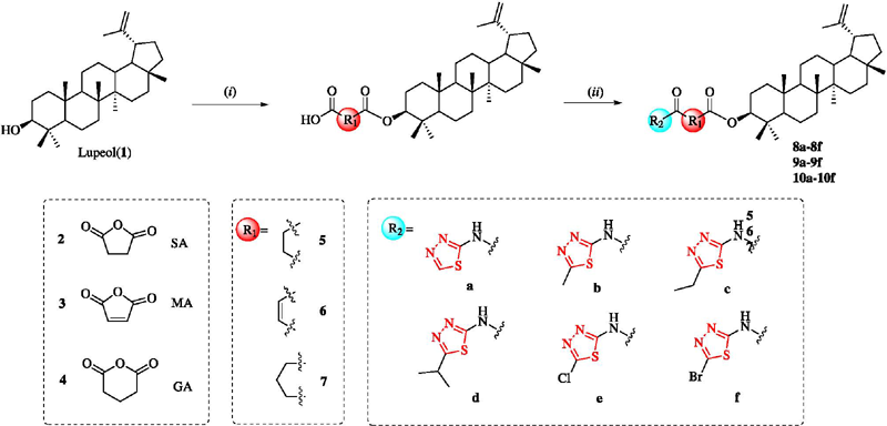
The synthetic routes for the preparation of intermediates 5, 6, and 7 and the target compounds 8a-8f, 9a-9f, and 10a-10f are illustrated in Scheme 1. Starting from lupeol (1), acylation reactions with succinic anhydride (SA), maleic anhydride (MA), or glutaric anhydride (GA) were catalyzed by triethylamine, successively yielding intermediates 5, 6, and 7. In the presence of the coupling reagent PyBOP and triethylamine, intermediates 5, 6, and 7 were reacted with a series of thiazolyl-amino substituents in dichloromethane to afford the three series of corresponding target compounds 8a-8f, 9a-9f, and 10a-10f.

Synthesis of lupeol derivatives 8a-8f, 9a-9f, and 10a-10f. Reagents and conditions: (i) 2 (SA), 3 (MA), 4 (GA), DCM, Et3N, room temperature, 48 h, 63-78%; (ii) thiazolyl-amino substituents, Et3N, PyBOP, room temperature, 4 h, 65-77%.
All synthesized compounds were structurally characterized using HRMS, 1H and 13C NMR spectroscopy. Taking compound 8a as an exemplar, the hydrogen atoms of the lupeol skeleton were predominantly observed within the 1H NMR spectrum ranging from 0.70 to 2.40 ppm. Four sets of peaks between 0.76 and 1.02 ppm corresponded to the hydrogen atoms of the methyl groups on the lupeol, while the peak at 4.49 ppm represented the hydrogen atom at the C-3 position of the lupeol. The presence of these peaks confirmed the structure of the lupeol. Additionally, two sets of peaks at 2.83 and 3.10 ppm corresponded to the four hydrogen atoms of the succinic anhydride side chain, and the peak at 8.78 ppm represented a hydrogen atom on the thiadiazole ring of the side chain. The existence of these peaks verified the successful conjugation of a succinic anhydride side chain, containing a thiadiazole moiety, to the C-3 skeleton of lupeol. Concurrently, in the corresponding 13C NMR spectrum, peaks at 147.33 and 160.54 ppm represented the two carbon atoms within the thiadiazole structure, while peaks at 29.82, 30.97, 170.68, and 171.84 ppm corresponded to the four carbon atoms of the succinic anhydride side chain. Furthermore, HRMS analysis of the synthesized product revealed a molecular weight of [M + Na]+ 632.3862, which was consistent with the predicted value. Based on this information, it could be ascertained that the synthesized product corresponded to the target compound 8a.
In vitro anti-proliferative activity
The anti-proliferative activity of eighteen derivatives (8a-8f, 9a-9f, and 10a-10f) was evaluated using the MTT assay in a panel of four cancer cell lines: A549, Hep G2, MCF-7, and MDA-MB-231. Cisplatin was employed as the positive control drug, while the parent drug was lupeol. The results are presented in Table 1.
Compared to lupeol, the majority of the compounds demonstrated stronger inhibitory effects against the four types of cancer cells, with particularly notable activity against the A549 cell line. The anti-proliferative activity of compounds 9a-9f against the A549 cell line was significantly higher than that of compounds 8a-8f and 10a-10f. Notably, compound 9c showed excellent inhibitory activity (IC50 = 4.12 µM), making it 8.7 times more active than lupeol (IC50 = 35.86 µM). Importantly, compound 9c displayed a favorable selectivity against MRC-5 (IC50 = 75.29 µM), resulting in a selectivity index of 18.27 (IC50MRC-5/IC50A549).
The current data suggest that the majority of lupeol derivatives with an ester chain at the C-3 of lupeol exhibit improved inhibitory activity against tumor cell lines, as compared to lupeol. The structure-activity relationship analysis indicated that compounds featuring a single thiadiazole ring (8a, 9a, 10a) showed enhanced inhibitory activity, with IC50 values ranging from 5.81 to 8.99 µM. In contrast, the introduction of a chlorine atom into the thiadiazole compounds (8e, 9e, 10e) resulted in weaker inhibitory activity, with IC50 values between 10.34 and 12.48 µM. The introduction of methyl, ethyl, or propyl groups into the thiadiazole compounds (8c, 8d, 9b, 9c, 10b, 10d) also enhanced anti-proliferative activity, indicating that the presence of electron-donating groups could effectively increase the inhibitory effect.
Furthermore, the linker influenced the anti-proliferative activity of the compounds. Lupeol derivatives with an MA linker (9a-9f) exhibited stronger inhibitory effects than those with SA (8a-8f) or GA (10a-10f), suggesting that the enone structure could also enhance anti-proliferative activity.
Compound 9c inhibits GLS1 activity
Based on the strong inhibitory effect of compound 9c on A549 cells, it was necessary to explore its inhibitory effect on GLS1. The inhibitory activity of compound 9c on GLS1 was assessed by employing a GLS1 inhibitor screening kit. Lupeol and BPTES were utilized as references. Both compound 9c and lupeol were found to suppress the activity of GLS1 (Figure 3a). But, the inhibitory effect of compound 9c on GLS1 in A549 cells (IC50 = 6.71 μM) was significantly enhanced compared with that of lupeol (IC50 = 49.74 μM). It is worth noting that compound 9c has an inhibitory effect similar to that of BPTES (IC50 = 4.35 µM). Meanwhile, Western blot analysis also obtained the similar results (Figures 3b and 3c). According to the results, as the concentration of compound 9c increased, the level of GLS1 expression gradually decreased, indicating that the inhibitory effect on GLS1 was dose-dependent. Therefore, as a new antitumor candidate drug, compound 9c is worthy of further study.

Effect of compound 9c on GLS1 activity in A549 cells. (a) The levels of GLS1 for lupeol, compound 9c, and BPTES were determined by utilizing a GLS1 inhibitor screening kit. (b) A549 cells were treated with various concentrations of compound 9c for 48 h, and the expression level of GLS1 protein was detected through western blot analysis. (c) Quantitative analysis was performed. The data were presented as the mean ± SD (n = 3), *p < 0.05, **p < 0.01, ***p < 0.001, compared with the control group.
Compound 9c arrests cell cycle
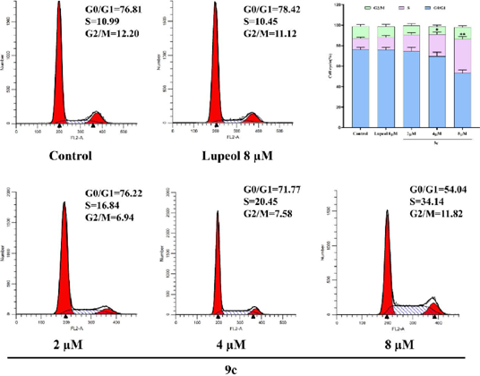
Glutamine is an anaplerotic carbon source that plays a significant role in the cell cycle, and cancer cells lacking glutamine undergo cell cycle arrest.46 The impact of compound 9c on the cell cycle distribution of A549 cells was determined through flow cytometry. The results, as depicted in Figure 4, demonstrate that compound 9c induces S-phase cell cycle arrest in a dose-dependent manner. The percentage of cells in the S phase ranges from 10.99 to 34.14% as the concentration of compound 9c increases from 0 to 8 μM, while the percentage of cells in the G0/G1 phase concomitantly decreased.

Effect of compound 9c and lupeol on cell cycle distribution in A549 cells. The data were expressed as the mean ± SD (n = 3); *p < 0.05, **p < 0.01, compared with the control group.
Compound 9c inhibits colony formation and induces apoptosis in A549 cells
To confirm the effects of compound 9c on the inhibition of A549 cells proliferation, we used different concentrations of compound 9c and lupeol to treat A549 cells. The results are shown in Figure 5a, where it can be seen that compound 9c markedly inhibited the proliferation of A549 cells, with the inhibitory capacity being much greater than that of lupeol. Compared to lupeol (8 μM), the proliferation inhibition rate of compound 9c (8 μM) against A549 cells reached 81.21%. The effect of compound 9c on the apoptotic rate of the A549 cells was assessed by employing an AV/PI kit (Figure 5b). The percentage of apoptotic A549 cells treated with the compound increased from 3.49% in the blank group to 17.8, 24.76, and 57.6% at concentrations of 2, 4, and 8 μM, respectively. In conclusion, compound 9c induced cell apoptosis in a manner that is dose-dependent.

Effect of compound 9c on cell proliferation of A549 cells. (a) Compound 9c inhibited the formation of A549 cell colony. (b) Compound 9c promoted the apoptosis of A549 cells, detected by flow cytometry. Data are expressed as average ± SD (n = 3); *p < 0.05, **p < 0.01, ***p < 0.001, compared with the control group.
Compound 9c inhibits glutamate production in A549 cells
The first step in glutamine metabolism is the GLS1-catalyzed conversion of glutamine to glutamate, which subsequently enters the tricarboxylic acid cycle. Therefore, we used the Chekine™ glutamate assay kit to detect the glutamate content in A549 cells. The experimental findings indicate that compound 9c decreased the production of glutamate in A549 cells in a dose-dependent manner (as shown in Figure 6). When compared with lupeol, compound 9c demonstrated a more potent inhibitory effect on glutamate in A549 cells. These results imply that compound 9c can effectively inhibit GLS1, consequently blocking the glutamine hydrolysis pathway and inducing cell death.

Effect of compound 9c and lupeol on glutamate levels in A549 cells. Data are expressed as average ± SD (n = 3); **p < 0.01, compared with the lupeol group.
Compound 9c induces ROS generation
Tumor cells often have their ROS levels influenced by glutamine. Consequently, the regulation of glutamine metabolism is of crucial importance for maintaining ROS homeostasis. Thus, DCFH-DA staining was employed to examine changes of ROS content in A549 cells. The confocal microscopy results (Figure 7a) revealed that the brightness of the ROS fluorescence progressively increased in the group treated with compound 9c when compared to the control group. This was further corroborated by the FCM results (Figure 7b), which demonstrated an increase in the intracellular ROS content as the drug concentration rose. These findings suggest that compound 9c, functioning as a GLS1 inhibitor, can effectively inhibit glutamine hydrolysis, thereby resulting in an elevation of ROS.

Effect of compound 9c on ROS content of A549 cells. (a) Fluorescence microscopy image of intracellular ROS production in A549 cells stained with DCFH-DA (green). (b) Flow cytometry analysis. (c) Merge. (d) Quantitative analysis. The data were expressed as the mean ± SD (n = 3); *p < 0.05, **p < 0.01, ***p < 0.001, compared with the control group.
Molecular docking studies and binding pattern analysis
The experimental results clearly indicate that compound 9c is capable of serving as a novel inhibitor of GLS1. On this basis, we carried out a molecular docking study for a more in-depth exploration of the potential binding mode between compound 9c and GLS1 (PDB: 3UO9). The analysis showed that, from the carbonyl group of compound 9c, oxygen forms a hydrogen bond with the hydroxyl group of Ser314, and the amino group of the substituents forms a hydrogen bond with the carbonyl group of Asp467, further stabilizing compound 9c in the binding pocket of GLS1 (Figure 8). It is noteworthy that the binding energy of the protein-ligand complex amounted to -9.65 kcal mol-1. Compared with lupeol (−7.76 kcal mol-1), compound 9c has a higher binding affinity, fully demonstrating its high activity.

Docking results of compound 9c with GLS1 (PDB ID: 3UO9): (a) 9c shown with the protein surface representation of GLS1, (b) key interactions of 9c with GLS1, (c) lupeol shown with the protein surface representation of GLS1, (d) key interactions of lupeol with GLS1. Here, 9c and lupeol were rendered as sticks and were colored according to atomic type, with yellow indicating carbon, blue indicating nitrogen, and red indicating oxygen. The key residual atoms in GLS1 that interact with the compound were represented by green bars. The red dotted line represents the hydrogen bonds.
Conclusions
In conclusion, a series of potential GLS1 inhibitors based on lupeol were designed, and synthesized. These compounds display high anti-proliferative activity against A549 cells. The inhibitory effect of compound 9c on GLS1 is stronger than that of lupeol and similar to that of BPTES. Compound 9c inhibits glutamine metabolism, resulting in a deficiency in GSH levels, the accumulation of ROS, and cell cycle arrest in the S phase, ultimately leading to cell apoptosis. In molecular docking studies, compound 9c binds to GLS1, indicating a good binding capability and having significantly lower binding energy than lupeol. Therefore, as a novel GLS1 inhibitor, compound 9c holds good application prospects in the treatment of NSCLC and is worthy of further development.
Supplementary Information
Supplementary file (containing the NMR and HRMS charts for the synthesized compounds) is available free of charge at https://jbcs.sbq.org.br as PDF file.PDF
Acknowledgments
This work was funded by Heilongjiang Province Postdoctoral Research Fund Project (LBH-QY24003).
References
-
1 Stine, Z. E.; Schug, Z. T.; Salvino, J. M.; Dang, C. V.; Nat. Rev. Drug Discovery 2022, 21, 141. [Crossref]
» Crossref -
2 Campos-Contreras, A. D. R.; Díaz-Muñoz, M.; Vázquez-Cuevas, F. G.; Cells 2020, 9, 1612. [[Crossref]
» Crossref -
3 Bergers, G.; Fendt, S. M.; Nat. Rev. Cancer 2021, 21, 162. [Crossref]
» Crossref -
4 Jin, J.; Byun, J. K.; Choi, Y. K.; Park, K. G.; Exp. Mol. Med. 2023, 55, 706. [Crossref]
» Crossref -
5 Yuneva, M.; Zamboni, N.; Oefner, P.; Sachidanandam, R.; Lazebnik, Y.; J. Cell. Biol. 2007, 178, 93. [Crossref]
» Crossref -
6 Lacey, J. M.; Wilmore, D. W.; Nutr. Rev. 1990, 48, 297. [Crossref]
» Crossref -
7 Mayers, J. R.; Heiden, M. G. V.; Trends Biochem. Sci. 2015, 40, 130. [Crossref]
» Crossref -
8 Cyriac, R.; Lee, K.; J. Enzyme Inhib. Med. Chem. 2024, 39, 2290911. [Crossref]
» Crossref -
9 Zhu, L.; Zhu, X.; Wu, Y.; Biomolecules 2022, 12, 580. [Crossref]
» Crossref -
10 Jewell, J. L.; Kim, Y. C.; Russell, R. C.; Yu, F. X.; Park, H. W.; Plouffe, S. W.; Tagliabracci, V. S.; Guan, K. L.; Science 2015, 347, 194. [Crossref]
» Crossref -
11 Saha, S. K.; Islam, S. M. R.; Abdullah-Al-Wadud, M.; Islam, S.; Ali, F.; Park, K. S.; J. Clin. Med. 2019, 8, 355. [Crossref]
» Crossref -
12 Katt, W. P.; Lukey, M. J.; Cerione, R. A.; Future Med. Chem. 2017, 9, 223. [Crossref]
» Crossref -
13 Xu, X.; Meng, Y.; Li, L.; Xu, P.; Wang, J.; Li, Z.; Bian, J.; J. Med. Chem. 2019, 62, 1096. [Crossref]
» Crossref -
14 Shukla, K.; Ferraris, D. V.; Thomas, A. G.; Stathis, M.; Duvall, B.; Delahanty, G.; Alt, J.; Rais, R.; Rojas, C.; Gao, P.; Xiang, Y.; Dang, C. V.; Slusher, B. S.; Tsukamoto, T.; J. Med. Chem. 2012, 55, 10551. [Crossref]
» Crossref -
15 Zhou, Y.; Eid, T.; Hassel, B.; Danbolt, N. C.; Neurochem. Int. 2020, 140, 104809. [Crossref]
» Crossref -
16 Jiang, B.; Zhang, J.; Zhao, G.; Liu, M.; Hu, J.; Lin, F.; Wang, J.; Zhao, W.; Ma, H.; Zhang, C.; Wu, C.; Yao, L.; Liu, Q.; Chen, X.; Cao, Y.; Zheng, Y.; Zhang, C.; Han, A.; Lin, D.; Li, Q.; Mol. Cell 2022, 82, 1821. [Link] accessed in February 2025
» Link -
17 Gross, M. I.; Demo, S. D.; Dennison, J. B.; Chen, L.; Chernov-Rogan, T.; Goyal, B.; Janes, J. R.; Laidig, G. J.; Lewis, E. R.; Li, J.; Mackinnon, A. L.; Parlati, F.; Rodriguez, M. L.; Shwonek, P. J.; Sjogren, E. B.; Stanton, T. F.; Wang, T.; Yang, J.; Zhao, F.; Bennett, M. K.; Mol. Cancer Ther. 2014, 13, 890. [Crossref]
» Crossref -
18 Soth, M. J.; Le, K.; Di Francesco, M. E.; Hamilton, M. M.; Liu, G.; Burke, J. P.; Carroll, C. L.; Kovacs, J. J.; Bardenhagen, J. P.; Bristow, C. A.; Cardozo, M.; Czako, B.; de Stanchina, E.; Feng, N.; Garvey, J. R.; Gay, J. P.; Do Geck, M. K.; Greer, J.; Han, M.; Harris, A.; Herrera, Z.; Huang, S.; Giuliani, V.; Jiang, Y.; Johnson, S. B.; Johnson, T. A.; Kang, K.; Leonard, P. G.; Liu, Z.; McAfoos, T.; Miller, M.; Morlacchi, P.; Mullinax, R. A.; Palmer, W. S.; Pang, P.; Rogers, N.; Rudin, C. M.; Shepard, H. E.; Spencer, N. K.; Theroff, J.; Wu, Q.; Xu, A.; Yau, Y. A.; Draetta, G.; Toniatti, C.; Heffernan, T. P.; Jones, P.; J. Med. Chem. 2020, 63, 12957. [Crossref]
» Crossref -
19 Shapiro, R. A.; Morehouse, R. F.; Curthoys, N. P.; Biochem J. 1982, 207, 561. [Crossref]
» Crossref -
20 Xu, X.; Wang, J.; Wang, M.; Yuan, X.; Li, L.; Zhang, C.; Huang, H.; Jing, T.; Wang, C.; Tong, C.; Zhou, L.; Meng, Y.; Xu, P.; Kou, J.; Qiu, Z.; Li, Z.; Bian, J.; J. Med. Chem. 2021, 64, 4588. [Crossref]
» Crossref -
21 Lee, E. J.; Duggirala, K. B.; Lee, Y.; Yun, M. R.; Jang, J.; Cyriac, R.; Jung, M. E.; Choi, G.; Chae, C. H.; Cho, B. C.; Lee, K.; Bioorg. Med. Chem. Lett. 2022, 75, 128956. [Crossref]
» Crossref -
22 Shen, Y. A.; Hong, J.; Asaka, R.; Asaka, S.; Hsu, F. C.; Rahmanto, Y. S.; Jung, J. G.; Chen, Y. W.; Yen, T. T.; Tomaszewski, A.; Zhang, C.; Attarwala, N.; DeMarzo, A. M.; Davidson, B.; Chuang, C. M.; Chen, X.; Gaillard, S.; Le, A.; Shih, I. M.; Wang, T. L.; Cancer Res. 2020, 80, 4514. [Crossref]
» Crossref -
23 Reckzeh, E. S.; Karageorgis, G.; Schwalfenberg, M.; Ceballos, J.; Nowacki, J.; Stroet, M. C. M.; Binici, A.; Knauer, L.; Brand, S.; Choidas, A.; Strohmann, C.; Ziegler, S.; Waldmann, H.; Cell Chem. Biol. 2019, 26, 1214. [Link] accessed in February 2025
» Link -
24 Okabe, S.; Tanaka, Y.; Moriyama, M.; Gotoh, A.; Cancer Drug Resist. 2024, 7, 25. [Crossref]
» Crossref -
25 Luo, R.; Zhao, H.; Deng, S.; Wu, J.; Wang, H.; Guo, X.; Han, C.; Ren, W.; Han, Y.; Zhou, J.; Lin, Y.; Bu, M.; Molecules 2024, 29, 4375. [Crossref]
» Crossref -
26 Luo, Z.; Yin, F.; Wang, X.; Kong, L.; Chin. J. Nat. Med. 2024, 22, 195. [Crossref]
» Crossref -
27 Ferreira, M. J.; Int. J. Mol. Sci. 2023, 24, 14321. [Crossref]
» Crossref -
28 Shen, Y.; Hao, X.; Sci. China: Life Sci. 2020, 63, 1634. [Crossref]
» Crossref -
29 Wang, L.; Tian, S.; Deng, S.; Wu, J.; Wang, H.; Guo, X.; Han, C.; Ren, W.; Han, Y.; Zhou, J.; Lin, Y.; Bu, M.; Bioorg. Chem. 2024, 153, 107862. [Crossref]
» Crossref -
30 Duan, M.; Mahal, A.; Mohammed, B.; Zhu, Y.; Tao, H.; Mai, S.; Al-Haideri, M.; Zhu, Q.; Nat. Prod. Res. 2022, 36, 5268. [Crossref]
» Crossref -
31 Mahal, A.; Wu, P.; Jiang, Z. H.; Wei, X.; ChemistrySelect 2019, 4, 366. [Crossref]
» Crossref -
32 Mahal, A.; Wu, P.; Jiang, Z. H.; Wei, X.; Nat. Prod. Bioprospect. 2017, 7, 461. [Crossref]
» Crossref -
33 Duan, M.; Mahal, A.; Alkouri, A.; Wang, C.; Zhang, Z.; Ren, J.; Obaidullah, A. J.; Molecules 2024, 29, 3010. [Crossref]
» Crossref -
34 Saleem, M.; Cancer Lett. 2009, 285, 109. [Crossref]
» Crossref -
35 Cho, W. C.; Leung, K. N.; Cancer Lett. 2007, 252, 43. [Crossref]
» Crossref -
36 Jian, B.; Zhang, H.; Han, C.; Liu, J.; Molecules 2018, 23, 387. [Crossref]
» Crossref -
37 Park, J. S.; Rehman, I. U.; Choe, K.; Ahmad, R.; Lee, H. J.; Kim, M. O.; Nutrients 2023, 15, 3059. [Crossref]
» Crossref -
38 Bhatt, M.; Patel, M.; Adnan, M.; Reddy, M. N.; Anticancer Agents Med. Chem. 2021, 21, 201. [Crossref]
» Crossref -
39 Bociort, F.; Macasoi, I. G.; Marcovici, I.; Motoc, A.; Grosu, C.; Pinzaru, I.; Petean, C.; Avram, S.; Dehelean, C. A.; Curr. Oncol. 2021, 28, 5054. [Crossref]
» Crossref -
40 Eldohaji, L. M.; Fayed, B.; Hamoda, A. M.; Ershaid, M.; Abdin, S.; Alhamidi, T. B.; Mohammad, M. G.; Omar, H. A.; Soliman, 5. S. M.; Arch. Pharm. 2021, 354, e2100120. [Crossref]
» Crossref -
41 Chen, Z.; Luo, R.; Xu, T.; Wang, L.; Deng, S.; Wu, J.; Wang, H.; Lin, Y.; Bu, M.; Bioorg. Med. Chem. 2024, 113, 117934. [Crossref]
» Crossref -
42 Tian, S.; Zhao, Y.; Deng, S.; Hou, L.; Song, J.; Wang, M.; Bu, M.; Molecules 2024, 29, 3990. [Crossref]
» Crossref - 43 IBM SPSS Statistics, version 24.0; SPSS Inc., Chicago, IL, USA, 2016.
-
44 Morris, G. M.; Huey, R.; Lindstrom, W.; Sanner, M. F.; Belew, R. K.; Goodsell, D. S.; Olson, A. J.; J. Comput. Chem. 2009, 16, 2785. [Crossref]
» Crossref -
45 Schrödinger, L.; DeLano, W.; PyMOL; DeLano Sciences, San Francisco, CA, USA, 2020. [Link] accessed in February 2025
» Link -
46 Saqcena, M.; Mukhopadhyay, S.; Hosny, C.; Alhamed, A.; Chatterjee, A.; Foster, D. A.; Oncogene 2015, 34, 2672. [Crossref]
» Crossref
Edited by
-
Editor handled this article:
Albertina Moglioni (Associate)