 Natural Compounds of Curcumin Against the Main Protease and Papain like Protease of SARS-CoV-2, a Possible Anti-COVID-19 Drug: Docking and Molecular Dynamics Simulation Studies
Abstract
Coronavirus disease 2019 (COVID-19) is caused by severe acute respiratory syndrome coronavirus 2 (SARS-CoV-2). Computational techniques, such as molecular docking, are essential for drug repurposing and screening to identify potential treatments for the virus. This approach enables the investigation of interactions between a ligand and a key protein. In this study, in silico methods, including molecular docking and molecular dynamics (MD) simulations, were employed to evaluate the antiviral potential of 10 natural curcumins against SARS-CoV-2. Density functional theory (DFT) calculations were also performed to analyze the structural properties of the selected ligands. Our findings suggest that the curcumins demethoxycurcumin, bisdemethoxycurcumin, and cassumunin B are promising inhibitors of the SARS-CoV-2 3CLPro protein.
Keywords:
docking; molecular dynamics simulations; SARS-COV2; curcumin; DFT
Introduction
Coronavirus disease 2019 (COVID-19), caused by severe acute respiratory syndrome coronavirus 2 (SARS CoV-2), was first reported in the Chinese province of Wuhan and has spread rapidly around the world.1,2 On February 11, 2020, this virus strain, formerly referred to as 2019-nCoV (or Wuhan virus), was officially designated severe acute respiratory syndrome coronavirus 2 (SARS CoV-2)3 due to the similarity of its ribonucleic acid (RNA) genome to that of the previously identified SARS coronavirus (SARS-CoV).4,5 On March 11, 2020, the World Health Organization6 (WHO) recognized the pandemic. In addition, the WHO played an important role in suggesting provisional guidelines for laboratories carrying out tests for the new outbreak, as well as guidelines for the infection prevention and control.7 This new virus belongs to the coronaviridae family and has a spherical shape. It mainly spreads through the saliva, droplets or nasal secretions of an infected individual after coughing or sneezing.8 These droplets can spread to 1-2 m and can remain viable on surfaces for days.9
Since its emergence in late 2019, more than 200 countries face a health crisis due to the epidemiological disease COVID-19 caused by the SARS-CoV-2 virus.1 It is having a very high impact on the world economy and the global health sector. Computational techniques such as molecular docking are essential for the reuse and planning of drugs for the treatment of the virus. This technique allows and offers an investigation of the interaction between a ligand and a key protein.10-13 In addition to being a tool that promotes an efficient search process, greatly reducing expenses with equipment and reagents, it also obtains short-term results from the biological action of a particular compound.14,15 Molecular dynamics (MD), on the other hand, provides a pathway to dynamical properties of a system; transport coefficients, time-dependent responses to perturbations, spectra, rheological properties and others.16,17
Curcumins, substances produced by the plant species Curcuma longa, have received great attention in traditional medicines and science due to their antioxidant, anti-inflammatory, antidiabetic, antiviral, antimicrobial, anticancer, immunoregulatory, cardio and hepatoprotective as well as neuroprotective properties.18-21 In particular, curcumin can not only interact with the spike proteins but also inhibit important signaling pathways in the viral infection. It also appears to regulate inflammatory response.21
This study aims to elucidate the optimal binding orientation and molecular affinity of ten curcumin derivatives against the 3CLpro22 (PDB ID: 5R84) and PLpro23 (PDB ID: 7NFV) proteases of SARS-CoV-2 through molecular docking and MD simulations. For the 3CLpro analyses, PDB ID 5R84 was selected as the structural model. Although more recent crystallographic entries are available, 5R84 was chosen because the objective of this work is to conduct an exploratory docking analysis, focusing on binding orientations and molecular interactions within the protease active site. Its established use in the literature ensures methodological consistency and facilitates comparison with published results.24 These proteases are recognized as key therapeutic targets, and notably, Pfizer Nirmatrelvir, a 3CLpro inhibitor, has already advanced to clinical trials.
Methodology
DFT calculations
The ligands were geometrically optimized using the density functional theory (DFT) method with the exchange-correlation functional B3LYP with the 6-311G(d,p) basis set. The calculations were carried out using the Gaussian 09 software.25
Protein preparation
The proteins of interest in this study were the main protease (3CLpro) and papain-like protease (PLPro). The 3D structures of these proteins were obtained from the RCSB Protein Data Bank. Visualization and preprocessing of the protein structures, including the removal of water molecules, ions, and ligands, were carried out using UCSF Chimera.26
Molecular docking, binding affinity, and Ki values
Molecular docking procedure: the PDB files of both ligands and proteins were converted into an extended PDB format called PDBQT to perform molecular docking analysis using AutoDock 4.0 (Molecular Graphics Laboratory, The Scripps Research Institute; MGLTools 1.5.6, 2009). The docking process was performed with the grid box size of 60 × 60 × 60 and spacing of 0.375 Å. The output pdbqt files were written to a config. (configuration) file. The conformation with the lowest binding energy was considered as the most stable conformations of the ligand relative to the macromolecule. The results were analyzed by using the free version of the Biovia Discovery Studio 2020 client.27,28 Molecular docking was applied to analyze the binding affinity and calculate the theoretical inhibitory constant Ki (µM) of the screened ligand with the target proteins.29
Protein preparation and grid generation to docking study
The 3D structure of the proteins was downloaded from RCSB: Protein Databank. Then polar hydrogen bonds and missing hydrogen atoms were added, while water molecules and heteroatoms were excluded.15,29 Energy minimization was performed with a default constraint of 0.3 Å root mean square (RMS), and charges were assigned. After protein preparation, the clean structure was saved as a pdbqt file. A grid box of 60 × 60 × 60 and 0.375 Å spacing was generated around the centroid of inhibitor/drug compounds with assigned x, y, and z-axis.
Protein-ligand interaction studies
MD simulation
The ligand-receptor complexes with the best docking results were used to construct molecular dynamics simulation systems using the CHARMM-GUI server30,31 with the CHARMM36m force field.32 The structure was then placed in a cubic box, keeping the protein in the center at a distance of 7.0 Å from the edges to avoid interactions between the protein and itself. Subsequently, the box was solvated with water using the TIP3P model,33 to which 0.15 mol L-1 KCl was added to neutralize the complex charge. The minimization, equilibration, and production run steps were performed using GROMACS 2024.3 software.34 Energy minimization was performed using the steepest-descent algorithm with positional constraints on hydrogen bonds and sufficient steps to achieve an energy lower than 100 kJ mol-1 nm-1. The equilibration step, corresponding to the canonical NVT ensemble,35 was performed, in which the number of molecules, volume, and system temperature were kept constant.36 The system temperature was controlled using a V-rescale thermostat,37 which applies a velocity scaling factor based on the difference between the current system temperature and the desired temperature, adjusting the kinetic energy of the particles and maintaining a constant temperature of 310.15 K. Finally, all MD simulations were conducted for 150 ns.
Drug likeliness
The physicochemical parameters of the most promising natural spices such as SARS-CoV-238 Mpro inhibitors were predicted using the online Molinspiration Cheminformatics software.39 The predicted parameters included the number of rotatable bonds (nrotb), number of hydrogen bond acceptors (nON), number of hydrogen bond donors (nOHNH), n-octanol/water partition coefficient (P), molecular weight (MW), molecular volume (MVol), topological polar surface area (TPSA) and percent absorption (ABS). The ABS value in percentage was estimated as follows:19,40
Distance analysis
To investigate the stability of ligand binding within the active site, a distance analysis was performed following MD simulations. The approach consisted of monitoring the time-dependent variation in the distance between key catalytic residues and the bound ligands throughout the simulation trajectory.
Molecular mechanics Poisson-Boltzmann surface area (MMPBSA) analysis
To evaluate the thermodynamic stability of the simulated models, the MMPBSA approach was applied. Specifically, the gmx_MMPBSA tool was utilized to process trimmed trajectories derived from the GROMACS molecular dynamics simulations.41 This analysis yielded detailed estimates for several thermodynamic terms, including van der Waals and electrostatic interactions, the internal energy of the complex, and both polar and non-polar solvation free energies.42 The overall binding affinity of the protein-ligand complexes was determined using the following standard thermodynamic equation:
In this equation, ΔGbind represents the total binding free energy, Gcomplex the total energy of the bound protein-ligand system, Greceptor the energy of the isolated protein, and Gligand the energy of the free small molecule. The analysis was performed over the last 100 ns of the simulation in a 5-frame interval, totaling 200 frames, with the complex trajectory being fitted in GROMACS, using both the receptor and the complete ligand as a reference.43,44
Results and Discussion
Optimized molecular structures of the ligands
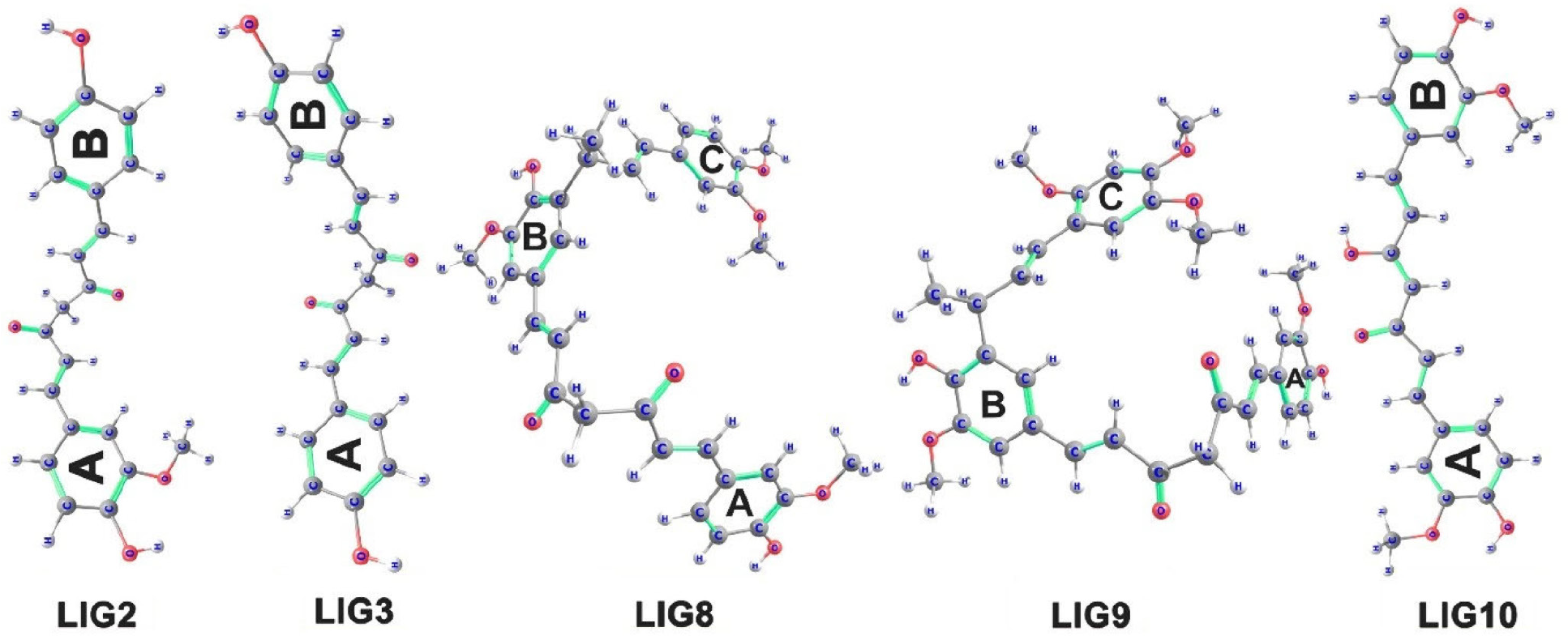
Ten natural curcumins were evaluated against the SARS CoV-2 virus (Table S1, in the Supplementary Information (SI) section). However, only five ligands showed interactions with the active sites of the target proteins. Figure 1 illustrates the optimized molecular structures of these ligands:
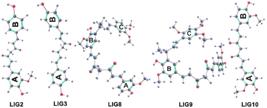
Optimized structures of the ligand molecules that interacted with the binding site of the target proteins.
(i) LIG2 (demethoxycurcumin): (1E,6E)-1-(4-hydroxy-3-methoxyphenyl)-7-(4-hydroxyphenyl)hepta-1,6-diene-3,5-dione (C19H16O4).
(ii) LIG3 (bisdemethoxycurcumin): (1E,6E)-1,7-bis(4-hydroxyphenyl)hepta-1,6-diene-3,5-dione (C20H15O5).
(iii) LIG8 (cassumunin A): (1E,6E)-1-{3-[(2R,3E)-4-(3,4-dimethoxyphenyl)but-3-en-2-yl]-4-hydroxy-5-methoxyphenyl}-7-(4-hydroxy-3-methoxyphenyl)hepta-1,6-diene-3,5-dione (C33H34O8).
(iv) LIG9 (cassumunin B): (1E,6E)-1-{3-[(2R,3E)-4-(3,4-dimethoxyphenyl)but-3-en-2-yl]-4-hydroxy-5-methoxyphenyl}-7-(4-hydroxy-3-methoxyphenyl)hepta-1,6-diene-3,5-dione (C34H36O9).
(v) LIG10 (curcumin PE): (1E,4Z,6E)-5-hydroxy-1,7-bis(4-hydroxy-3-methoxyphenyl)hepta-1,4,6-trien-3-one (C21H20O6).
Protein-ligand interaction studies
Screening of inhibitors for main protease
According to Jin et al.,45 the main protease 3CLpro is a cysteine protease containing three domains, i.e., domains I (8-101 residues) and II (102-184 residues) with β-barrel antiparallel structure and the domain III (201-303 residues), consisting of five α-helices linked to domain II by a long loop (185-200 residues).45,46 The catalytic dyads CYS145 and HIS41, and the residue GLU166 are involved in protein dimerization, substrate cleaving through catalysis present in the cleft between domains I and II. The enzyme active site consists of six subunits (S1-S6), and the active site residues 140-145 and 163-166 are present in the domain II.
PLpro, on the other hand, folds in a right-handed architecture with features named fingers, thumb and palm domains. More importantly, it displays a catalytic triad consisting of residues Cys111, His272 and Asp286. There is also an N-terminal domain that is similar to ubiquitin.47
Protein and ligand interactions
Main protein 3CLpro (ID = 5r84) and ligand interactions
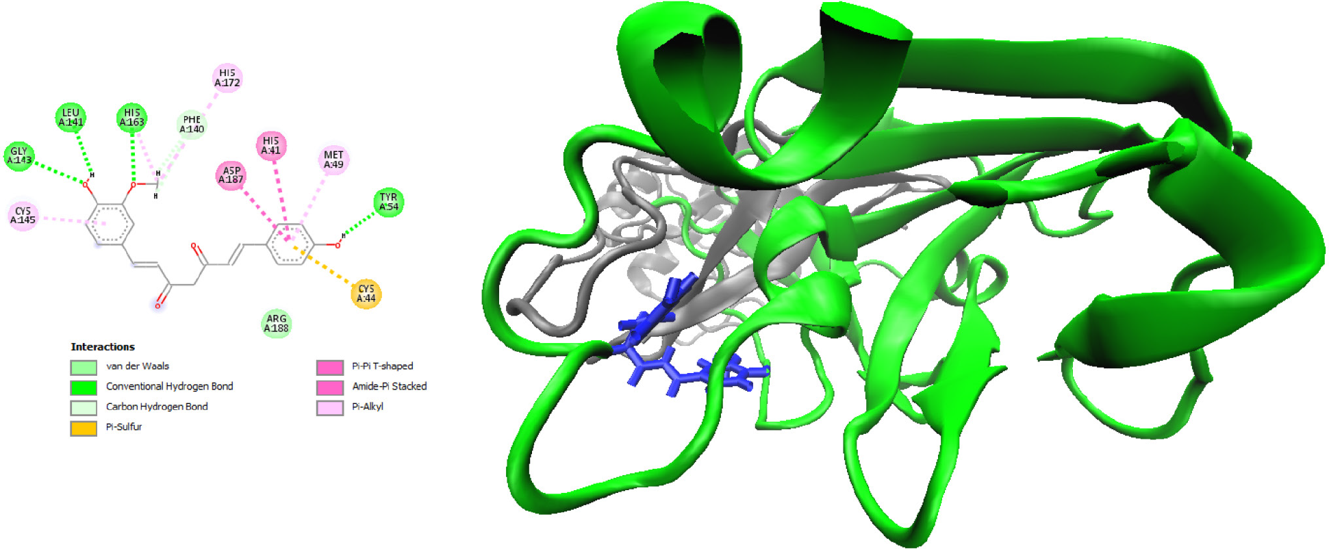
To analyze the detailed interaction of the best poses, the free version of the Biovia Discovery Studio Visualizer27,28 was used. The interaction of LIG2 with 3CLpro showed the highest affinity interaction in chain A (Figure 2) with ∆Gbiding and ∆Gdocking equal to -9.0 and -13.23 kcal mol-1, respectively. The ligand makes hydrogen bonding with TYR54, GLY143, LEU141 and HIS163 residues of the target protein. This binding affinity can also be attributed to interactions such as T-shaped π interactions with HIS41, amide π stacked with ASP 187 and π-sulfur with CYS44. The results on Table S1 (SI section) showed that all selected ligands exhibit good binding affinity with the target protein.
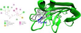
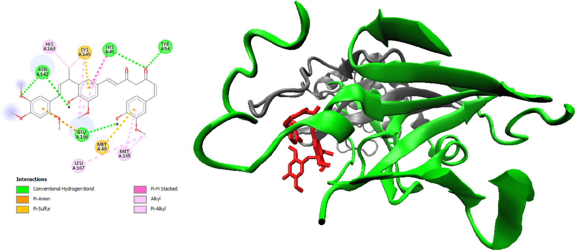
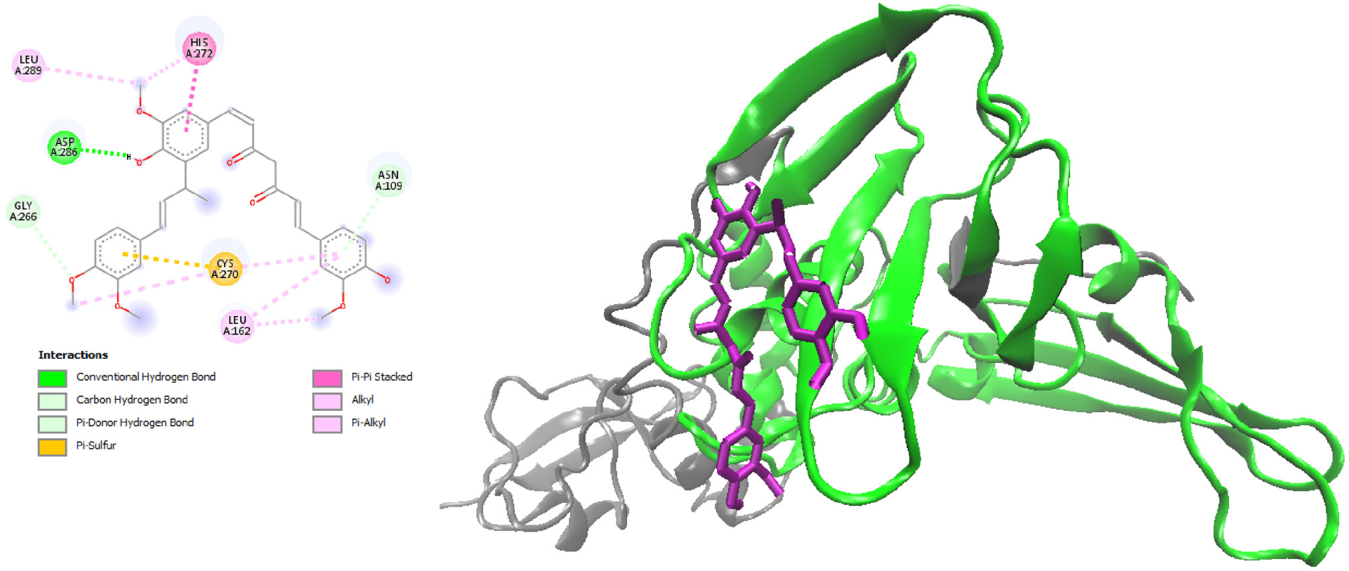
Regarding binding affinity, LIG2 showed the highest affinity, followed by LIG9 and LIG3. The interaction of LIG9 with 3CLpro exhibited the second-highest affinity interaction in chain A (Figure 3), with ∆Gbinding and ∆Gdocking values of -8.95 and -18.52 kcal mol-1, respectively. LIG9 forms hydrogen bonds with the HIS41, TYR54, ASN142, and GLU166 residues of the 3CLpro protease. This binding affinity is further supported by various interactions, including π-π stacking and π-alkyl interactions with HIS41, π-sulfur interactions with CYS145, and interactions with MET49. Figure 3 provides additional details on the other interactions between LIG9 and 3CLpro.
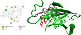
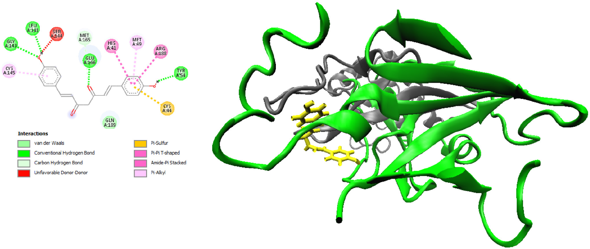
LIG3 exhibited the third-highest affinity interaction with chain A, with ∆Gbinding and ∆Gdocking values of -8.29 and -11.44 kcal mol-1, respectively (Figure 4). It forms hydrogen bonds with the TYR54, LEU141, GLY143, and GLU166 residues of 3CLpro. This strong binding affinity can also be attributed to additional interactions, including π-π stacking (ARG188), π-π alkyl (MET49, CYS145), π-π shaped (HIS41), and π-sulfur with CYS44. Figure 4 provides further details on the interactions between LIG3 and the target protein.
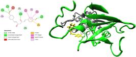
LIG8 interacted with the active site residue CYS145 through a π -sulfur interaction. It also formed hydrogen bonds with GLY143, HIS163, and ARG188, while interacting with MET49 and GLU166 via π-sulfur and π-anion interactions, respectively. LIG10 formed conventional hydrogen bonds with HIS41, GLU166, and ASP187, and π-sulfur interactions with CYS145. Additional details on interactions between ligands 1, 4, 5, 6, 7, 8, and 10 and the 3CLpro receptor are provided in the Supplementary Information. Table S2 (SI section) summarizes the interactions between these ligands and the 3CLpro protein.
As can be seen from these results, the best poses of these three ligands bind to residues on the active site of 3CLpro. More importantly, all ligands make close contacts or interactions with residues CYS145 and HIS41 involved in the catalytic function, that is: they could, in principle, act as 3CLpro inhibitors. To further investigate this possibility, several MD runs were performed to examine the stability of this protein-ligand complex. The docking data for 3CLpro indicated that the curcumins had a docking performance in the region of the active binding site. The results indicate that curcumins have a good affinity for the 3CLpro receptor and that the estimated binding values are similar to other ligands. In the works carried out by Reza et al.48 and Kushwaha et al.49 also found similar binding energies for various ligands and 3CLpro receptor interaction.
PLPro (ID = 7nfv and ligand interactions)
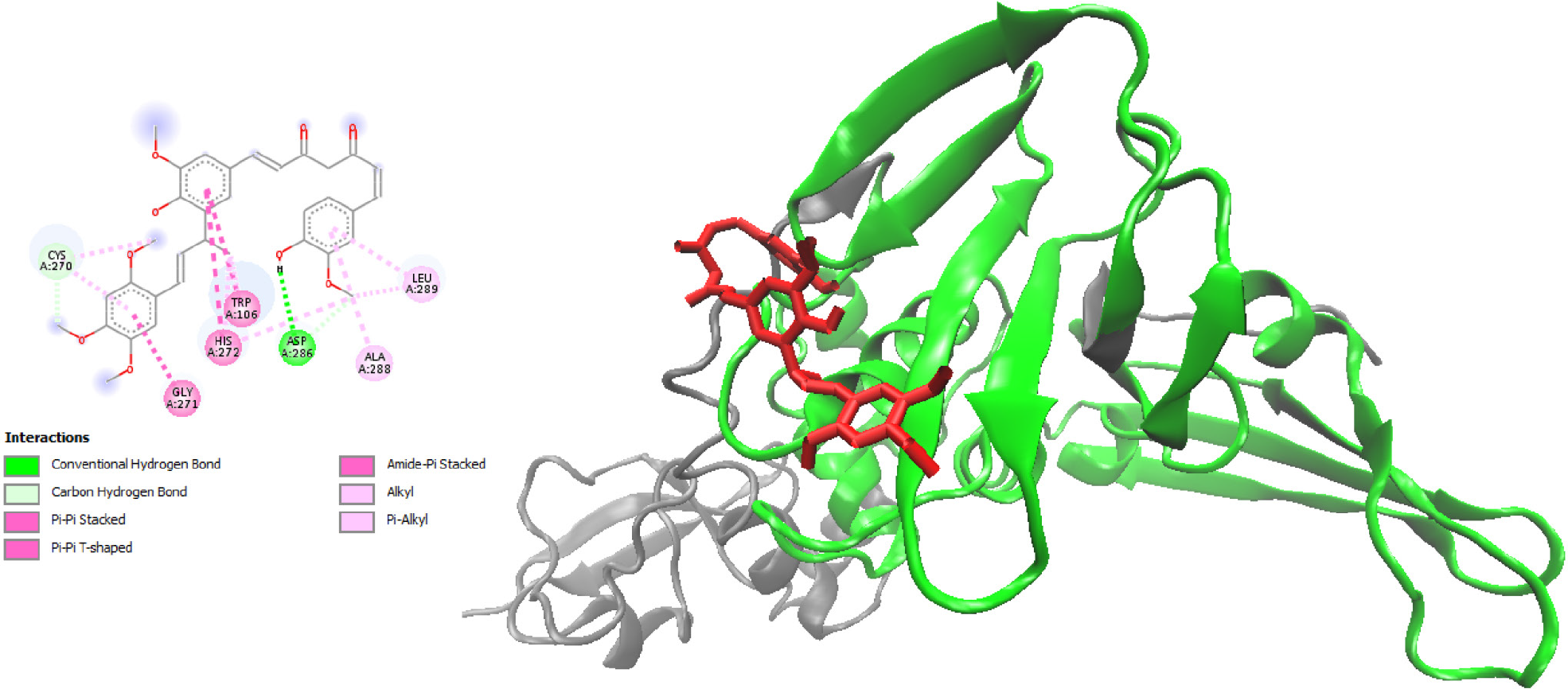
The interaction of LIG8 with PLPro exhibited the highest affinity interaction in chain A (Figure 5), with ∆Gbinding and ∆Gdocking values of -6.72 and -14.69 kcal mol-1, respectively. It forms a hydrogen bond with the ASP286 residue of the target protein. The interaction of cassumunin B (LIG9) with PLPro showed the second-highest affinity interaction in chain A (Figure 6), with ∆Gbinding and ∆Gdocking values of -6.67 and -15.35 kcal mol-1, respectively. The results in Table S3 (SI section) indicate that all selected ligands exhibit good binding affinity with the target protein PLPro.
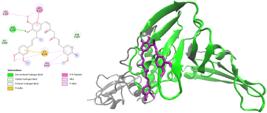
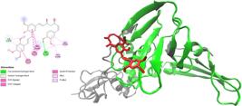
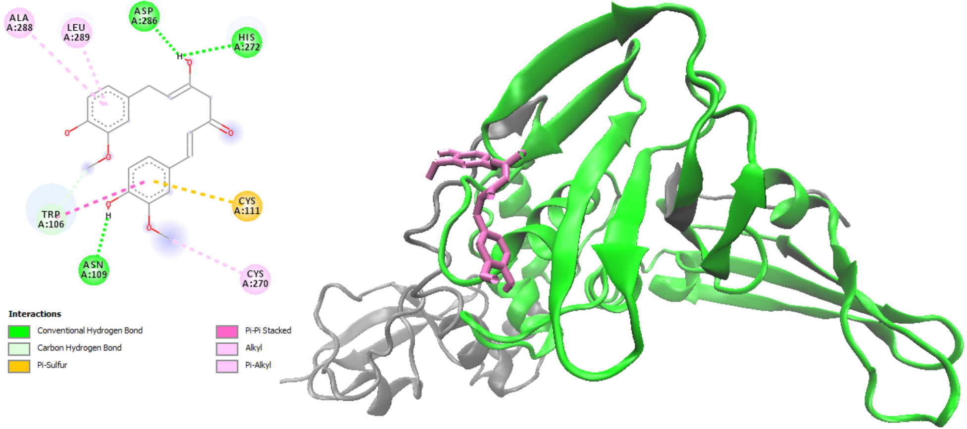
LIG10 showed the 3rd highest affinity interaction in the chain A with ∆Gbiding and ∆Gdocking equal to -6.26 and -11.08 kcal mol-1, respectively (Figure 6). It makes hydrogen bonding with the ASP286 residue of the protein (Figure 7). The interaction with HIS272 was via hydrogen bond and CYS111 was via π-sulfur. Table S4 (SI section) shows the interactions among ligands and the protein PLPro.
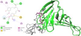
As before, the best poses of best three ligands bind to residues on the active site of PLPro and, in particular, to residues involved in the catalytic action and therefore can also act as inhibitors of this protease. The docking data for PLpro indicated that the curcumins had a reasonable docking performance in the region of the active binding site. In this case, the results indicate that curcumins have a lower affinity for PLPro (ID = 7nfv) than 3CLpro (ID = 5r84). Other authors as Baildya et al.,38 Reza et al.48 and Peele et al.50 also found similar behavior with other ligands.
Drug-likeness
Drug-likeness is a qualitative measure utilized in drug discovery to evaluate pharmacokinetic properties, such as oral bioavailability.51,52 Physicochemical parameters were evaluated using Molinspiration Cheminformatics,39 an online software calculation toolkit. The predicted parameters are summarized on Table 1. The permeability across the cell membrane, as measured by the Moriguchi logP value (MlogP), was less than five, indicating that these components have satisfactory membrane permeability. In addition, with the exception of ligands 7, 8 and 9, the other ligands had MW = 500 (molecular weight), suggesting that all of them must be promptly transferred, diffused, and absorbed. Another parameter indicating molecular bioabsorption is the topological polar surface area (TPSA), calculated as the sum of the surface of polar atoms or molecules, including oxygen, nitrogen, and bonded hydrogens. Molecules with a TPSA greater than 140 Å2 tend to be poor at permeating cell membranes, whereas a TPSA less than 90 Å2 is usually highly favorable. Except for ligand 7, the ligands have good cell membrane permeability and also a good level of oral bioavailability.
Predicted physiochemical parameters of the two identified natural spices as putative SARS-CoV-2 main protease (Mpro) inhibitors and their different structural descriptors
Molecular dynamics study
Root mean square fluctuation (RMSF) study
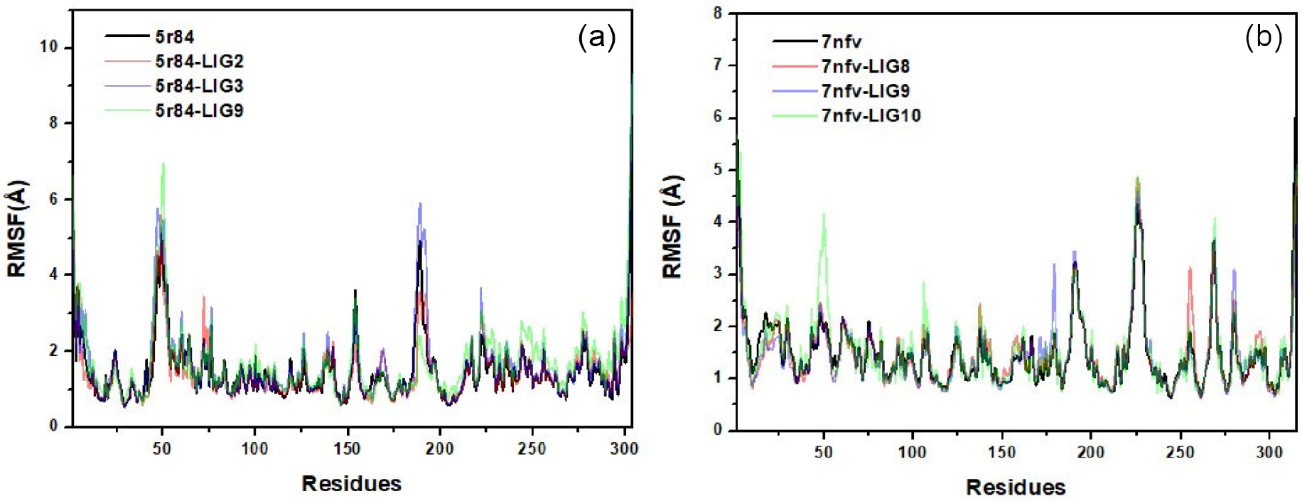
The RMSF analysis provides residue-level insights into the flexibility and dynamic behavior of the receptor and its ligand-bound complexes. For the unbound receptor 5r84, the RMSF profile revealed moderate fluctuations across several loop regions, consistent with the intrinsic flexibility of the protein. Peaks in the RMSF curve correspond to solvent-exposed and structurally less constrained regions, while the core residues remained relatively stable.
The complex with ligand 2 exhibited RMSF values similar to the receptor baseline, but with slightly elevated fluctuations in specific regions. This pattern is consistent with the transient binding behavior previously (Figure S1 in the SI section) identified for ligand 2. The intermittent association of the ligand with the binding pocket fails to reduce local flexibility, allowing certain residues to maintain higher mobility.
In contrast, the complexes with ligands 3 and 9 (Figure 8) demonstrated reduced fluctuations across most residues, indicating enhanced stabilization of the protein structure. The ligand 3 complex showed a marked decrease in RMSF values in the binding site region, reflecting strong intermolecular interactions that restrict local flexibility. Similarly, the ligand 9 complex exhibited stabilization across the majority of residues, although minor peaks persisted in flexible loop regions. Importantly, the overall reduction in RMSF compared to the receptor baseline confirms that ligands 3 and 9 remain stably accommodated within the protein interior, thereby reinforcing conformational stability.
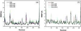
RMSF values of the (a) 3CLpro and (b) PLpro receptor residues from a 150 ns simulation. Black lines correspond to the receptor without a ligand, while colored lines correspond to the receptor with the ligand.
Root mean square deviation (RMSD) and radius of gyration (Rg)
RMSD is a quantitative measure of the average deviation between atomic positions of superimposed structures, commonly used to assess structural stability and conformational changes in MD simulations (e.g., atoms in the backbone of a protein). RMSD value is a measure of how much the protein conformation has changed.19,53 RMSD and Rg were used to evaluate the stability of the 3CLpro and PLPro proteins.
RMSD values
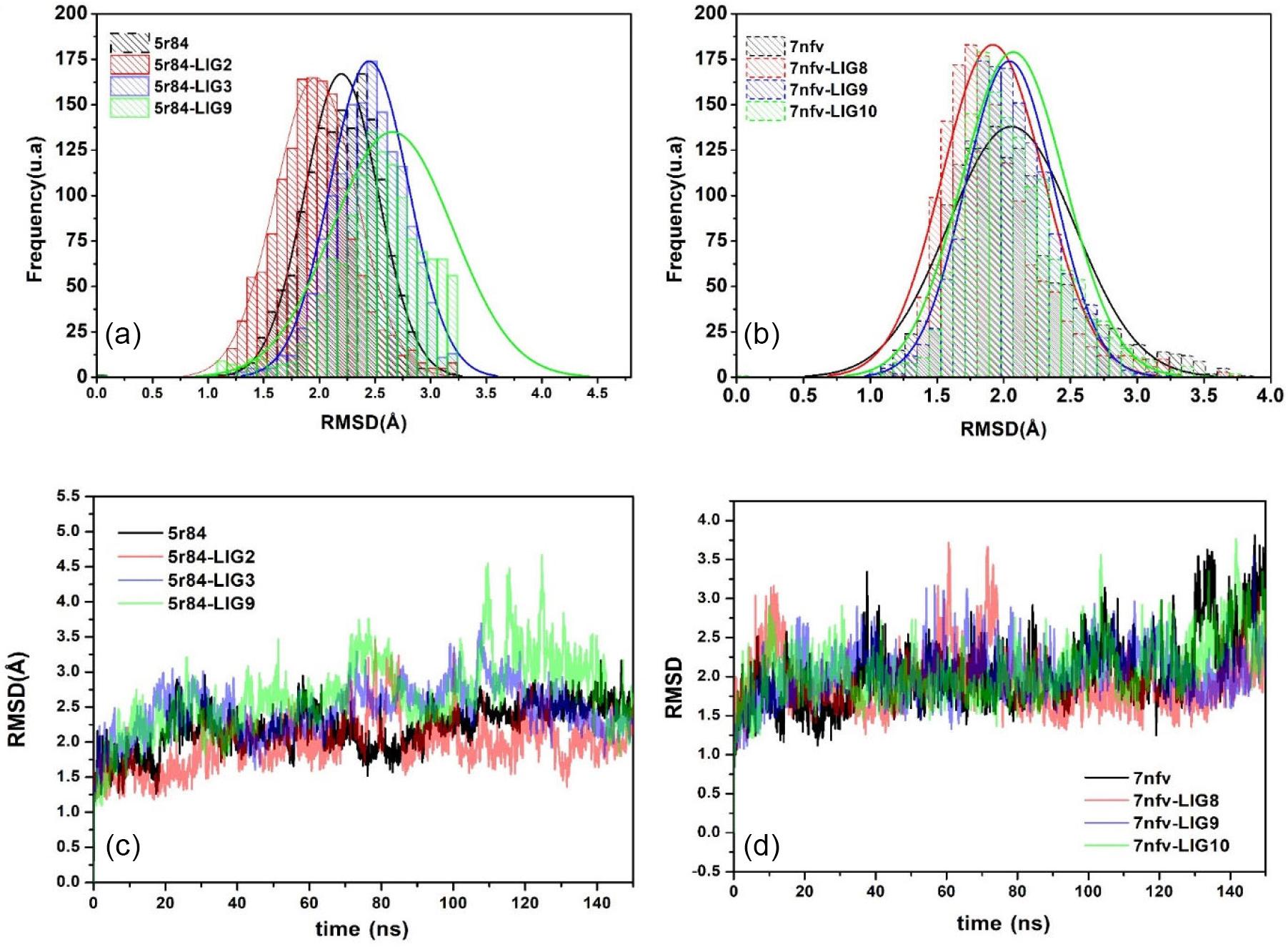
Ligand compounds 2, 3, and 9 were selected for the MD simulations because they performed better in the docking study for the 3CLpro receptor than other ligands. The docked complexes were subjected to RMSD analysis to assess the residual flexibility of 3CLpro. The experimental conditions were prepared using the CHARMM-GUI server30,31 with the CHARMM36m force field. In Figure 9, RMSD values for the Cα of 3CLpro protein complexes were evaluated to analyze conformational stability during the 150 ns period of MD simulations.
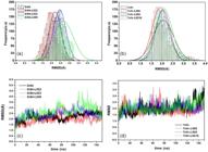
RMSD and RMSD distribution values: (a) and (d) for the Cα of 3Clpro, and (b) and (e) for the PLpro receptors, respectively, in a 150 ns simulation with and without the ligands.
The receptor (5r84) exhibited a mean RMSD of 2.20 Å, reflecting intrinsic stability and serving as a baseline for comparison. The complex with ligand 2 displayed a mean RMSD of 1.94 Å, slightly lower than the receptor baseline. This reduction suggests moderate stabilization; however, the distribution revealed broader fluctuations, consistent with a transient binding behavior. Ligand 2 intermittently associates with the binding pocket but fails to achieve persistent stabilization.
The complex with ligand 3 showed a mean RMSD of 2.48 Å, higher than the receptor baseline. Despite this increase, the distribution reached a plateau after equilibration, indicating that ligand 3 remains stabilized within the protein interior. The elevated RMSD reflects conformational adjustments of the receptor-ligand complex but does not imply dissociation.
The complex with ligand 9 exhibited the highest mean RMSD, 2.66 Å, suggesting greater deviation compared to both the receptor and the other complexes. Nevertheless, the distribution analysis confirmed that ligand 9 remains stably accommodated within the binding site, maintaining persistent interactions throughout the simulation. The higher RMSD values likely reflect structural rearrangements required to accommodate ligand 9, rather than instability or dissociation.
Comparative analysis
(i) ligand 2: transient binding, moderate instability, mean RMSD 1.94 Å;
(ii) ligand 3: stable binding, conformational adaptation, mean RMSD 2.48 Å;
(iii) ligand 9: stable binding, highest conformational deviation, mean RMSD 2.66 Å.
This comparative assessment highlights divergent binding behaviors: ligand 2 exhibits transient and unstable binding, while ligands 3 and 9 remain consistently stabilized within the protein interior, despite higher RMSD values.
In Figure 9b, RMSD values for the Cα of PLPro protein complexes were evaluated to analyze conformational stability during the 150 ns time period of MD simulations. The experimental conditions were prepared using the CHARMM-GUI server30,31 with the CHARMM36m force field. Mean RMSD values were 1.94 Å (7nfv_LIG8), 2.03 Å (7nfv_LIG9), and 2.11 Å (7nfv_LIG10).
The receptor alone (7nfv) exhibited a mean RMSD of 2.03 Å, reflecting intrinsic stability and serving as a baseline for comparison. The complex with ligand 8 showed a slightly lower mean RMSD (1.94 Å), suggesting moderate stabilization. However, the distribution revealed broader fluctuations, consistent with a transient binding behavior, where the ligand intermittently associates with the binding pocket but fails to achieve persistent stabilization.
The complex with ligand 9 displayed a mean RMSD equal to the receptor baseline (2.03 Å). Despite this apparent similarity, trajectory analysis confirmed that ligand 9 does not remain stabilized within the protein interior. Instead, it exits the binding site and does not return, reflecting complete dissociation rather than stable binding.
The complex with ligand 10 exhibited the highest mean RMSD (2.11 Å), indicating greater deviation compared to the receptor and the other complexes. This increase is consistent with transient binding behavior, where ligand 10 partially interacts with the receptor but fails to maintain persistent stabilization.
Rg values
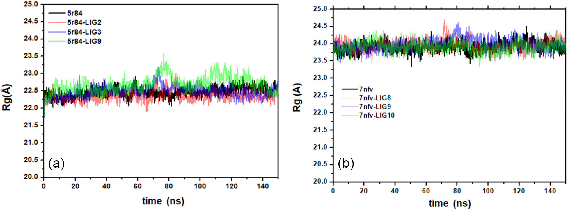
Rg provides a quantitative measure of the compactness and overall structural stability of protein-ligand complexes during MD simulations. For the receptor 5r84, the Rg values remained relatively stable throughout the 140 ns trajectory, fluctuating within a narrow range around ca. 22 Å (Figure 10). This stability reflects the intrinsic conformational robustness of the receptor in the absence of ligands.
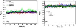
Radius of gyration values of docked and undocked receptor proteins in a 150 ns simulation: (a) for the 3CLpro receptor and (b) for the PLpro receptor.
The complex with ligand 2 exhibited Rg values comparable to the receptor baseline, but with slightly broader fluctuations. This behavior is consistent with the transient binding mode previously identified for ligand 2. The ligand intermittently associates with the binding pocket, leading to modest variations in protein compactness without achieving persistent stabilization.
The complex with ligand 3 maintained values around ca. 23 Å, with limited fluctuations after equilibration, indicating that the ligand remains stably accommodated within the protein interior. Importantly, at 73.5 ns, a rapid increase in Rg was observed, followed by a subsequent decrease that restored stability. This transient expansion suggests a temporary conformational adjustment of the protein-ligand complex, after which the system returned to a stable compact state.
The complex with ligand 9 showed stable Rg values overall, but with a distinctive dynamic pattern. Around 80 ns, the Rg increased and then decreased, followed by another rise beginning at ca. 100 ns, reaching a maximum near 110 ns. After this peak, the system gradually returned to stability during the final stages of the simulation. This sequence of fluctuations suggests that ligand 9 remains stabilized within the receptor interior, but induces conformational rearrangements that temporarily alter protein compactness before equilibrium is re-established.
DFT study
Frontier molecular orbitals
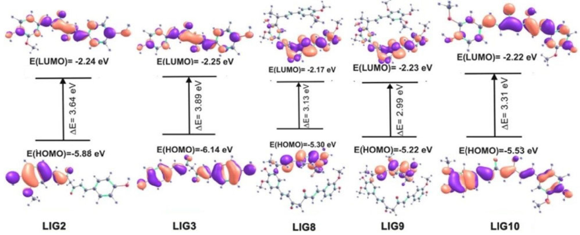
Frontier molecular orbitals (FMO) consist of the highest occupied molecular orbital (HOMO) and the lowest unoccupied molecular orbital (LUMO). The HOMO-LUMO energy gap indicates the susceptibility of electron transfer from the HOMO to LUMO and are used as parameters to calculate the reactivity of molecules. More importantly, FMOs have been taken into consideration in the structure-activity relationships of molecules54,55 and are therefore important to evaluate the bioactivity of small molecules. The isodensity surface plots of the FMO of the ligands are shown in Figure 11.
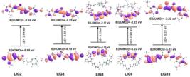
Frontier molecular orbitals studies, showing HOMO and LUMO energy values of the LIG2, LIG3, LIG8, LIG9 and LIG10, as well as the energy gap (ΔE)
As can be seen in Figure 11, FMO analysis reveals that LIG2 and LIG3 have the lowest HOMO energies, at -6.14 and -5.88 eV, respectively. LIG3 display the largest energy gap at 3.89 eV, while LIG9 exhibits the smallest, at 2.99 eV. Interestingly, the three ligands with the highest LUMO energies are LIG2, LIG3 and LIG9, the same ligands chosen for the 3CLpro MD runs based on the docking results. Their LUMO energies are: -2.24, -2.25 and -2.23eV, respectively.
LIG2, LIG3, and LIG9 are identified as the most relevant compounds based on results from docking and MD analyses. These ligands exhibit favorable frontier orbital characteristics, which directly affect their interactions with the receptors of the 3CLpro protein of the new coronavirus. The combination of strong docking scores and advantageous FMO descriptors (Figure 11) indicates that their electronic structures enhance binding affinity and stabilization within the active site. The HOMO reflects the ability of a molecule to donate electron density to electrophilic regions of a receptor, while the LUMO represents the tendency to accept electron density from nucleophilic residues in the protein, thereby facilitating molecular interactions. As result, the HOMO-LUMO energy gap correlates with molecular reactivity, and the ease of charge-transfer processes involved in ligand-protein binding. The relatively high LUMO energies observed for LIG2, LIG3 and LIG9 that these ligands may efficiently participate in intermolecular orbital overlap with electron-rich amino acid residues.
The possible reaction mechanism associated with these interactions can be interpreted in terms of donor-acceptor stabilization. In particular, π-sulfur interactions observed experimentally in docking (Figures 2-4) may arise from overlap between sulfur lone-pair orbitals and ligand π* orbitals. This interaction mechanism is especially relevant because CYS145 is one of the catalytic residues directly involved in peptide bond cleavage by 3CLpro. LIG9 displayed the smallest HOMO-LUMO gap (ca. 2.99 eV), indicating higher electronic polarizability. Molecules with smaller energy gaps generally undergo charge redistribution more easily during intermolecular interactions, which may facilitate adaptation to the electrostatic environment of the receptor pocket. In the context of 3CLpro, whose catalytic machinery involves the nucleophilic cysteine residue CYS145 and the proton-transfer partner HIS41, this enhanced polarizability may favor transient charge-transfer interactions between the ligand π-system and catalytic residues. Such behavior may strengthen noncovalent stabilization mechanisms such as π-sulfur and π-π stacking interactions observed during docking. The strong interaction of LIG9 with both HIS41 and CYS145 is therefore not only geometrically favorable but also electronically consistent with its reduced HOMO-LUMO gap.
In summary, the FMO results provide a quantum-chemical explanation for why LIG2, LIG3, and LIG9 consistently emerged as the strongest candidates throughout the docking and MD analyses.
Molecular electrostatic potential map
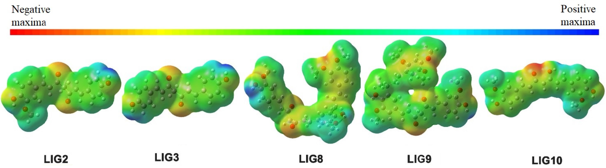
Molecular electrostatic potential (MEP) provides insights into the overall size of a molecule and highlights regions with more positive or negative charges. It serves as a valuable tool for predicting whether a molecule is likely to interact with positively or negatively charged binding sites. The MEPs of the studied ligands are shown in Figure 12. The active site of 3CLpro is negatively charged, suggesting that, from an electrostatic perspective, a ligand with more positively charged regions would bind more favorably to the target. Conversely, the binding site of PLPro is nearly neutral.
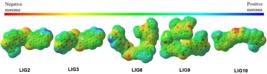
Molecular electrostatic potential (MEP) map of the ligands LIG2, LIG3, LIG8, LIG9 and LIG10. Cooler colors (blue and green) correspond to positive regions, while warmer colors (yellow and red) correspond to negative regions.
As illustrated in Figure 12, both LIG2 and LIG3 have positively charged ends, making them well-suited for interactions with the 3CLpro active site. LIG8 also creates a positive potential around one side of the molecule, while LIG9 appears to be mostly neutral. Notably, the negatively charged C=O groups are prominent across all ligands. In the case of LIG10, the C=O group adjacent to an OH group renders this region of the molecule highly negative, which could influence its binding affinity for 3CLpro.
MEP analysis is valuable for identifying electron-rich and electron-deficient regions of a molecule, helping predict preferred sites for electrophilic or nucleophilic interactions during receptor binding. The interactions between LIG2 and 3CLpro protein residues predicted through molecular docking (Figure 2) indicate that LIG2 contains regions of moderate electron density capable of favorable interactions with HIS41, and forms hydrogen bonds with TYR54, GLY143, LEU141, and HIS163. Furthermore, the electrostatic potential localized around the carbonyl oxygen atoms of LIG2 makes these sites efficient hydrogen-bond acceptors for polar residues within the oxyanion stabilization region of 3CLpro. The MEP map (Figure 12) and the interactions between LIG9 and 3CLpro protein residues predicted through molecular docking (Figure 3) display strong interactions with HIS41, TYR54, ASN142, GLU166, and CYS145, interacting by means of binding mechanism that depends more on induced polarization than on long-range electrostatic attraction.
The MEP map of LIG3 reveals electron-rich carbonyl oxygen atoms and positively polarized peripheral regions that are well suited for electrostatic complementarity with the receptor pocket (Figure 4), where the interactions between LIG3 and 3CLpro protein residues show that hydrogen bonding occur with TYR54, LEU141, GLY143, and GLU166. In particular, the polarized structure of LIG3 facilitates precise electrostatic alignment with the GLU166 residue, contributing to stabilization of the active site geometry in 3CLpro. As seen in Figures 5-7, the interactions between LIG8, LIG9, and LIG10 and PLpro protein residues present patterns that differ somewhat from those observed for 3CLpro. As seen, the ligands LIG8, LIG9, and LIG10 interact with the PLpro active site, mainly with residues ASP286, HIS272, and CYS111 through hydrogen bonds and π-sulfur interactions. Consequently, ligand binding in PLpro appears to depend more strongly on local hydrogen bonding and induced polarization effects.
The MEP map of LIG10 shows strongly negative regions surrounding the diketone and hydroxyl groups, particularly near the carbonyl adjacent to the hydroxyl substituent. This high electron density facilitates hydrogen-bond formation with proton-donating residues inside the PLpro active site. In summary, the MEP maps presented in this work provide valuable information regarding the electronic distribution of the curcumin derivatives and their possible interaction mechanisms with the active sites of SARS-CoV-2 proteases.
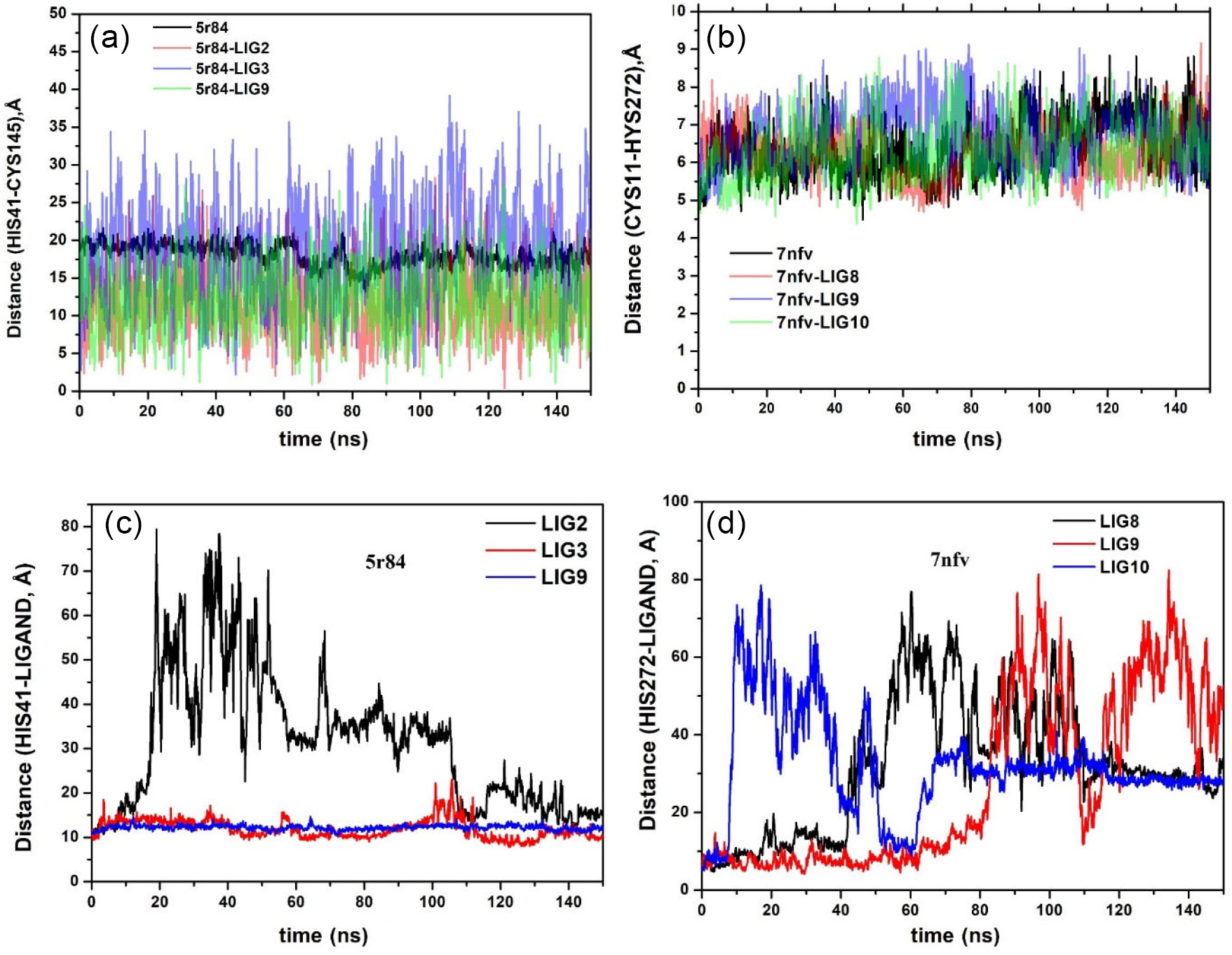
Distance variation between HIS41 and CYS145 (ID: 5r84)
The catalytic dyad formed by HIS41 and CYS145 is essential for the proteolytic activity of 3CLpro, and monitoring their inter-residue distance provides insight into the conformational stability of the active site (Figure 13). For the unbound receptor 5r84, the HIS41-CYS145 distance remained relatively stable throughout the 150 ns trajectory, reflecting the intrinsic robustness of the catalytic geometry.
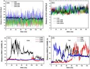
Time-dependent distance analysis during molecular dynamics simulations of SARS-CoV-2 proteases: (a) distance between catalytic residues His41-Cys145 in 3CLpro (PDB ID: 5R84) and its complexes with LIG2, LIG3, and LIG9; (b) distance between catalytic residues Cys111-His272 in PLpro (PDB ID: 7NFV) and its complexes with LIG8, LIG9, and LIG10; (c) distance between His41 and ligands (LIG2, LIG3, LIG9) in 3Clpro; and(d) distance between His272 and ligands (LIG8, LIG9, LIG10) in PLpro.
The complex with ligand 2 exhibited moderate fluctuations, consistent with its transient binding behavior. Notably, the variation in distance suggests a tendency to compress the two catalytic residues, perturbing the active-site geometry without achieving persistent stabilization. The complex with ligand 3 showed larger fluctuations compared to the receptor baseline, reflecting conformational adjustments of the active site. Despite these deviations, ligand 3 remained stably accommodated within the protein interior, indicating that the observed dynamics represent structural adaptation rather than destabilization. The complex with ligand 9 displayed smaller variations overall, but the pattern of distance change also indicated a compressive effect on HIS41 and CYS145, reinforcing the catalytic geometry while maintaining stability. This suggests that ligand 9 contributes to preserving the compactness of the active site, despite transient adjustments.
The catalytic dyad formed by CYS111 and HIS272 is fundamental to the enzymatic activity of PLpro, and monitoring their inter-residue distance provides a direct measure of active-site stability. As illustrated in Figure 13b, the unbound receptor 7nfv maintained a relatively stable CYS111-HIS272 distance throughout the 150 ns trajectory, reflecting the intrinsic robustness of the catalytic geometry in the absence of ligands.
The complex with ligand 8 exhibited moderate fluctuations in the dyad distance, consistent with its transient binding behavior. The ligand intermittent association perturbed the catalytic residues, producing temporary compressions and expansions without achieving persistent stabilization. In contrast, the complex with ligand 9 showed pronounced instability. The trajectory in Figure 13b clearly demonstrates that the ligand failed to remain accommodated within the protein interior, eventually dissociating from the binding pocket and not returning. This dissociation disrupted the catalytic geometry, highlighting the inability of ligand 9 to stabilize the active site.
The complex with ligand 10 displayed fluctuations similar to ligand 8, also consistent with transient binding behavior. While the ligand occasionally influenced the dyad distance, it did not maintain long-term stabilization, resulting in moderate perturbations of the active-site residues.
Comparative analysis
In Figure 13a, ligand 2 displayed transient binding behavior, leading to high distance variation and partial compression of the catalytic residues. This instability suggests that the ligand was unable to maintain persistent interactions with the active site, resulting in conformational fluctuations that may compromise its binding affinity. In contrast, ligands 3 and 9 remained stabilized within the protein interior, with ligand 3 inducing broader conformational adjustments and ligand 9 reinforcing a more compact geometry. In Figure 13b, which illustrates the CYS111-HIS272 dyad of 7nfv, the ligand-dependent effects were less favorable. In Figure 13d, both ligands 8 and 10 exhibited transient binding behavior, producing moderate perturbations without achieving persistent stabilization, while ligand 9 failed to remain accommodated in the binding pocket, dissociating completely and not returning, thereby disrupting the catalytic geometry.
MMPBSA analysis
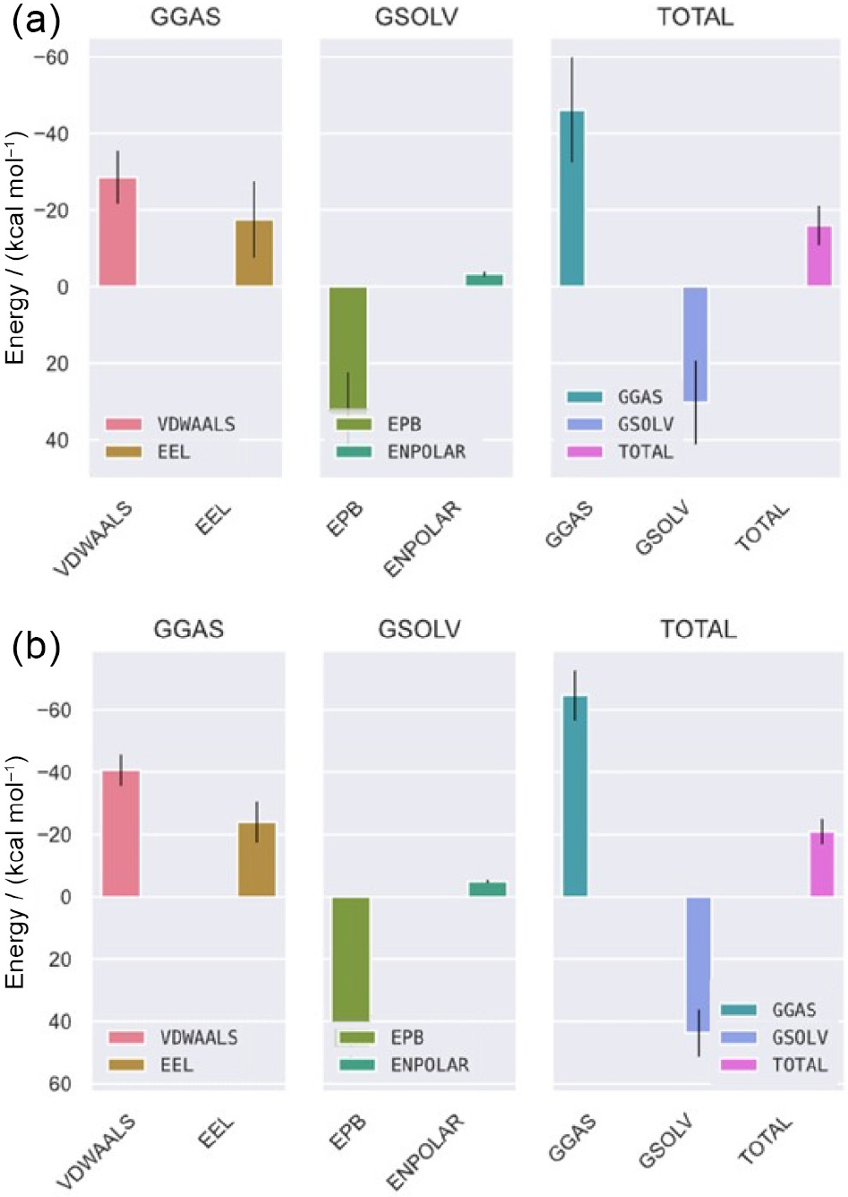
When analyzing the molecular dynamics of the ligands and receptors, it was observed that LIG3 and LIG9 remained in the active site of 5r84 throughout the simulation. Therefore, MMPBSA analyses were performed on these complexes. Table 2 and Figure 14 show the calculated energy values. A comparative analysis of the thermodynamic profiles reveals that ligand 9 exhibits a more favorable overall binding affinity to the 5r84 than LIG3, with total binding free energies of approximately -21 and -16 kcal mol-1, respectively. This enhanced stability in the Ligand 9 complex is primarily driven by significantly stronger gas-phase interactions (GGAS), particularly a substantial improvement in Van der Waals forces, which suggests superior shape complementarity and hydrophobic packing within the binding pocket. Although LIG9 incurs a higher polar desolvation penalty (GSOLV) to displace water molecules upon binding, the overwhelming energetic gains from its direct intermolecular interactions with the protein more than compensate for this energetic cost, ultimately making it the thermodynamically superior binder. Furthermore, the values were close to and in some cases more negative than other SARS-CoV-2 protein inhibitors.56-58
Free energy values calculated by molecular mechanics Poisson-Boltzmann surface area (MMPBSA) analysis of the 5r84-ligand 3/ligand 9 complex calculated over the last 100 ns with 5-frame intervals, totaling 200 frames
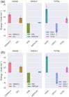
Free energy values calculated by MMPBSA analysis of complexes (a) 5r84-ligand 3 and (b) 5r84-ligand 9. The columns on the left show Van der Waals energy values (pink) and electrostatic energy (brown), the middle columns the Poisson-Boltzmann electrostatic energy (moss green) and non-polar energy (green) and the columns on the right show solvation free energy (blue), gas phase free energy (blue-green) and total energy (purple).
Conclusions
The results of this study suggest that demethoxy-curcumin (LIG2), bisdemethoxycurcumin (LIG3), and cassumunin B (LIG9) bind to the active site of the 3CLpro protein and may serve as promising inhibitors of SARS CoV-2. Cassumunin B showed strong interactions with catalytic residues HIS41 via a single hydrogen bond and CYS145 through π-sulfur interactions, along with additional binding interactions. The ligands cassumunin A (L), cassumunin B (ligand 9), and curcumin PE (LIG10) exhibited stronger interactions with the PLPro receptor. Curcumin PE, for instance, interacted with catalytic residue HIS272 via a single strong hydrogen bond and with CYS111 through π-sulfur interactions. Drug-likeness analysis (excluding ligand 7) indicated that the ligands have good cell membrane permeability and oral bioavailability.
The molecular dynamics simulations revealed distinct conformational behaviors among the receptor-ligand complexes. For 5r84, ligand 2 exhibited transient binding with moderate instability, while ligands 3 and 9 remained stabilized, despite higher RMSD and dynamic Rg fluctuations, ultimately reinforcing structural persistence. For 7nfv, ligands 8 and 10 also showed transient binding, whereas ligand 9 dissociated completely, failing to remain stabilized within the protein interior. RMSF analysis confirmed that stable ligands restricted local residue flexibility, while transient or dissociating ligands allowed higher mobility.
Overall, ligands 3 and 9 in 5r84 emerged as promising candidates, while ligands 8 and 10 in 7nfv require optimization, and ligand 9 proved unsuitable. The MD simulations revealed that ligands 3 and 9 act as promising stabilizers of the 5r84 active site, whereas ligand 2 remains only partially effective due to its transient binding behavior. In the case of 7nfv, the CYS111-HIS272 distance analysis demonstrated that ligands 8 and 10 were unable to achieve stable interactions, while ligand 9 proved unsuitable due to complete dissociation from the binding pocket. Overall, these comparative results underscore the stronger stabilizing potential of ligands in 5r84 compared to those tested in 7nfv, reinforcing the importance of ligand-specific optimization for robust active-site stabilization.
The frontier molecular orbital analysis provided quantum chemical parameters to evaluate the chemical reactivity of the ligands. Interestingly, the ligands with the highest LUMO energies, LIG2, LIG3, and LIG, were the same selected for 3CLpro MD simulations based on docking results. The molecular electrostatic potential maps further identified the regions most susceptible to electrophilic and nucleophilic attack. Nevertheless, in vitro and in vivo studies are necessary to confirm the potential of these six natural compounds as repurposed treatments for 2019-nCoV.
Supplementary Information
Supplementary information is available free of charge at http://jbcs.sbq.org.br as PDF file.
Supplementary PDF
Acknowledgments
G. D. S. acknowledges FUNCAP (DEP-0164-00350.01.00/19), MCTI/CNPq Universal 28/2018 (426995/2018-0), and CNPq PQ 09/2020 (311898/2020-3); F. F. S. CAPES and MCT/CNPq (438753/2018-6 and 308789/2022-9); C. E. S. N. FUNCAP (BP4 00172-00065.01.01/20); A. M. R. T. CNPq PQ (308178/2021 1); A. J. R. C., Ph.D., MCTI/CNPQ PQ-09/2022 (310440/2022-0). All authors acknowledge MCTI/CNPq/CAPES for financial support and thank CENAPAD (Universidade Federal do Ceará) for the computational resources and facilities used in this research.
Data Availability Statement
All data are available in the text and at the link: https://shre.ink/3u5A.
References
-
1 Sahoo, S.; Mehra, A.; Suri, V.; Malhotra, P.; Yaddanapudi, L. N.; Dutt Puri, G.; Grover, S.; Asian J. Psychiatr. 2020, 53, 102187. [Crossref]
» Crossref -
2 Cascella, M.; Rajnik, M.; Aleem, A.; C.; D. S.; Di Napoli, R.; Stat Pearls Publishing 2023 [Link] accessed in June 2026
» Link -
3 Gorbalenya, A. E.; Baker, S. C.; Baric, R. S.; de Groot, R. J.; Drosten, C.; Gulyaeva, A. A.; Haagmans, B. L.; Lauber, C.; Leontovich, A. M.; Neuman, B. W.; Penzar, D.; Perlman, S.; Poon, L. L. M.; Samborskiy, D. V.; Sidorov, I. A.; Sola, I.; Ziebuhr, J.; Nat. Microbiol. 2020, 5, 536. [Crossref]
» Crossref -
4 Baig, A. M.; Khaleeq, A.; Ali, U.; Syeda, H.; ACS Chem. Neurosci. 2020, 11, 995. [Crossref]
» Crossref -
5 Wu, A.; Peng, Y.; Huang, B.; Ding, X.; Wang, X.; Niu, P.; Meng, J.; Zhu, Z.; Zhang, Z.; Wang, J.; Sheng, J.; Quan, L.; Xia, Z.; Tan, W.; Cheng, G.; Jiang, T.; Cell Host Microbe 2020, 27, 325. [Crossref]
» Crossref -
6 The World Health Organization (WHO), WHO Director-General’s Opening Remarks at the Media Briefing on COVID19, 2020. [Link] accessed in June 2026
» Link -
7 Hui, D. S.; Clin. Chest. Med. 2017, 38, 71. [Crossref]
» Crossref -
8 Pant, S.; Singh, M.; Ravichandiran, V.; Murty, U. S. N.; Srivastava, H. K.; J. Biomol. Struct. Dyn. 2021, 39, 2904. [Crossref]
» Crossref -
9 Kampf, G.; Todt, D.; Pfaender, S.; Steinmann, E.; J. Hosp. Infect. 2020, 104, 246. [Crossref]
» Crossref -
10 Talluri, S.; Comb. Chem. High Throughput Screening 2021, 24, 716. [Crossref]
» Crossref -
11 Cosconati, S.; Forli, S.; Perryman, A. L.; Harris, R.; Goodsell, D. S.; Olson, A. J.; Expert. Opin. Drug. Discovery 2010, 5, 597. [Crossref]
» Crossref -
12 Ziebuhr, J.; Snijder, E. J.; Gorbalenya, A. E.; J. Gen. Virol. 2000, 81, 853. [Crossref]
» Crossref -
13 Xu, Z.; Peng, C.; Shi, Y.; Zhu, Z.; Mu, K.; Wang, X.; Zhu, W.; bioRxiv, 2020, 921627. [Crossref]
» Crossref -
14 Wang, X.; Yang, Y.; Gao, Y.; Niu, X.; Int. J. Mol. Sci. 2020, 21, 3567. [Crossref]
» Crossref -
15 Kim, J.; Kwon, J.; Kim, M.; Do, J.; Lee, D.; Han, H.; Poly. J. 2016, 48, 829. [Crossref]
» Crossref -
16 Hollingsworth, S. A.; Dror, R. O.; Neuron 2018, 99, 1129. [Crossref]
» Crossref -
17 Ahamad, S.; Kanipakam, H.; Birla, S.; Ali, M. S.; Gupta, D.; Eur. J. Pharmacol. 2021, 890, 173664. [Crossref]
» Crossref -
18 Viana, J. O.; Félix, M. B.; Maia, M. S.; Serafim, V. L.; Scotti, L.; Scotti, M. T.; Braz. J. Pharm. Sci. 2018, 54, e01010. [Crossref]
» Crossref -
19 Ibrahim, M. A. A.; Abdelrahman, A. H. M.; Hussien, T. A.; Badr, E. A. A.; Mohamed, T. A.; El-Seedi, H. R.; Pare, P. W.; Efferth, T.; Hegazy, M. F.; Comput. Biol. Med. 2020, 126, 104046. [Crossref]
» Crossref -
20 Kataria, R.; Khatkar, A.; Curr. Pharm. Biotechnol. 2019, 20, 410. [Crossref]
» Crossref -
21 Urosevic, M.; Nikolic, L.; Gajic, I.; Nikolic, V.; Dinic, A.; Miljkovic, V.; Antibiotics (Basel) 2022, 11, 135. [Crossref]
» Crossref -
22 Douangamath, A.; Fearon, D.; Gehrtz, P.; Krojer, T.; Lukacik, P.; Owen, C. D.; Resnick, E.; Strain-Damerell, C.; Aimon, A.; Abranyi-Balogh, P.; Brandao-Neto, J.; Carbery, A.; Davison, G.; Dias, A.; Downes, T. D.; Dunnett, L.; Fairhead, M.; Firth, J. D.; Jones, S. P.; Keeley, A.; Keseru, G. M.; Klein, H. F.; Martin, M. P.; Noble, M. E. M.; O’Brien, P.; Powell, A.; Reddi, R. N.; Skyner, R.; Snee, M.; Waring, M. J.; Wild, C.; London, N.; von Delft, F.; Walsh, M. A.; Nat. Commun. 2020, 11, 5047. [Crossref]
» Crossref -
23 Srinivasan, V.; Brognaro, H.; Prabhu, P. R.; de Souza, E. E.; Gunther, S.; Reinke, P. Y. A.; Lane, T. J.; Ginn, H.; Han, H.; Ewert, W.; Sprenger, J.; Koua, F. H. M.; Falke, S.; Werner, N.; Andaleeb, H.; Ullah, N.; Franca, B. A.; Wang, M.; Barra, A. L. C.; Perbandt, M.; Schwinzer, M.; Schmidt, C.; Brings, L.; Lorenzen, K.; Schubert, R.; Machado, R. R. G.; Candido, E. D.; Oliveira, D. B. L.; Durigon, E. L.; Niebling, S.; Garcia, A. S.; Yefanov, O.; Lieske, J.; Gelisio, L.; Domaracky, M.; Middendorf, P.; Groessler, M.; Trost, F.; Galchenkova, M.; Mashhour, A. R.; Saouane, S.; Hakanpaa, J.; Wolf, M.; Alai, M. G.; Turk, D.; Pearson, A. R.; Chapman, H. N.; Hinrichs, W.; Wrenger, C.; Meents, A.; Betzel, C.; Commun. Biol. 2022, 5, 805. [Crossref]
» Crossref -
24 Cree, B.; Bieniek, M. K.; Amin, S.; Kawamura, A.; Cole, D. J.; Digital Discovery 2025, 4, 438. [Crossref]
» Crossref -
25 Frisch, M. J.; Trucks, G. W.; Schlegel, H. B.; Scuseria, G. E.; Robb, M. A.; Cheeseman, J. R.; Scalmani, G.; Barone, V.; Mennucci, B.; Petersson, G. A.; Nakatsuji, H.; Caricato, M.; Li, X.; Hratchian, H. P.; Izmaylov, A. F.; Bloino, J.; Zheng, G.; Sonnenberg, J. L.; Hada, M.; Ehara, M.; Toyota, K.; Fukuda, R.; Hasegawa, J.; Ishida, M.; Nakajima, T.; Honda, Y.; Kitao, O.; Nakai, H.; Vreven, T.; Montgomery Jr., J. A.; Peralta, J. E.; Ogliaro, F.; Bearpark, M.; Heyd, J. J.; Brothers, E.; Kudin, K. N.; Staroverov, V. N.; Kobayashi, R.; Normand, J.; Raghavachari, K.; Rendell, A.; Burant, J. C.; Iyengar, S. S.; Tomasi, J.; Cossi, M.; Rega, N.; Millam, J. M.; Klene, M.; Knox, J. E.; Cross, J. B.; Bakken, V.; Adamo, C.; Jaramillo, J.; Gomperts, R.; Stratmann, R. E.; Yazyev, O.; Austin, A. J.; Cammi, R.; Pomelli, C.; Ochterski, J. W.; Martin, R. L.; Morokuma, K.; Zakrzewski, V. G.; Voth, G. A.; Salvador, P.; Dannenberg, J. J.; Dapprich, S.; Daniels, A. D.; Farkas, Ö.; Foresman, J. B.; Ortiz, J. V.; Cioslowski, J.; Fox, D. J.; Gaussian 09, Revision D.01; Gaussian, Inc., Wallingford CT, 2009. [Link] accessed in June 2026
» Link -
26 Pettersen, E. F.; Goddard, T. D.; Huang, C. C.; Couch, G. S.; Greenblatt, D. M.; Meng, E. C.; Ferrin, T. E.; J. Comput. Chem. 2004, 25, 1605. [Crossref]
» Crossref -
27 Dassault Systèmes; BIOVIA Discovery Studio Visualizer version 22.1, 2022. [Link]accessed in June 2026
» Link -
28 Dassault Systèmes; BIOVIA Discovery Studio Visualizer version 17.2.0, 2017. [Link] accessed in June 2026
» Link -
29 Forli, S.; Huey, R.; Pique, M. E.; Sanner, M. F.; Goodsell, D. S.; Olson, A. J.; Nat. Protoc. 2016, 11, 905. [Crossref]
» Crossref -
30 Jo, S.; Kim, T.; Iyer, V. G.; Im, W.; J. Comput. Chem. 2008, 29, 1859. [Crossref]
» Crossref -
31 Lee, J.; Cheng, X.; Swails, J. M.; Yeom, M. S.; Eastman, P. K.; Lemkul, J. A.; Wei, S.; Buckner, J.; Jeong, J. C.; Qi, Y.; Jo, S.; Pande, V. S.; Case, D. A.; Brooks III, C. L.; MacKerell Jr., A. D.; Klauda, J. B.; Im, W.; J. Chem. Theory Comput. 2016, 12, 405. [Crossref]
» Crossref -
32 Huang, J.; Rauscher, S.; Nawrocki, G.; Ran, T.; Feig, M.; de Groot, B. L.; Grubmuller, H.; MacKerell Jr., A. D.; Nat. Methods 2017, 14, 71. [Crossref]
» Crossref -
33 Jorgensen, W. L.; Chandrasekhar, J.; Madura, J. D.; Impey, R. W.; Klein, M. L.; Chem. Phys. 1983, 79, 926. [Crossref]
» Crossref -
34 Abraham, M. J.; Murtola, T.; Schulz, R.; Páll, S.; Smith, J. C.; Hess, B. L., E.; SoftwareX 2015, 1-2, 19. [Crossref]
» Crossref -
35 Andersen, H. C.; J. Chem. Phys. 1980, 72, 2384. [Crossref]
» Crossref -
36 Bussi, G.; Donadio, D.; Parrinello, M.; J. Chem. Phys. 2007, 126, 014101. [Crossref]
» Crossref -
37 Bussi, G.; Mol. Phys. 2013, 112, 379. [Crossref]
» Crossref -
38 Baildya, N.; Khan, A. A.; Ghosh, N. N.; Dutta, T.; Chattopadhyay, A. P.; J. Mol. Struct. 2021, 1227, 129390. [Crossref]
» Crossref -
39 Molinspiration Cheminformatics. [Link] accessed in June 2026.
» Link -
40 Zhao, Y. H.; Abraham, M. H.; Le, J.; Hersey, A.; Luscombe, C. N.; Beck, G.; Sherborne, B.; Cooper, I.; Pharm. Res. 2002, 19, 1446. [Crossref]
» Crossref -
41 Valdes-Tresanco, M. S.; Valdes-Tresanco, M. E.; Valiente, P. A.; Moreno, E.; J. Chem.Theory Comput. 2021, 17, 6281. [Crossref]
» Crossref -
42 Choudhury, A.; Prabha, S.; Saeed, M. U.; Mohammad, T.; Hussain, A.; AlAjmi, M. F.; Khan, F. I.; Islam, A.; Hassan, M. I.; Int. J. Biol. Macromol. 2025, 320, 145785. [Crossref]
» Crossref -
43 Genheden, S.; Ryde, U.; Expert Opin. Drug. Discovery 2015, 10, 449. [Crossref]
» Crossref -
44 Kumari, R.; Kumar, R.; Open Source Drug Discovery Consortium; Lynn, A.; J. Chem. Inf. Model 2014, 54, 1951. [Crossref]
» Crossref -
45 Jin, Z.; Du, X.; Xu, Y.; Deng, Y.; Liu, M.; Zhao, Y.; Zhang, B.; Li, X.; Zhang, L.; Peng, C.; Duan, Y.; Yu, J.; Wang, L.; Yang, K.; Liu, F.; Jiang, R.; Yang, X.; You, T.; Liu, X.; Yang, X.; Bai, F.; Liu, H.; Liu, X.; Guddat, L. W.; Xu, W.; Xiao, G.; Qin, C.; Shi, Z.; Jiang, H.; Rao, Z.; Yang, H.; Nature 2020, 582, 289. [Crossref]
» Crossref -
46 Vardhan, S.; Sahoo, S. K.; Comput. Biol. Med. 2020, 124, 103936. [Crossref]
» Crossref -
47 Shin, D.; Mukherjee, R.; Grewe, D.; Bojkova, D.; Baek, K.; Bhattacharya, A.; Schulz, L.; Widera, M.; Mehdipour, A. R.; Tascher, G.; Geurink, P. P.; Wilhelm, A.; van der Heden van Noort, G. J.; Ovaa, H.; Muller, S.; Knobeloch, K. P.; Rajalingam, K.; Schulman, B. A.; Cinatl, J.; Hummer, G.; Ciesek, S.; Dikic, I.; Nature 2020, 587, 657. [Crossref]
» Crossref -
48 Reza, R.; Dutta, T.; Baildya, N.; Ghosh, N. N.; Khan, A. A.; Das, R. K.; Microb. Pathog. 2022, 169, 105615. [Crossref]
» Crossref -
49 Kushwaha, P. P.; Singh, A. K.; Prajapati, K. S.; Shuaib, M.; Gupta, S.; Kumar, S.; Microb. Pathog. 2021, 157, 104954. [Crossref]
» Crossref -
50 Peele, K. A.; Durthi, C. P.; Srihansa, T.; Krupanidhi, S.; Ayyagari, V. S.; Babu, D. J.; Indira, M.; Reddy, A. R.; Venkateswarulu, T. C.; Inf. Med. Unlocked 2020, 19, 100345. [Crossref]
» Crossref -
51 Lipinski, C. A.; Lombardo, F.; Dominy, B. W.; Feeney, P. J.; Adv. Drug. Delivery Rev. 2001, 46, 3. [Crossref]
» Crossref -
52 Mannhold, R.; Poda, G. I.; Ostermann, C.; Tetko, I. V.; J. Pharm. Sci. 2009, 98, 861. [Crossref]
» Crossref -
53 Zhang, H.; Zhang, L.; Gao, C.; Yu, R.; Kang, C.; J. Mol. Struct. 2021, 1245, [Crossref]
» Crossref -
54 Odabasoglu, H. Y.; Erdogan, T.; Karci, F.; J. Mol. Struct. 2022, 1252, 131066. [Crossref]
» Crossref -
55 Majumdar, D.; pal, T. k.; Singh, D. K.; Pandey, D. K.; Parai, D.; Bankura, K.; Mishra, D.; J. Mol. Struct. 2020, 1209, 127936. [Crossref]
» Crossref -
56 Kumar, K. A.; Sharma, M.; Dalal, V.; Singh, V.; Tomar, S.; Kumar, P.; J. Mol. Graph. Model. 2021, 107, 107969. [Crossref]
» Crossref -
57 Sang, P.; Tian, S.; Meng, Z.; Yang, L.; ChemRxiv 2020, 04 [Crossref]
» Crossref -
58 Kumari, R.; Kumar, V.; Dhankhar, P.; Dalal, V.; J. Biomol. Struct. Dyn. 2023, 41, 4650. [Crossref]
» Crossref
Edited by
-
Editor handled this article:
Paulo Augusto Netz (Associate)