 First record of natural infection by Angiostrongylus cantonensis (Nematoda: Metastrongyloidea) in Tanychlamys indica (Godwin-Austen, 1883) in the city of São Paulo, Brazil
Abstract
BACKGROUND Tanychlamys indica (Godwin-Austen, 1883) was reported as a serious pest in India. The snails are voracious and feed on a wide range of commercial crops. It has also been identified as an intermediate nematode host of Angiostrongylus cantonensis in Bombay, India. T. indica was recently introduced in Brazil by international trade of citrus fruit seedlings. First in the State of Santa Catarina and then in Paraná. Recently, it has been detected in the city of São Paulo threatening to spread to other Brazilian states.
OBJECTIVES We report the first record, in Brazil, of the natural infection by L3 larvae of A. cantonensis isolated from T. indica collected in the Vila Leopoldina neighbourhood, located in the west zone of São Paulo city.
METHODS In January 2023, a team from LABFAUNA and UVIS Lapa collected 36 molluscs identified as T. indica in Vila Leopoldina, São Paulo city. Of these, 20 molluscs were subjected to individual parasitological analysis at the Instituto Adolfo Lutz, using the modified Rugai methodology.
FINDINGS A total of 145 larvae were identified morphologically and classified according to Ash’s criteria. These larvae were identified as third - stage larvae (L3) of A. cantonensis by real time polymerase chain reaction (PCR).
MAIN CONCLUSIONS It is evident that further research is imperative to map the distribution of T. indica in Brazil and to assess its potential as an intermediate host for the nematode A. cantonensis, as well as the economic risks to agriculture. Over the past two decades, human cases of neuroangiostrongyliasis have been documented in the Southeast, North, Northeast, and South regions of Brazil. Additionally, there are records of natural infection with A. cantonensis in molluscs and rodents.
Key words:
Angiostrongylus cantonensis
;
Macrochlamys indica
; exotic species; eosinophilic meningitis; neuroangiostrongyliasis
Terrestrial gastropods are agricultural pest species with significant importance in the world. They cause significant economic damage in a range of agricultural crops, including vegetables, fruit trees, field crops, ornamentals and medicinal plants.1,2
The human activities caused the dispersion of the numerous species of terrestrial molluscs across a vast geographical area. As a consequence, they adapted to new habitats, where they are now regarded as exotic species.3,4
Synanthropic gastropods are species capable of long-distance dispersal. Thus, they had become invasive in urban and agricultural environments around the world.5
Due to their abundance and biomass, terrestrial gastropods are capable of depositing considerable amounts of mucus and faecal material on crops. This process results in reduction of crop value and quality. In addition, terrestrial gastropods cause wounds in plants, facilitating the entry of pathogenic microbes.6,7 Furthermore, various terrestrial gastropods act as intermediate hosts for parasites and pathogens that pose risks to animal and human health.8
Angiostrongylus cantonensis (Chen, 1935) is a parasite that affects the pulmonary arteries and right ventricle of rats and was described in Canton in China. Since then, they have been identified in various locations as the Pacific Islands, Asia, Australia, Africa, and some Caribbean islands. Recently, it has been detected in the Americas.9 The definitive hosts for A. cantonensis are Rattus norvegicus (Berkenhout, 1769) and Rattus rattus (Linnaeus, 1758).10
Accidental infection in humans by A. cantonensis occurs when they consume raw or undercooked snails, slugs, and paratenic hosts (e.g., shrimps, crabs, lizards, frogs, land planarians, and centipedes) or vegetables contaminated with mucus from infected molluscs.9,11,12 The increase of larvae release occurs when these are exposed to stressful situations.13 In contrast to the natural definitive host, A. cantonensis does not complete its life cycle in humans. Instead, the larvae die when they reach in the brain, causing an intense inflammatory reaction that result in neuroangiostrongyliasis.14,15
The territorial molluscs, as intermediate hosts are responsible for transmission of angiostrongyliasis to humans. A. cantonensis has already been identified in all Brazilian regions as in Southeast,16,17,18,19 Northeast,20,21,22 North23,24,25 and South.11,15,16 Therefore, distant regions from one another can exhibit comparable terrestrial mollusc faunas comprising species of exotic nature.5 In Brazil, as in other countries, the increase of human activities has resulted in the rapid introduction of mollusc species. Thereby, an in-depth study of their fauna is necessary.3Tanychlamys (Benson, 1834) is a genus that comprises more than 100 species that are widely distributed in India and Southeast Asia.26,27Tanychlamys indica (Godwin-Austen, 1883) is one most common species in India.26,28 Previously this species was known as Macrochlamys indica (Godwin-Austen, 1883).29 The horntail snail, T. indica, is a voracious herbivore with the capacity to completely devour young seedlings. It is a pervasive pest in vegetable and ornamental plant nurseries in India and Bangladesh with potential to become invasive.30,31,32
In the USA, T. indica is included on the list of quarantine plant pests, due to the potential threat it poses to agricultural interests.33 Singh et al.30 documented the presence of T. indica on citrus and guava seedlings in Punjab, India. The snails exhibit a high degree of trophic specialisation, feeding on a diverse array of commercially cultivated plants, including cabbage, beans, lettuce, moringa, yams, chrysanthemums, and cucurbits.34,35,36 The T. indica introduction in Brazil is a recent phenomenon, potentially linked to the trade in citrus fruit seedlings in the State of Santa Catarina and, subsequently, in Paraná.37,38
On 29 November 2022, the Vigilância Ambiental da Unidade de Vigilância em Saúde Lapa (UVIS Lapa) conducted surveillance and control actions on synanthropic animals. The results produced a documentation of T. indica specimen in a small wood/garden located in a municipal school in Vila Leopoldina neighbourhood, in the western zone of the city São Paulo. Subsequently, on 19 January 2023, an investigation joint with the Laboratório de Identificação e Pesquisa de Fauna Sinantrópica (Labfauna) DVZ and UVIS Lapa resulted in a capture of 36 specimens of T. indica, comprising juveniles and adults.
This report constitutes the first description of T. indica naturally infected by A. cantonensis, L3 larvae in Brazil. The infections were identified in four molluscs. The specimens were analysed in Instituto Adolfo Lutz.
MATERIALS AND METHODS
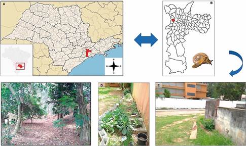
Study areas - The malacological research was conducted at a municipal school situated on the west side of the municipality of São Paulo, SP, with geographical coordinates 23°32’9.708” S, 46°43’27.743”. The site encompasses an area of 7.557.36 m2. The educational establishment is a primary and elementary school, comprising grades 1 to 9, with a capacity of 755 places for children and young people aged between seven and 15 years. The outdoor area of the school includes a vegetable garden, a playground, a sports court, a garden, and a green area (2.897.07 m²), which was the site of the active searches for molluscs. The mollusc collection was extended to encompass a garden area and the graves of the Cemitério da Lapa, due to its proximity to the wall of the school under investigation (Fig. 1).
(A-B) map of São Paulo State, with São Paulo city and the administrative district of Vila Leopoldina highlighted in red. The blue arrow indicates a general view of the Tanychlamys indica collection areas in the Vila Leopoldina neighbourhood in January 2023. (C-D) woods and school garden. (E) a general view of the collection point near a grave in the Cemitério da Lapa- São Paulo/SP.
Molluscs collected and Angiotrongylid identification - In November 2022 occurred by UVIS Lapa, the first record of a land snail specimen collected in the educational establishment. Subsequently, on 19 January 2023 a grouped action was undertaken by LABFAUNA/DVZ and UVIS Lapa, whereby the school’s garden, flowerbeds, vegetable garden, sports court, playground, and woods, as well as a garden area near the cemetery graves, were inspected.
The investigation was conducted during the day in locations where gastropods could be found, including toys in the playground, vegetable garden beds, soil under leaves, branches, plants, stones, mud bricks, cement blocks, roof tiles, pieces of wood, rubble, cracks and crevices in the masonry of plant beds and walls, shaded environments under tree canopies, and recesses in the masonry wall and graves. The 36 gastropods were placed in plastic containers lined with moist paper towels for subsequent analysis. The containers were stored in a polystyrene box to ensure the maintenance of optimal humidity levels. Next, the snails were conveyed to LABFAUNA, where they were accommodated in a terrarium and nourished with lettuce leaves that had been sanitised in a 1% sodium hypochlorite solution. Taxonomic identification was based on an analysis of the snail’s shell and body morphology.37,39 Shell diameter measurements were obtained utilising a Digimess Stainless Hardened digital calliper, with a precision of 0.05mm/1/128 inch.
In February 2023, 20 specimens of terrestrial gastropods were collected and sent to Instituto Adolfo Lutz for nematode larval extraction using the modified Rugai methodology.40 A total of 20 molluscs were individually digested as previously described by Mota et al.19 The L3 larvae, which were recovered alive, were morphologically identified and classified based on Ash’s criteria.41 Next, the specimens were transferred to an excavated plate utilising a Carl Zeiss stereomicroscope (Stemi 500). Concurrently, 30 larvae were fixed in 70% alcohol and photographed using a digital camera attached to a standard optical microscope (Jenaval). The images were analysed using the AxioVision 4.8 software. Furthermore, a group of 20 L3 larvae recovered from natural mollusc infections was frozen at -20ºC for molecular analysis.
Angiostrongylus species: molecular identification - The L3 larvae samples were molecularly analysed to identify Angiostrongylus species. Prior to DNA extraction, the larvae were placed in 1.5 mL tubes, crushed, and digested in a lysis buffer (Tris-HCl, 10 mM, pH 8.0; EDTA, 10 mM; SDS, 0.5%; N-laurylsarcosyl, 0.01%; proteinase K, 100 μg/mL). The larvae were disrupted by shaking with glass beads in a single cycle of 50 oscillations per second for five minutes with the aid of the TissueLyser LT apparatus (Qiagen). The mixtures were incubated at 56ºC for 20 min, or until complete tissue lysis. Next, DNA samples were extracted using the QIAamp DNA Mini Kit (Qiagen), in accordance with the manufacturer’s instructions. The DNA concentrations and purity were determined by measuring the ratio of the optical density (OD) at 260 and 280 nanometres (nm) in a NanoDrop ONE spectrophotometer (Thermo Scientific).
DNA molecules were submitted to conventional polymerase chain reaction (cPCR) using the molecular markers NC1 (5’ ACGTCTGGTTCAGGGTTGTT 3’) and NC2 (5’ TTAGTTTCTTTTCCTCCGCT 3’), which amplify a specific region of the internal transcribed spacer 2 (ITS2) of the rDNA.42,43 The amplifications were performed using a mixture containing 25 pmol of each molecular marker, 12.5 μL of Go Taq Green Master Mix (Promega), which comprises 1 unit of Taq DNA polymerase, 10 mM Tris-HCl (pH 8.5), 50 mM KCl, 1.5 mM MgCl2, and 200 mM of each dNTP. In each amplification reaction were included a negative control (ultrapure water) and two positive DNA controls: from A. cantonensis, Hawaiian strain and A. costaricensis, Crissiumal/RS strain. The thermal cycles were conducted in a Veriti®-96 Well Thermal Cycler/Applied Biosystems, with the following conditions: an initial denaturation at 94ºC for 5 min; 30 cycles of denaturation at 94ºC for 60 s, annealing at 58ºC for 60 s, and extension at 72ºC for 60 s; and a final extension at 72ºC for 10 min.
Angiostrongylus cantonensis was further identified by real-time PCR (qPCR) using the molecular marker AcantITS1, which amplifies a region of the ITS1 of A. cantonensis.44 The sequence designers were: forward (5′ TTCATGGATGGCGAACTGATAG 3′); reverse (5′ GCGCCCATTGAACATTATACTT 3′) and probe (5′ ATCGCATATCTACTATACGCATGTGACACCTG 3′) labelled with FAM (6-carboxyfluorescein) and BHQ1 (Black Hole Quencher 1) at the 5′ and 3′ ends, respectively. The reactions were performed in a final volume of 20 µL, comprising DNA samples (3 µL of DNA up to 100 ng/µL), and 10 µL of 2 X TaqMan Universal PCR Master Mix, 18 µM of molecular markers and 5 µM of the probe. Amplifications were conducted on an Agilent AriaMax Real-Time PCR system, employing a thermal profile comprising two minutes at 50ºC and 10 min at 95ºC. Subsequently, 40 cycles were conducted at 95ºC for 15 s and 60ºC for 1 min. The results were automatically determined by the equipment and expressed as a cycle threshold value (CT), which indicated the quantity of the target gene at which the fluorescence exceeded a present threshold.
RESULTS
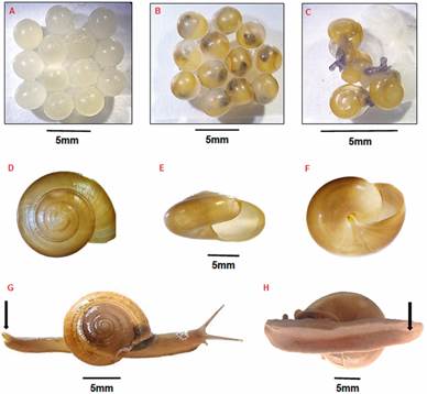
The 36 molluscan specimens collected in the school garden and areas in Cemitério da Lapa exhibited a depressed, fragile, smooth, thin, translucent, and perforated shell. Their surface displayed fine growth lines, a light brown periostrum, a demarcated suture, and a slightly elevated spiracle. The aperture is dextral, semilunar and oblique, with a thin peristome, a curved columellar lip (which is oblique, vertical and slightly reflexed, partially covering the umbilicus), and an outer lip that is sharp and turns varied from five to six (Fig. 2D-F). The shell diameter ranged from 7.4 mm to 21 mm. The body was characterised by a reticulated, greyish epidermis, which became darker in the cephalic region and exhibited more intense pigmentation along the tentacles, while the colouration was lighter near the sole of the foot. In response to disturbance, the animal released a yellowish-green mucus. The sole was tripartite, while the posterior dorsal end of the foot was longitudinally bipartite, forming a raised protuberance known as the caudal horn (Fig. 2G-H). This is a characteristic feature of the species T. indica.
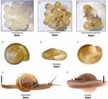
(A) whitish eggs of Tanychlamys indica in the first days of laying. (B) light brown eggs days after laying. (C) newly hatched specimens. (D) shell of T. indica in apical view. (E) shell opening. (F) basal view. (G) live specimen of T. indica collected in Vila Leopoldina neighbourhood arrow indicates caudal horn. (H) preserved specimen of T. indica collected in Vila Leopoldina neighbourhood. The arrows show in details the tripartite sole.
In February 2023, 16 specimens of T. indica were formally catalogued in the malacological collection of the LABFAUNA, bearing the designation 40215-1/2023. At the LABFAUNA collection, one of the molluscan species with a shell diameter of 18.4 mm was observed to lay a clutch of 53 eggs. The eggs were observed to be round, translucent, and whitish in colour. It was observed that the eggs underwent a change in colour, becoming light brown as the embryos developed. The hatching process occurred between 18 and 21 days after the initial deposition of the eggs (Fig. 2A-C).
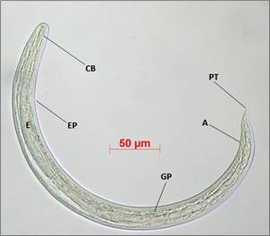
A total of 145 L3 larvae were obtained from a batch of 20 T. indica specimens that were individually digested, with an infection rate of 20% (n = 4). In the gastropods that exhibited natural infection by Angiostrongylid larvae, the shell diameter ranged from 19 to 21 mm, with the number of larvae recovered varying from two to 92. Specifically, two, 21, 30, and 92 larvae were isolated from four specimens of T. indica. The 30 L3 larvae employed for the morphometric study exhibited a filiform body, furrowed cuticle, rounded anterior end, elongated buccal capsule, and a claviform bulb situated at the terminus of a lengthy oesophagus. The excretory pore was identified in the mid-region of the oesophagus. Furthermore, a genital primordium was observed in the posterior third of the intestine, and the larvae exhibited a pointed tail (Fig. 3). The results of the morphometric analyses of the 30 specimens are presented in Table I.
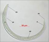
light microscopy of a third-stage larvae (L3) of Angiostrongylus cantonensis isolated from Tanychlamys indica collected at a municipal school in the Vila Leopoldina neighbourhood in February 2023. At the anterior end of the L3 larvae (CB) buccal capsule, (EP) excretory pore, (E) oesophagus. Posterior end of larva showing pointed tail (PT), genital primordium (GP) and anus (A).
Table II presents the results of the molecular analysis of L3 larvae naturally infecting four specimens of T. indica. The results are presented as Ct values obtained for each DNA concentration. Control samples were prepared using standard strains of A. costaricensis and A. cantonensis. The results demonstrated that all samples obtained from T. indica in qPCR were positive for A. cantonensis.
DISCUSSION
Tanychlamys indica is a species of land snail belonging to the family Ariophantidae. The species is native to the Indian subcontinent, including India, Sri Lanka, and parts of Nepal and Bangladesh. It inhabits moist and shaded areas, such as forests, gardens, and agricultural fields.39 The accidental introduction of T. indica to other regions of the world has occurred as a consequence of human activities, including the trade in plants and agricultural products. The species’ adaptability and rapid reproduction have facilitated its expansion into new areas, particularly in tropical and subtropical regions, where it has found favourable conditions for survival and proliferation.32
In addition to the Indian subcontinent, T. indica has been documented in a number of other Asian countries, including Saudi Arabia, the Philippines, Indonesia, Malaysia, Japan and Thailand.27-31,39 In these regions, the species has successfully established itself in habitats that are similar to those of its native range. While less prevalent, there are also documented instances of T. indica in select regions of South America, the Caribbean, and the United States. In Brazil, the introduction of T. indica is associated with the trade in citrus fruit seedlings, initially in the State of Santa Catarina and subsequently in the State of Paraná.37
The molluscan specimens were identified as belonging to the species T. indica, which represents the inaugural record of this gastropod in the municipality of São Paulo, specifically in the Vila Leopoldina neighbourhood. The area in question covers 7.2 km², has a population of 49.020 inhabitants, a density of 41.93 inhabitants/ha, and a very high Human Development Index (HDI) of 0.907. The district is an important economic hub in the city of São Paulo and is home to the Companhia de Entrepostos e Armazéns Gerais de São Paulo (CEAGESP), which is the largest fruit, vegetable, and flower wholesale centre in Latin America.
The discovery of T. indica in the garden area of a municipal school and in the vicinity of the tombs of the Cemitério da Lapa indicates that the species may have been introduced into the municipality via the trade of flowers, grass, ornamental plants, and fruit plant seedlings. This hypothesis is corroborated by the proximity of these businesses to the school and the Cemitério da Lapa, as well as their location just 2 km away from CEAGESP. The larvae of A. cantonensis that were recovered from T. indica were visually classified as belonging to the larval stage L3 based on their morphometry and morphology. While the tail ending in a fine tip (lanceolate tail) is a typical feature of the species, it is not a precise taxonomic identification factor. However, the L3 measurements were compatible with those obtained by Ash41 Thiengo et al.20 and Mota et al.19
Molecular analysis confirmed the presence of A. cantonensis larvae in a population of T. indica in an urban area of the city of São Paulo, with an infection rate of 20.0%. In Brazil, the distribution of A. cantonensis is closely associated with the dispersal of the African snail Achatina fulica Bowdich, 1822, which is considered one of the main potential transmitters of human neural angiostrongyliasis. This is due to the snail’s wide distribution, high population densities and proximity to humans. It is among the top 100 most invasive species globally and represents a significant economic burden as an agricultural pest. Furthermore, it is acknowledged as an urban pest, with documented occurrences in all Brazilian states.15-25,45 The variation in the rate of infection by A. cantonensis in different populations of A. fulica has been considerable in other studies conducted in Brazil. The prevalence was found to be 21.7% in nine municipalities within the Baixada Santista region46 in two other municipalities in Rio de Janeiro, the prevalence was 35.4% in São Gonçalo,17 and 10.3% in Barra do Piraí.17 In Joinville, SC, the prevalence was 27.4%.17 In São Bernardo do Campo/SP, the prevalence was 75%.47
At present, over 140 species of mollusc have been identified as natural or experimental intermediate hosts for A. cantonensis.48 This suggests that A. cantonensis has a wide range of intermediate hosts.
Natural infection by A. cantonensis in T. indica had not been recorded since its introduction into the Southern region of Brazil. This is the first report of A. cantonensis infection of T. indica in Brazil, thus extending the list of molluscs considered to be intermediate hosts of this nematode in the country to nine species.11,15-20,22,23 However, T. indica had already been reported as an intermediate host of A. cantonensis in Bombay, India, together with the slug Laevicaulis alte (Férrusac, 1822).49
In light of the data presented here, it is imperative to implement a surveillance programme for land molluscs, with particular focus on T. indica, in order to gain insight into epidemiological patterns and to facilitate the control of diseases transmitted by this gastropod. Furthermore, this study highlights the necessity for social programmes aimed at raising awareness among local human populations, with the objective of reducing the incidence of human infection, given that cases of neuroangiostrongyliasis have already been documented in the city of São Paulo and in other regions of the country.15,50,51,52
It is noteworthy that the area in which T. indica was identified as naturally infected is in close proximity (approximately 2 km) to CEAGESP, which oversees the largest public network of warehouses, silos, and granaries in the State of São Paulo, comprising 12 active units distributed across the state. Additionally, the company maintains a network of warehouses (deposits or sales of goods) comprising 13 active units, which are also distributed throughout the State of São Paulo. Among these warehouses is the Entreposto Terminal São Paulo (ETSP), which is the largest supply centre for fruit, vegetables, flowers, fish, and miscellaneous products (garlic, potatoes, onions, dried coconuts, and eggs) in South America. ETSP, where approximately 48.000 individuals and 14.000 vehicles traverse the site on a daily basis,53 a scenario that may facilitate the unintentional dispersal of T. indica infected with A. cantonensis to other regions of the State of São Paulo and the country through the land transportation of fruit, vegetables, flowers and ornamental plants.2,3
In conclusion, T. indica is a species with a complex history of global dispersal, which has been influenced by both human activity and the species’ own adaptive capacity. The advent of this species in new regions presents challenges for both agriculture and public health, underscoring the necessity for efficacious management and control strategies to mitigate its impacts.
It is evident that further research is imperative to map the distribution of T. indica in Brazil and to ascertain its potential as an intermediate host of the nematode A. cantonensis, as well as to evaluate the economic risks it poses to agriculture. Over the past two decades, there have been reports of human cases of neuroangiostrongyliasis in Brazil, specifically in the states of Rio de Janeiro, Espírito Santo, São Paulo, Minas Gerais, Amazonas, Amapá, Pará, Paraná, Rio Grande do Sul, and Santa Catarina. Furthermore, cases of natural infection of A. cantonensis have been documented in molluscs and rodents in various regions of the country.
ACKNOWLEDGEMENTS
To Osvaldo Rodrigues de Souza and Rodrigo Ribeiro Gaspar de Almeida, endemic disease control agents (ACS) from the Unidade de Vigilância em Saúde (UVIS Lapa/Pinheiros), for their technical support during the field collections. Their expertise and dedication were invaluable to the success of this study.
REFERENCES
- 1 Godan D. Pest slugs and snails: biology and control. Springer-Verlag; 1983. 448 pp.
- 2 Barker GM. Molluscs as crop pests. New York: CABI publishing; 2002. 400 pp.
- 3 Cowie RH, Robinson DG. Pathways of introductions of nonindigenous land and freshwater snails and slugs. In: Carlton J, Ruiz GM, editors. Invasive species vectors and management strategies. Washington, DC: Island Press; 2003. p. 93-122.
- 4 Mc Donnell MJ, Hahs AK. Adaptation and adaptedness of organisms to urban environments. Annu Rev Ecol Evol Syst. 2015(46): 261-80.
- 5 Robinson DG. Alien invasions: the effects of the global economy on nonmarine gastropod introductions into the United States. Malacologia. 1999(41): 413-38.
- 6 Michailides TJ, Morgan DP. Effect of snail (Helix aspersa) damage on Botrytis gray mold caused by Botrytis cinerea in kiwifruit. Plant Dis. 1996(80): 1141-6.
- 7 Borkakati RN, Gogoi R, Borah BK. Snail: from present perspective to the history of assam. Asian Agri-Hist. 2009(13): 227-37.
- 8 Valente R, Robles MR, Diaz JI. Gastropods as intermediate hosts of Angiostrongylus spp. in the Americas: bioecological characteristics and geographical distribution. Mem Inst Oswaldo Cruz. 2020; 115: e200236.
- 9 Foronda P, López-González M, Miquel J, Torres J, Segovia M, Abreu-Acosta N, et al. Finding of Parastrongylus cantonensis (Chen, 1935) in Rattus rattus in Tenerife, Canary Islands (Spain). Acta Trop. 2010; 114: 123-7.
- 10 Lunn JA, Lee R, Smaller J, MacKay BM, King T, Hunt GB, et al. Twenty-two cases of canine neural angiostrongyliosis in eastern Australia (2002-2005) and a review of the literature. Parasit Vectors. 2012; 5: 70.
- 11 Carvalho OS, Scholte RGC, Mendonça CLF, Passos LKJ, Caldeira RL. Angiostrongylus cantonensis (Nematode: Metastrongyloidea) in molluscs from harbour areas in Brazil. Mem Inst Oswaldo Cruz. 2012; 107(6): 740-6).
- 12 Martin-Alonso A, Foronda P, Quispe-Ricalde MA, Feliu C, Valladares B. Seroprevalence of Angiostrongylus cantonensis in wild rodents from the Canary Islands. PLoS One. 2011; 6(11): e27747.
- 13 Rollins RL, Medeiros MCI, Cowie RH. Stressed snails release Angiostrongylus cantonensis (rat lungworm) larvae in their slime. One Health. 2023; 17: 100658.
- 14 Wang QP, Lai DH, Zhu XQ, Chen XG, Lun ZR. Human angiostrongyliasis. Lancet Infect Dis. 2008; 10: 621-6.
- 15 Morassutti AL, Thiengo SC, Fernandez M, Sawanyawisuth K, Graeff-Teixeira C. Eosinophilic meningitis caused by Angiostrongylus cantonensis: an emergent disease in Brazil. Mem Inst Oswaldo Cruz. 2014; 109(4): 399-407.
- 16 Caldeira RL, Mendonça CLGF, Goveia CO, Lenzi HL, Graeff-Teixeira C, Lima WS, et al. First record of molluscs naturally infected with Angiostrongylus cantonensis (Chen, 1935) (Nematoda: Metastrongylidae) in Brazil. Mem Inst Oswaldo Cruz. 2007; 102(7): 887-9.
- 17 Maldonado Jr A, Simões R, Oliveira APM, Motta EM, Fernandez MA, Pereira ZM, et al. First report of Angiostrongylus cantonensis (Nematoda: Metastrongylidae) in Achatina fulica (Mollusca: Gastropoda) from Southeast and South Brazil. Mem Inst Oswaldo Cruz. 2010; 105(7):938-41.
- 18 Orico LD, Barbosa CM, Luca LR, Soares RM, Gregori F. Angiostrongylus cantonensis and Ancylostoma caninum detection in snails of São Paulo city (2016-2017), Brazil. Ars Vet. 2019; 35(3): 115-21.
- 19 da Mota DJG, de Melo LCV, Pereira-Chioccola VL, Gava R, Pinto PLS. First record of natural infection by Angiostrongylus cantonensis (Nematoda: Metastrongyloidea) in Belocaulus willibaldoi and Rattus norvegicus in an urban area of São Paulo city, SP, Brazil. Heliyon. 2020; 8: e05150.
- 20 Thiengo SC, Maldonado A, Mota EM, Torres EJ, Caldeira R, Carvalho OS, et al. The giant African snail Achatina fulica as natural intermediate host of Angiostrongylus cantonensis in Pernambuco, northeast Brazil. Acta Trop. 2010; 115: 194-9.
- 21 Ramos-de-Souza J, Thiengo SC, Fernandez MA, Gomes SR, Antônio JC, Clímaco MC, et al. First records of molluscs naturally infected with Angiostrongylus cantonensis (Nematoda: Metastrongyloidea) in Northeastern Brazil, including new global records of natural intermediate hosts. Rev Inst Med Trop São Paulo. 2018; 60: e51.
- 22 Souza FN, Santos MA, Alves DA, de Melo LCV, da Mota DJG, Pertile AC, et al. Angiostrongylus cantonensis in urban populations of terrestrial gastropods and rats in an impoverished region of Brazil. Parasitology. 2021; 148: 994-1002.
- 23 Moreira VL, Giese EG, Melo FT, Simões RO, Thiengo SC, Maldonado Jr A, et al. Endemic angiostrongyliasis in the Brazilian Amazon: natural parasitism of Angiostrongylus cantonensis in Rattus rattus and R. norvegicus, and sympatric giant African land snails, Achatina fulica. Acta Trop. 2013; 125: 90-7.
- 24 Barbosa TA, Thiengo SC, Fernandez MA, Graeff-Teixeira C, Morassutti AL, Mourão FRP, et al. Infection by Angiostrongylus cantonensis in both humans and the snail Achatina (Lissachatina) fulica in the city of Macapá, in the Amazon Region of Brazil. Mem Inst Oswaldo Cruz. 2020; 115: e200115.
- 25 Ramos-de-Souza J, Gomes SR, de Mattos AC, de Sousa AKP, da Silva EF, Maldonado Jr A, et al. Achatina fulica infected by Angiostrongylus cantonensis in Manaus, Brazilian Amazon Region, and the risk of transmission of eosinophilic meningitis. J Trop Pathol. 2023; 52(4): 295-303.
- 26 Ramakrishna S, Mitra SC, Dey A. Annotated checklist of Indian land molluscs. Rec Zool Surv India. 2010; 306: 1-359.
- 27 Kudo K, Kagawa O, Ito S, Wada S, Nishi H, Wada S, et al. Species identification and invasion pathways of an introduced snail Macrochlamys sp. in Japan. BioInvasions Records. 2022; 11(4): 839-54.
- 28 Sajan SK, Tripathy B, Biswas T. Species inventory of land and freshwater molluscs from Andhra Pradesh and Telangana states of India. Rec Zool Surv India. 2018; 118: 141-55.
-
29 Blanford WT, Godwin-Austen HH. The fauna of British India, including Ceylon and Burma. Mollusca. Testacellidae and Zonitidae. London: Taylor y Francis; 1908. p. 95-96. Available from: https://archive.org/stream/molluscatestacel00blaniala#page/94/mode/2up
» https://archive.org/stream/molluscatestacel00blaniala#page/94/mode/2up - 30 Singh S, Sandhu RK, Aravind NA. Record of pestiferous land snail, Macrochlamys indica Godwin-Austen 1883 (Gastropoda: Ariophantidae), on citrus and guava plants in Punjab, India. Rec Zool Surv India. 2020; 120: 293-6.
- 31 Sajan S, Tripathy B, Chandra K, Sivakumar K. A new species of the genus Macrochlamys Gray, 1847 (Stylommatophora: Ariophantidae) from western Himalaya. Indian J Nat Hist. 2019; 53: 797-813.
- 32 Raut SK, Ghose KC. Pestiferous land snails of India, Zoological Survey of India, Calcutta. Technical Monologue. 1984; 11: 1-151.
- 33 Cowie RH, Dillon RT, Robinson DG, Smith JW. Alien non-marine snails and slugs of priority quarantine importance in the United States: a preliminary risk assessment. Am Malacol Bull. 2009; 27: 113-32.
- 34 Jayashankar M, Reddy MS, Ramakrishna S. Incidence of the common garden snail, Macrochlamys indica Benson, 1832 (Gastropoda: Ariophantidae) in Bangalore region. Bioscan. 2015; 10: 1003-6.
-
35 Talamas EJ. Pest alert. The horntail snail, Macrochlamys indica Benson, detected in South Florida. FDACS-P-01925. 2020. Available from: https://ccmedia.fdacs.gov/content/download/93400/file/horntail-snail-pest-alert.pdf
» https://ccmedia.fdacs.gov/content/download/93400/file/horntail-snail-pest-alert.pdf -
36 Revinthy AM, Carrilo D, Seal DR, Vassilaros EV, Kendra PE. The horntail snail (Macrochlamys indica): a new invasive pest in Florida. IFAS Extension. 2022; 2022(2). https://doi.org/10.32473/edis-in1355-2022
» https://doi.org/10.32473/edis-in1355-2022 - 37 Agudo-Padrón I, Luz JS. First confirmed occurrence record of a Indo-Asiatic land snail in Brazil and the Americas. Rev Minerva. 2017; 1: 19-27.
- 38 Salvador RB, Miranda MS, Silva FS, Oliveira CDC, Arruda JO, Cavallari DC, et al. Checklist of the terrestrial gastropods of Brazil. J Conchology. 2024; 45: 142-85.
- 39 Abobakr Y, Al-Sarar AS, Alzabib AA, Saleh AA. Morphological and molecular characterization of the invasive pestiferous land snail Macrochlamys indica Godwin-Austen, 1883 (Gastropoda: Ariophantidae) from Saudi Arabia. Agriculture. 2022; 12: 1756.
- 40 Rugai E, Mattos T, Brisola A. Nova técnica para isolar larvas de nematóides das fezes - modificação do método de Baermann. Rev Ins Adolfo Lutz. 1954; 14: 5-8.
- 41 Ash LR. Diagnostic morphology of the third-stage larvae of Angiostrongylus cantonensis, Angiostrongylus vasorum, Angiostrongylus abstrusus and (Nematoda: Metastrongyloidea) Anafilaroides rostratus. J Parasitol. 1970; 56: 249-53.
- 42 Gasser RB, Chilton NB, Hoste H, Beveridge I. Rapid sequencing of rDNA from single worms and eggs of parasitic helminths. Nucleic Acids Res. 1993; 21: 2525-26.
- 43 Caldeira RL, Carvalho OS, Mendonça CLFG, Graeff-Teixeira C, Silva MCF, Ben R, et al. Molecular differentiation of Angiostrongylus costaricensis, A. cantonensis, and A. vasorum by polymerase chain reaction-restriction fragment length polymorphism. Mem Inst Oswaldo Cruz. 2003; 98(8): 1039-43.
- 44 Qvarnstrom Y, da Silva AC, Teem JL, Hollingsworth R, Bishop H, Graeff-Teixeira C, et al. Improved molecular detection of Angiostrongylus cantonensis in mollusks and other environmental samples with a species-specific internal transcribed spacer 1-based TaqMan assay. Appl Environ Microbiol. 2010; 76: 5287-9.
- 45 Graeff-Teixeira C. Expansion of Achatina fulica in Brazil and potential increased risk for angiostrongyliasis. Trans R Soc Trop Med Hyg. 2007; 101: 743-4.
- 46 Guerino LR, Pecora IL, Miranda MS, Aguiar-Silva C, Carvalho OD, Caldeira RL, et al. Prevalence and distribution of Angiostrongylus cantonensis (Nematoda, Angiostrongylidae) in Achatina fulica (Mollusca, Gastropoda) in Baixada Santista, São Paulo, Brazil. Rev Soc Bras Med Trop. 2017; 50: 92-8.
- 47 Cardoso CV, Vaccas DC, Bondan EF, Martins MFM. Prevalence of Angiostrongylus cantonensis and Angiostrongylus costaricensis in Achatina fulica snails in the municipality of São Bernardo do Campo (SP, Brazil). Arq Bras Med Vet Zootec. 2020; 72: 273-6.
- 48 Kim JR, Hayes KA, Yeung NW, Cowie RH. Diverse gastropod hosts of Angiostrongylus cantonensis, the rat lungworm, globally and with a focus on the Hawaiian Islands. PLoS One. 2014; 9(5): e94969.
- 49 Limaye LS, Pradhan VR, Bhopale MK, Renapurkar DM, Sharma KD. Angiostrongylus cantonensis study of intermediate paratenic and definitive hosts in greater Bombay India. Helminthologia. 1988; 25: 31-5.
- 50 Espírito-Santo MC, Pinto PLS, Mota DJG, Gryschek RCB. The first case of Angiostrongylus cantonensis eosinophilic meningitis diagnosed in the city of São Paulo, Brazil. Rev Inst Med Trop. 2013; 55: 129-32.
- 51 Andrade GC, Dias JRO, Maia A, Kanecadan LA, Moraes NSB, Belfort Jr R, et al. Intravitreal Angiostrongylus cantonensis: first case report in South America. Arq Bras Oftalmol. 2018; 81(1): 63-5.
- 52 Melo LVC. Guia informativo: Neuroangiostrongíliase/Angiostrongylus cantonensis - Informações e Diagnóstico Laboratorial. São Paulo: SES/CCD/IAL; 2024. 34 pp.
-
53 CEAGESP [Homepage on Internet]. [cited 2024 Aug 20]. Available from: https://ceagesp.gov.br/acesso-a-informacao/institucional/a-ceagesp
» https://ceagesp.gov.br/acesso-a-informacao/institucional/a-ceagesp
-
1
How to cite: da Mota DJG, Rocco SC, Di Luca LR, dos Santos JA, Werneck EFP, Baccin AO, et al. First record of natural infection by Angiostrongylus cantonensis (Nematoda: Metastrongyloidea) in Tanychlamys indica (Godwin-Austen, 1883) in the city of São Paulo, Brazil. Mem Inst Oswaldo Cruz. 2025; 120: e240192.