Assessing parasitic contamination in Katsuwonus pelamis
ABSTRACT
Katsuwonus pelamis is a fish resource of high economic value and widely used in the fish processing industry, mainly for canned products. This study aimed to identify the parasitic fauna of Katsuwonus pelamis by morphological methods, to analyze the potential impact on industry and consumers. For the evaluation of parasitological contamination, fifty-three animals were visually inspected and the structures with morphology consistent with parasitic forms were analyzed stereoscopically, microscopically, and by Scanning Electron Microscopy (SEM). Two genera of zoonotic parasites have been found: Anisakis sp. (Nematoda) and Trypanorhyncha gen. sp. (Cestoda). Of the 53 samples analyzed, 96% showed parasite contamination. The most affected organs were the intestine, muscle tissue, and stomach, with 596, 441, and 408 parasites, respectively. These results highlight the importance of sanitary inspection and emphasize the importance of immediate evisceration to avoid the migration of larvae from the viscera to the musculature.
Keywords:
Seafood; Food safety; Zoonosis; Pelagic fish; Anisakis
Katsuwonus pelamis, also known as katsuo, oceanic bonito, or striped tuna, is a pelagic fish belonging to the Scombridae family. This migratory species is distributed and caught in tropical and temperate waters worldwide (Aoki et al., 2017). Highly commercially valuable, it ranked third among the most captured marine species globally in 2022, with a total catch of 3.1 million tons (FAO, 2024). In Brazil, it is the most abundant tuna species, with 31,435 tons captured in 2023 (ICCAT, 2025), and of great commercial importance, primarily used in the fish canning industry. It is also widely marketed fresh or frozen globally.
The presence of parasites in fish poses a serious health problem. Some parasites exhibit zoonotic behavior, potentially harming human health if ingested. Furthermore, the presence of parasites in fish reduces the commercial value of the product, leading to considerable economic losses (Dias et al., 2010). Zoonoses occur when humans accidentally consume infested seafood (Fæste et al., 2015). Among zoonoses transmitted through the ingestion of fish parasites, anisakiasis is particularly important due to its global distribution and potential impact on human health. This zoonosis is caused by ingesting the marine parasite of the genus Anisakis. Anisakis uses cetaceans as its definitive host and small crustaceans and fish as intermediate hosts. In fish, most nematode larvae are located within and on the visceral organs, mesenteries, and peritoneum. However, larvae can migrate to surrounding tissues, with some penetrating deep into the fish muscle (Levsen and Lunestad, 2010). Human anisakiasis manifests in ectopic, gastric, intestinal, and gastro-allergic clinical forms (Audicana and Kennedy, 2008; Caldeira et al., 2020). The nematode can penetrate the human intestinal wall, causing an acute, transient infection characterized by symptoms such as abdominal pain, vomiting, nausea, and/or diarrhea (Audicana and Kennedy, 2008). The clinical presentation of anisakiasis extends beyond gastrointestinal symptoms, with reports of associated allergic reactions in some individuals (Ivanovic et al., 2015).
Considering its importance to fisheries worldwide, studies on the parasites of Katsuwonus pelamis are still scarce. However, the existing research is of fundamental importance for identifying the parasitic fauna and infection sites in this fish species. Table 1 describes some of the parasites already found in Katsuwonus pelamis, their infection sites, and fish origin, as reported in the literature.
As can be observed, most infection sites of parasites in Katsuwonus pelamis are located in the gills or are part of the gastrointestinal system. This highlights the importance of performing evisceration as quickly as possible, thus preventing the migration of parasites into the fish’s muscular tissue (Aquino et al., 2021; Magalhães et al., 2012).
This study aimed to investigate the parasitological contamination in Katsuwonus pelamis using morphological methods and scanning electron microscopy (SEM).
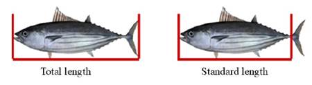
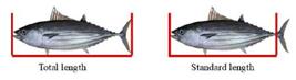
Fifty-three Katsuwonus pelamis samples were captured in 2019 and 2021 in FAO fishing areas 2.2, 2.3, and 3.1. The tuna was packed in insulated boxes and transported to the Aquatic Organisms Health Laboratory (AQUOS), Federal University of Santa Catarina (UFSC), Florianópolis, Brazil, and stored frozen at -18°C. The sex, weight (kg), and total and standard lengths (cm) of the animals were recorded. Total length (cm) was measured from the tip of the mouth to the tip of the caudal fin, and standard length (cm) was measured from the tip of the mouth to the end of the caudal peduncle (Figure 1).
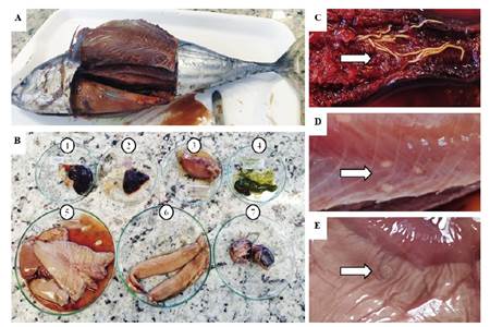
The specimens underwent necropsy for parasitological evaluation of the musculature and internal organs. For this purpose, the eyes, gills, gastrointestinal tract, and viscera were removed and examined. Evisceration was performed via longitudinal abdominal ventral incision from the operculum to the cephalic region. To investigate parasitism in the flesh (muscle), two incisions were made longitudinally on both sides, extending from the insertion of the tail towards the head to the level of the operculum, thus obtaining two fillets per fish (Figure 2).
The samples were visually inspected, and structures with morphology suggestive of parasites were analyzed using both stereoscopic microscopy and an Olympus CX22 microscope (Tokyo, Japan). The parasites found were fixed in 70% alcohol for subsequent identification according to Eiras et al. (2006). Following parasite collection, a clearing process was performed using beech creosote to enhance the visibility of internal structures. Subsequently, the parasites were mounted on permanent slides with Canada balsam and then analyzed microscopically to identify structures and species using taxonomic keys (Moravec, 1998).
Scanning electron microscopy (SEM) was performed on collected parasite samples. Samples were fixed onto stubs and sputter-coated with a 40 nm layer of gold (Au) under vacuum in a planetary coater (LEICA EM, model SCD500, Wetzlar, Germany), following the methodology described by Bozzola and Russell (1999). This study was conducted at the Central Laboratory of Electron Microscopy (LCME) at UFSC. Samples were then imaged using a JEOL JSM-6390LV scanning electron microscope (Peabody, MA, USA) using an accelerating voltage of 15 kV up to 500× magnification.
The specimens included 24 males, 25 females, and 4 individuals of undetermined sex. The average weight was 3.430 ± 1.41 kg, the average total length was 39.06 ± 17.39 cm, and the average standard length was 33.48 ± 15.37 cm.
Necropsy was performed on 10 organs, i.e., spleen, heart, intestine, gallbladder, stomach, liver, gonad, eyes, muscle (flesh), and gills (Figure 2b). Only the kidney and heart were free of parasitic contamination in all samples. In Table 2, the prevalence and intensity of parasite infection of the examined organs is reported.
Parasites detected in Katsuwonus pelamis, infection sites, and fish origin reported in the literature.
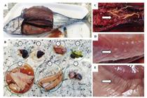
(A) Necropsied tuna; (B) Internal Organs: (1) spleen, (2) heart, (3) intestine, (4) gallbladder, (5) liver, (6) gonad, (7) eyes; Infected tissues: (C) Rhadinorhynchus sp.; (D) Trypanorhyncha; and (E) Anisakis sp.
The results indicated that 58.5% of the specimens exhibited parasitic contamination in the muscle tissue (Figure 2D), while 41.5% presented contamination in other organs (Figure 2 C and E). Parasitic contamination typically originates in the viscera, subsequently spreading to other organs and muscle tissue (Levsen and Lunestad, 2010). In fact, the intestine was the most heavily infested organ, with 96% of the samples contaminated.
In natural environments, fish and parasites coexist in equilibrium. However, environmental disturbances and alterations (typically associated with human activities), such as reduced dissolved oxygen levels, elevated carbon dioxide (CO2), ammonia (NH3), and nitrite (NO2) concentrations, high stocking densities, and intensive feeding practices, among other factors, can induce stress, reduce host resistance, promote injuries, and facilitate parasite development (Pakdeenarong et al., 2014).
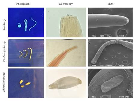
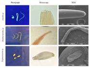
The parasites encountered were identified as belonging to the genera Anisakis sp. (Nematoda) and Rhadinorhynchus sp. (Acanthocephala), and the order Trypanorhyncha (Cestoda). Microscopic observation and scanning electron microscopy (SEM) imaging, crucial for identification, are shown in Figure 3.
Ferreira et al. (2006) emphasized the importance of recognizing Trypanorhyncha parasites during fish inspection, noting their repulsive appearance and the need for sanitary inspectors to be aware of them. Mattos et al. (2015) suggested that these parasites might elicit hypersensitivity reactions in humans, potentially due to toxins produced by Trypanorhyncha larvae within fish musculature. While studies on allergic reactions to fish parasite antigens frequently focus on Anisakis nematodes, research has also explored this potential in other parasites, including the cestode Trypanorhyncha. Although accidental human infection with Trypanorhyncha larvae is rare, some species within this group have demonstrated allergenic potential (Gòmez-Morales et al., 2008; Mattos et al., 2015; Rodero and Cuéllar, 1999; Vázquez-López et al., 2001, 2002). Rhadinorhynchus sp. is not reported in the literature as zoonotic.
Pelayo et al. (2009) conducted the first study on the seroprevalence of anti-Gymnorhynchus gigas antibodies in a Spanish population, demonstrating the existence of a specific Trypanorhyncha antigen in humans. Their research indicated that parasite antigens remain active even after fish are frozen, highlighting a potential human health risk. In Brazil, studies using a murine model have shown that Protoperidinium heteracanthum and Pterobothrium crassicolle antigens can induce the production of specific IgG and IgE immunoglobulins through various immunization routes, suggesting that these parasites could potentially induce allergic reactions in humans (Mattos et al., 2015).
Marine nematodes of the family Anisakidae are known to be harmful to human health, causing zoonoses known as anisakiasis (Bao et al., 2019; Fæste et al., 2014, 2015). Both live and dead Anisakis nematodes can elicit allergic reactions, sometimes with serious consequences, including anaphylactic shock (Audicana et al., 2002; Fæste et al., 2015). Several species of Anisakis transmitted by fish are recognized as causative agents of human disease; within the family Anisakidae, human contamination has been reported for Anisakis, Pseudoterranova, Contracaecum, Hysterothylacium spp., and Gnathostoma spp. (Arizono et al., 2011; Herman and Chiodini, 2009; Nawa et al., 2010; Torres et al., 2007).
Concerns regarding food allergies resulting from Anisakis sp. ingestion are prominent in the scientific community, leading to numerous publications on the topic. Although Anisakis simplex larvae are inactivated by heating above 60°C for at least 10 minutes or freezing at -20°C for 24 hours, some allergens are resistant to both cooking and freezing (Bahna, 2016). This resistance to thermal treatment was addressed by Caballero and Moneo (2004), who studied patients exhibiting reactions and symptoms after ingesting cooked or canned fish. Vidaček et al. (2009) and Audicana et al. (2002) also reported the resistance of Anisakis allergens to heat and freezing in their studies, noting that even inactivated larvae can be hazardous.
Literature reports suggest that cooking and/or freezing seafood may not be sufficient to prevent hypersensitivity reactions to Anisakis sp. Thermostable allergens have been detected in Anisakis simplex extracts, raising questions about the safety of consuming these parasites in cooked fish (Caballero and Moneo, 2004; Fæste et al., 2014; Moneret-Vautrin et al., 2005; Nieuwenhuizen and Lopata, 2009). As with other food allergens, small quantities can induce an allergic reaction in sensitized individuals. The presence of Anisakis proteins in fish and fish products poses a potential health risk for consumers allergic to this parasite; cross-reactive allergies are possible, for example, to dust mites or crustaceans (Aibinu et al., 2019).
Few articles report human infections with Anisakis sp. transmitted by fish (Chaves et al., 2016; Cruz et al., 2010; Eiras et al., 2016). However, as the number of infected fish species increases, particularly economically important species such as K. pelamis, so does the risk for humans ingesting nematode-infected fish.
Two genera of zoonotic parasites, Anisakis sp. and Trypanorhyncha, were identified in the analyzed K. pelamis specimens. The presence of Anisakis sp. in the viscera highlights the importance of immediate evisceration post-capture. The detection of Trypanorhyncha in the fish muscle reinforces the need for inspection by responsible agencies or the processing industry.
Beyond their potential impact on human health, parasites can reduce the marketability and value of seafood products due to implications for food safety and quality, decreasing consumer confidence and causing economic losses for the fishing industry. Therefore, knowledge of fish parasites is essential for proper sanitary inspection, as pathogenesis in humans can occur by exploitative, toxic, or mechanical action.
DATA AVAILABILITY STATEMENT
The raw data supporting the conclusions of this article will be made available by the authors on request.
SUPPLEMENTARY MATERIAL
No supplementary material is provided for this article.
ACKNOWLEDGMENTS
The authors thank the Central Laboratory of Electron Microscopy (LCME/UFSC) for the Scanning Electron Microscopy.
REFERENCES
-
Aibinu, I. E., Smooker, P. M. & Lopata, A. L. 2019. Anisakis nematodes in fish and shellfish-from infection to allergies. International Journal for Parasitology: Parasites and Wildlife, 9, 384-393. DOI: https://doi.org/10.1016/j.ijppaw.2019.04.007
» https://doi.org/10.1016/j.ijppaw.2019.04.007 -
Amim, O., Heckmann, R. A., Radwan, N., Anchundia, J. S. M. & Alcivar, M. A. Z. 2009. Redescription of Rhadinorhynchus ornatus (Acanthocephala: Rhadinorhynchidae) from skipjack tuna, Katsuwonus pelamis, collected in the Pacific Ocean off South America, with special reference to new morphological features. Journal of Parasitology, 95(3), 656-664. DOI: 10.1645/GE-1804.1.
» https://doi.org/10.1645/GE-1804.1 -
Anshary, H., Sriwulan Freeman, M. A. & Ogawa, K. 2014. Occurrence and molecular identification of Anisakis Dujardin, 1845 from marine fish in southern Makassar Strait, Indonesia. Korean Journal of Parasitology, 52(1), 9-19. DOI: 10.3347/kjp.2014.52.1.9
» https://doi.org/10.3347/kjp.2014.52.1.9 -
Aoki, Y., Kitagawa, T., Kiyofuji, H., Okamoto, S. & Kawamura, T. 2017. Changes in energy intake and cost of transport by skipjack tuna (Katsuwonus pelamis) during northward migration in the northwestern Pacific Ocean. Deep Sea Research Part II: Topical Studies in Oceanography, 140, 83-93. DOI: https://doi.org/10.1016/j.dsr2.2016.05.012
» https://doi.org/10.1016/j.dsr2.2016.05.012 - Aquino, C. M., Santos, G. G., Martins, M. L. & Scussel, V. M. 2021. Research of parasites with zoonotic potential for humans in skipjack tuna (Katsuwonus pelamis). Scientia Plena, 17(8), 1-7.
-
Arizono, A., Miura, T., Yamada, M., Tegoshi, T. & Onishi, K. 2011. Human infection with Pseudoterranova azari round worm. Emerging Infectious Diseases, 17(3), 555-556. DIO: https://doi.org/10.3201/eid1703.101350
» https://doi.org/10.3201/eid1703.101350 -
Audicana, M. T. & Kennedy, M. W. 2008. Anisakis simplex: From obscure infectious worm to inducer of immune hypersensitivity. Clinical Microbiology Reviews, 21(2), 360-379. DOI: https://doi.org/10.1128/cmr.00012-07
» https://doi.org/10.1128/cmr.00012-07 -
Audicana, M. T., Ansotegui, I. J., De Corres, L. F. & Kennedy, M. W. 2002. Anisakis simplex: Dangerous - dead and alive? Trends in Parasitology, 18(1), 20-25. DOI: https://doi.org/10.1016/S1471-4922(01)02152-3
» https://doi.org/10.1016/S1471-4922(01)02152-3 - Bahna, S. L. 2016. Seafood allergies: Fish and shellfish. Annals of Allergy, Asthma & Immunology, 117(5), 458-461.
-
Bao, M., Pierce, G. J., Strachan, N. J. C., Pascual, S., González-Muñoz, M. & Levsen, A. 2019. Human health, legislative and socioeconomic issues caused by the fish-borne zoonotic parasite Anisakis: Challenges in risk assessment. Trends in Food Science & Technology, 86, 298-310. DOI: https://doi.org/10.1016/j.tifs.2019.02.013
» https://doi.org/10.1016/j.tifs.2019.02.013 - Bozzola, J. J. & Russell, L. D. 1999. Electron microscopy: principles and techniques for biologists. Sudbury, Jones and Bartlett.
-
Caballero, M. L. & Moneo, I. 2004. Several allergens from Anisakis simplex are highly resistant to heat and pepsin treatments. Parasitology Research, 93(3), 248-251. DOI: https://doi.org/10.1007/s00436-004-1099-3
» https://doi.org/10.1007/s00436-004-1099-3 - Caldeira, A. J. R., Araújo, R. B. & Santos, M. J. 2020. É seguro consumir pescado cru? Uma perspectiva sobre a ocorrência de Anisakis spp. em produtos de pesca. Revista Anápolis Digital, 12(3), 1-12.
-
Cárdenas, M. Q., Moravec, F. & Kohn, N. 2009. First record of Philometra katsuwoni (Nematoda: Philometridae), a parasite of skipjack tuna Katsuwonus pelamis (Perciformes: Scombridae), off the South American Atlantic coast. Biota Neotrópica, 9(2), 263-266. DOI: 10.1590/s1676-06032009000200025
» https://doi.org/10.1590/s1676-06032009000200025 -
Chaves, C. M., Chaves, C., Zoroquiain, P., Belfort Jr., R. & Burnier Jr., M. N. 2016. Ocular gnathostomiasis in Brazil: A case report. Ocular Oncology and Pathology, 2(3), 194-196. DOI: https://doi.org/10.1159/000444259
» https://doi.org/10.1159/000444259 -
Crites, J. L., Overstreet, R. M. & Maung, M. 1993. Ctenascarophis lesteri n. sp. and Prospinitectus exiguous n. sp. (Nematoda: Cystidicolidae) from the skipjack tuna Katsuwonus pelamis Journal of Parasitology, 79, 847-859. DOI: 10.2307/3283721
» https://doi.org/10.2307/3283721 - Cruz, A. R., Souto, P. C. S., Ferrari, C. K. B., Allegretti, S. M. & Arrais-Silva, W. W. 2010. Endoscopic imaging of the first clinical case of anisakidosis in Brazil. Scientia Parasitologica, 11(2), 97-100.
-
Dias, F. J. E., São Clemente, S. C. & Knoff, M. 2010. Larvae of Anisakidae nematodes and Trypanorhyncha cestodes of public health importance in Aluterus monoceros (Linnaeus, 1758) in Rio de Janeiro State, Brazil. Revista Brasileira de Parasitologia Veterinária, 19(2), 94-97. DOI: https://doi.org/10.1590/S1984-29612010000200005
» https://doi.org/10.1590/S1984-29612010000200005 - Eiras, J. C., Takemoto, R. M. & Pavanelli, G. C. 2006. Métodos de estudo e técnicas laboratoriais em parasitologia de peixes Maringá, Eduem.
-
Eiras, J. C., Pavanelli, G. C., Takemoto, R. M., Yamaguchi, M. U., Karkling, L. C. & Nawa, Y. 2016. Potential risk of fish-borne nematode infections in humans in Brazil - Current status based on a literature review. Food and Waterborne Parasitology, 5, 1-6. DOI: https://doi.org/10.1016/j.fawpar.2016.08.002
» https://doi.org/10.1016/j.fawpar.2016.08.002 -
Fæste, C. K., Jonscher, K. R., Dooper, M. M. B. W., Egge-Jacobsen, W., Moen, A., Daschner, A., Egaas, E. & Christians, U. 2014. Characterisation of potential novel allergens in the fish parasite Anisakis simplex. EuPA Open Proteomics, 4, 140-155. DOI: https://doi.org/10.1016/j.euprot.2014.06.006
» https://doi.org/10.1016/j.euprot.2014.06.006 -
Fæste, C. K., Plassen, C., Lovberg, K. E., Moen, A. & Egaas, E. 2015. Detection of proteins from the fish parasite Anisakis simplex in Norwegian farmed salmon and processed fish products. Food Analytical Methods, 8(6), 1390-1402. DOI: https://doi.org/10.1007/s12161-014-0003-8
» https://doi.org/10.1007/s12161-014-0003-8 -
FAO (Food and Agriculture Organization). 2024. The state of world fisheries and aquaculture 202: blue transformation in action. Rome, FAO. DOI: https://doi.org/10.4060/cd0683en
» https://doi.org/10.4060/cd0683en -
Ferreira, M. F., São Clemente, S. C., Tortelly, T., Lima, F. C., Nascimento, E. R., Oliveira, G. A. & Lima, A. R. 2006. Parasitas da ordem Trypanorhyncha: sua importância na inspeção sanitária do pescado. Revista Brasileira de Ciências Veterinárias, 13(3), 190-193. DOI: http://dx.doi.org/10.4322/rbcv.2014.297
» https://doi.org/10.4322/rbcv.2014.297 - Freire, J. F., Filho, D. S. T., Souza, G. T. R., Madi, R. R., Alves, A. M. & Melo, C. M. 2016. Fauna parasitária de escombrídeos (Perciformes: Scombridae) desembarcados no Terminal Pesqueiro de Aracaju. In: Anais da 18ª Semana de Pesquisa da Universidade Tiradentes - "A prática interdisciplinar alimentando a Ciência", 2016. 1-4.
-
Gòmez-Morales, M. A., Ludovisi, A., Giuffra, E., Manfredi, M. T., Piccolo, G. & Pozio, E. 2008. Allergenic activity of Molicola horridus (Cestoda, Trypanorhyncha), a cosmopolitan fish parasite, in a mouse model. Veterinary Parasitology, 157(3-4), 314-320. DOI: https://doi.org/10.1016/j.vetpar.2008.07.010
» https://doi.org/10.1016/j.vetpar.2008.07.010 -
Herman, J. S. & Chiodini, P. L. 2009. Gnathostomiasis, another emerging imported disease. Clinical Microbiology Reviews, 22(3), 484-492. DOI: https://doi.org/10.1128/cmr.00003-09
» https://doi.org/10.1128/cmr.00003-09 -
Hermida, M., Cavaleiro, B., Gouveia, L. & Saraiva, A. 2018. Parasites of skipjack, Katsuwonus pelamis, from Madeira, Eastern Atlantic. Parasitology Research, 117(4), 1025-1033. DOI: 10.1007/s00436-018-5778-x
» https://doi.org/10.1007/s00436-018-5778-x -
ICCAT (International Commission for the Conservation of Atlantic Tunas). 2025. Available from: https://www.iccat.int/en Access date: 2025 Feb. 10.
» https://www.iccat.int/en -
Ivanovic, J., Baltic, M. Ž., Boškovic, M., Kilibarda, N., Dokmanovic, M., Markovic, R., Janjic, J. & Baltic, B. 2015. Anisakis infection and allergy in humans. Procedia Food Science, 5, 101-104. DOI: https://doi.org/10.1016/j.profoo.2015.09.028
» https://doi.org/10.1016/j.profoo.2015.09.028 -
Justo, M. C. N., Kohn, A., Pereira, C. S. & Flores-Lopes, F. 2013. Histopathology and autoecology of Didymocylindrus simplex (Digenea: Didymozoidae), a parasite of Katsuwonus pelamis (Scombridae) in the Southwestern Atlantic Ocean, off South America. Zoologia, 30(3), 312-316. DOI: https://doi.org/10.1590/S1984-46702013000300008
» https://doi.org/10.1590/S1984-46702013000300008 - Justo, M. C. N., Leão, M. S. L., Kohn, A. & Flores-Lopes, F. 2015. Pathological alterations induced by Pozdnyakovia gibsoni (Digenea: Didymozoidae), a parasite of the skipjack tuna Katsuwonus pelamis (Scombridae). Comparative Parasitology, 82(2), 301-303.
- Kohn, A., Santos, A. L. & Cohen, S. C. 2003. Report of two parasites from Brazilian tunas. Arquivos de Ciências do Mar, 36, 19-22.
-
Levsen, A. & Lunestad, B. T. 2010. Anisakis simplex third stage larvae in Norwegian spring spawning herring (Clupea harengus L.), with emphasis on larval distribution in the flesh. Veterinary Parasitology, 171(3-4), 247-253. DOI: https://doi.org/10.1016/j.vetpar.2010.03.039
» https://doi.org/10.1016/j.vetpar.2010.03.039 -
Magalhães, A. M. S., Costa, B. S., Tavares, G. C. & Carvalho, S. I. G. 2012. Zoonoses parasitárias associadas ao consumo de carne de peixe cru. Pubvet, 6(25). DOI: https://doi.org/10.22256/pubvet.v6n25.1416
» https://doi.org/10.22256/pubvet.v6n25.1416 -
Mattos, D. P. B. G., Verícimo, M. A., Lopes, L. M. S. & São Clemente, S. C. 2015. Immunogenic activity of the fish tapeworm Pterobothrium heteracanthum (Trypanorhyncha: Pterobothriidae) in BALB/c mice. Journal of Helminthology, 89(2), 203-207. DOI: https://doi.org/10.1017/s0022149x13000795
» https://doi.org/10.1017/s0022149x13000795 -
Mele, S., Macías, D., Gómez-Vives, M. J., Garippa, G., Alemany, F. & Merella, P. 2012. Metazoan parasites on the gills of the skipjack tuna Katsuwonus pelamis (Osteichthyes: Scombridae) from the Alboran Sea (western Mediterranean Sea). Diseases of Aquatic Organisms, 97(3), 219-225. DOI: https://doi.org/10.3354/dao02421
» https://doi.org/10.3354/dao02421 -
Moneret-Vautrin, D. A., Morisset, M., Flabbee, J., Beaudouin, E. & Kanny, G. 2005. Epidemiology of life-threatening and lethal anaphylaxis: A review. Allergy, 60(4), 443-451. DOI: https://doi.org/10.1111/j.1398-9995.2005.00785.x
» https://doi.org/10.1111/j.1398-9995.2005.00785.x - Moravec, F. 1998. Nematodes of freshwater fishes of the neotropical region Prague, Academia.
- Nawa, Y., Katchanov, J., Yoshikawa, M., Rojekittikhun, P., Decumyoy, P., Kusolusuk, T. & Watanakulpanich, D. 2010. Ocular gnathostomiasis: A comprehensive review. The Journal of Tropical Medicine and Parasitology, 33(2), 77-86.
- Nieuwenhuizen, N. & Lopata, A. L. 2009. Allergies in the workplace. Current Allergy and Clinical Immunology, 22, 132-138.
-
Pakdeenarong, N., Siribat, P., Chaisiri, K., Douangboupha, B., Ribas, A., Chaval, Y., Herbreteau, V. & Morand, S. 2014. Helminth communities in murid rodents from southern and northern localities in Lao PDR: The role of habitat and season. Journal of Helminthology, 88(3), 302-309. DOI: https://doi.org/10.1017/s0022149x13000187
» https://doi.org/10.1017/s0022149x13000187 -
Pelayo, V., García-Hernández, P., Puente, P., Rodero, M. & Cuéllar, C. 2009. Seroprevalence of Anti-Gymnorhynchus gigas (Trypanorhyncha, Gymnorhynchidae) Antibodies in a Spanish Population. Journal of Parasitology, 95(3), 778-780. DOI: https://doi.org/10.1645/ge-1894.1
» https://doi.org/10.1645/ge-1894.1 - Rodero, M. & Cuéllar, C. 1999. Humoral immune responses induced by Gymnorhynchus gigas extracts in BALB/c mice. Journal of Helminthology, 73(3), 239-243.
-
Santos, C. P., Gibson, D. I., Tavares, L. E. R. & Luque, J. L. 2008. Checklist of Acanthocephala associated with the fishes of Brazil. Zootaxa, 1938, 1-22. DOI: https://doi.org/10.11646/zootaxa.1938.1.1
» https://doi.org/10.11646/zootaxa.1938.1.1 - Sattari, A., Kheirandish, R., Fard, S. R. N., Shoaibi, B. & Sharifpour, I. 2014. Infection of skipjack tuna Katsuwonus pelamis (Linnaeus, 1758) of Oman Sea with cestode Trypanorhyncha (Diesing, 1863). Iranian Journal of Fisheries Sciences, 13(2), 469-476.
- Silva, C. G., Lima, J. T. A. X. & Figueiredo, N. C. 2023. First record of Gotocotyla acanthura on the gills of Katsuwonus pelamis in the southwestern Atlantic Ocean. Ciência Animal, 27(3), 80-88.
-
Torres, P., Jercic, M. I., Weitz, J. C., Dobrew, E. K. & Mercado, R. A. 2007. Human pseudoterranovosis, an emerging infection in Chile. Journal of Parasitology, 93(2), 440-443. DOI: https://doi.org/10.1645/ge-946r.1
» https://doi.org/10.1645/ge-946r.1 -
Vázquez-López, C., Armas-Serra, C., Bernardina, W. & Rodríguez-Caabeiro, F. 2001. Oral inoculation with Gymnorhynchus gigas induces anti-parasite anaphylactic antibody production in both mice and rats and adverse reactions in challenge mice. International Journal of Food Microbiology, 64(3), 307-315. DOI: https://doi.org/10.1016/s0168-1605(00)00477-3
» https://doi.org/10.1016/s0168-1605(00)00477-3 -
Vázquez-López, C., De Armas-Serra, C., Bernardina, W. & Rodríguez-Caabeiro, F. 2002. A 24-kDa collagenase from Gymnorhynchus gigas elicits rat ileum hyperreactivity and is a target of humoral responses in mice previously given a single oral dose of parasite extract. Digestive Diseases and Sciences, 47(4), 935-942. DOI: https://doi.org/10.1023/a:1014785210407
» https://doi.org/10.1023/a:1014785210407 -
Vidaček, S., de las Heras, C., Solas, M. T., Mendizábal, A., Rodriguez-Mahillo, A., González-Muñoz, M. & Tejada, M. 2009. Anisakis simplex allergens remain active after conventional or microwave heating and pepsin treatments of chilled and frozen L3 larvae. Journal of the Science of Food and Agriculture 89(12), 1997-2002. DOI: https://doi.org/10.1002/jsfa.3677
» https://doi.org/10.1002/jsfa.3677
-
AI USE DISCLOSURE
Artificial intelligence tools (ChatGPT, OpenAI) were used exclusively to refine the English language of this manuscript. The content was carefully reviewed by the authors to ensure consistency and correctness, and the authors are fully responsible for the final version of the manuscript.
-
FUNDING
This research was funded by the Coordination for the Improvement of Higher Education Personnel - Brazil (CAPES), Scholarship of authors 1 and 2 and by the Research Productivity Grant awarded to author 4 (CNPq 303822/2022-8).