 Clinical Longevity of Direct Resin Composite Restorations in Molar-Incisor Hypomineralisation: A Systematic Review
ABSTRACT
Objective: To systematically review the clinical longevity of direct resin composite (RC) restorations on teeth affected by Molar-Incisor Hypomineralisation (MIH).
Material and Methods: This review followed the PRISMA guidelines. Electronic databases (Cochrane Library, EMBASE, Medline/Pubmed, SCOPUS, Web of Science) and gray literature sources (Google Scholar, OpenGrey, ProQuest) were searched. Randomized clinical trials (RCTs) and cohort studies evaluating the longevity of direct RC restorations in MIH-affected molars in participants aged 6 years or older were included. Search covered database inception to November 2023 and was updated in November 2025. Risk of bias was assessed with the ROB-2 tool (RCTs) and the Newcastle-Ottawa Scale (cohort), and certainty of evidence was evaluated using GRADE.
Results: Nine studies (four RCTs and five cohort studies) were included, totaling 487 restorations in individuals aged 6 to 18 years. Overall longevity rates ranged from 49.2% at 24 months to 100% at 48 months, indicating substantial clinical and methodological heterogeneity. In cohort studies, the longevity ranged from 49.2% to 100%, whereas in RCTs, it ranged from 54% to 89.2%. Risk of bias was rated as uncertain, low, and moderate, and effect estimates as very low and moderate.
Conclusion: Longevity of direct RC restorations in MIH-affected teeth varies widely, ranging from 49.2% to 100%. Failures were mainly associated with loss of retention, marginal adaptation, secondary caries, and esthetic shortcomings.
Keywords:
Developmental Defects of Enamel; Composite Resins; Longevity; Evidence Synthesis.
Introduction
Molar-incisor hypomineralisation (MIH) is a qualitative developmental defect of the enamel in the first permanent molars, with or without incisors being affected [1,2]. These enamel defects are clinically characterized by demarcated white, brown, or yellow-brown opacities [3]. The disruption of the hypomineralised enamel increases when the tooth is under masticatory function [1,4]. The clinical management of MIH-affected teeth must consider the location, extension, and severity of the MIH lesion [5]. Other factors, such as tooth eruption stage, orthodontic status, socioeconomic conditions, behavior, and patient age, must also be considered during treatment planning for MIH cases [5,6]. A wide range of treatment modalities is available for MIH-affected teeth, including preventive approaches with remineralizing agents and, in severe cases, tooth extraction [1,5,7]. Restorative treatment is indicated to reestablish the form and function of teeth and protect the dentin-pulp complex, in moderate and severe cases when the hypomineralised enamel is disrupted [8,9]. Restorative materials that offer adequate longevity and minimal reintervention are desirable [10]. Glass ionomer cement, resin composite, amalgam, stainless-steel crowns, and ceramics have been used to restore the enamel defects [5,6,10]. Resin composite (RC) has been widely evaluated in clinical trials and is the material of choice for the direct restoration of MIH-affected molars [5,11].
Despite advances in the composition and properties of resin composites, the restoration of MIH-affected teeth remains a clinical challenge for dental clinicians [12]. The porosity of hypomineralised enamel hinders the material's performance and makes the restoration margins more prone to enamel breakdown [9]. Moreover, the longevity of restorations in MIH-affected teeth can be negatively affected by the decreased elasticity modulus of the enamel, due to the altered formation of tooth minerals, and the compromised performance of adhesive systems, justified by the high protein content in hypomineralised enamel matrix, being similar to dentin [13]. A previous study reported that children with MIH are three times more likely to undergo restorative retreatment than children without MIH [1]. Furthermore, a systematic review stated that MIH contributes to the development of carious lesions in individuals at high risk of caries [9]. The cavity formed by the post-eruptive breakdown of hypomineralised enamel, along with its surface characteristics, can facilitate biofilm accumulation and bacterial adhesion [2,9]. Several studies indicate that MIH-affected molars often receive multiple retreatments [1,2,14].
Although previous systematic reviews [5,6] have addressed different restorative approaches for molar-incisor hypomineralisation (MIH), including direct resin composite restorations, the clinical longevity of these restorations has not been explored as a primary outcome, nor has the certainty of evidence been formally assessed. Given that information on restoration survival is essential to support predictable, cost-effective clinical decision-making, a focused, up-to-date synthesis is needed. Therefore, this systematic review aims to evaluate the clinical longevity of direct resin composite restorations in MIH-affected teeth, providing evidence-based support for restorative planning and contributing to a clearer understanding of their clinical performance.
Material and Methods
Protocol and Registration
This systematic review was conducted according to the Preferred Reporting Items for Systematic Review and Meta-Analyses (PRISMA) guideline [15] and registered (CRD42022372914) on the International Prospective Register of Systematic Reviews (PROSPERO).
Eligibility Criteria
The PICOS acronym was used to formulate the main question of this systematic review, in which: P (population) - MIH-affected first permanent lower/upper molars, I (intervention) - direct resin composite restoration, C (comparison) - not applicable, O (outcome) - success rates/longevity, S (study design) - randomized clinical trials (RCTs) and retrospective/prospective cohort studies. Accordingly, the guiding research question was: “What is the clinical longevity of direct resin composite restorations in MIH-affected teeth?”. RCTs and cohort studies that evaluated direct resin composite restorations in MIH-affected molars with no restrictions regarding language or year of publication were considered eligible. Moreover, only studies involving participants aged 6 or older with MIH-affected molars were eligible.
Were excluded from this systematic review: a) observational cross-sectional studies, case reports, case series, narrative reviews, animal model studies, and in vitro studies; b) studies that indicated direct resin composite restorations but without a precise diagnosis of MIH; and c) studies without quantitative data or those whose data could not be estimated.
Databases and Search Strategy
MeSH descriptors, entry terms, and keywords related to the terms included in the PICOS question were used to formulate the search strategy on the following electronic databases: Cochrane Library, EMBASE, Medline/Pubmed, SCOPUS, and Web of Science (Table 1). Additional searches in the gray literature were also conducted, including Google Scholar, OpenGrey, ProQuest, and a hand search of the references from the included studies. The searches were conducted in November 2023 and updated in November 2025.
Search strategies defined appropriately for each database (Searches performed on November 12th, 2023, and updated on November 18, 2025).
Automated monthly search alerts were created in electronic databases to identify newly published studies potentially eligible for inclusion until the final search update. An online reference manager (https://www.endnoteweb.com/EndNoteWeb.html) was used to retrieve the references and remove duplicates.
Study Selection Process
Two independent reviewers (JFZ and JORS) selected the studies in two phases. In phase 1, studies retrieved from electronic databases were selected by reading their titles and abstracts using online software (Rayyan, https://www.rayyan.ai/). The studies that did not match the inclusion criteria were excluded. In phase 2, after reading the full texts of the articles, the same reviewers included the studies based on the eligibility criteria. Disagreements between reviewers were resolved by consensus, and when necessary, a third reviewer (AMS) was consulted. The use of Rayyan ensured a blinded and independent screening process.
Data Collection Process
The first reviewer (JFZ) collected the data from the included studies, which were individually checked by the second reviewer (JORS). In case of disagreements, both discussed the collected data and/or complemented the data. Initially, an electronic sheet was elaborated containing relevant information retrieved from the included studies, such as the study’s information (authors, year of publication, country), study design, sample size, participant’s age, number of restorations, calibration of the operator, type of restorative material and adhesive system, surface treatment of hard tissues, criteria, and period of evaluation of the restorations, and success rates.
Restoration success was defined according to the clinical evaluation criteria adopted in each original study (USPHS/modified USPHS, FDI, or Innes criteria). The success rates and longevity of direct resin composite restorations (main outcome) were extracted as reported in the original studies, in absolute or relative frequencies, and subsequently converted into relative frequencies (%) for each reported evaluation period. When multiple follow-up time points were available, the percentage corresponding to the final evaluation period of each study was considered representative of the reported longevity. No further temporal standardization or adjustment (such as annualized success rates or recalculated cumulative survival estimates) was performed, as the comparison across studies was descriptive in nature and substantial clinical and methodological heterogeneity was observed.
When some imperative information was unavailable or unclear in the article, the corresponding authors were contacted by e-mail or via a scientific social networking service (ResearchGate) to provide the required information.
Individual Risk of Bias
For RCTs, the assessment was performed following the Cochrane Collaboration risk-of-bias tool [16]. This tool is structured into domains: bias arising from the randomization process (bias of selection), missing allocation, blinding examiners, participants’ withdrawn, selective outcome reporting, and other possible biases. A judgment about the risk of bias arising from each domain is proposed by an algorithm, based on answers to the signaling questions. Judgments can be ‘Low’, or ‘High’ risk of bias, or can express ‘Some concerns’, according to the Handbook for Systematic Reviews of Interventions (https://handbook.cochrane.org).
An overall low risk of bias was scored if the study was judged to be at low risk of bias for all domains. If the trial was judged to raise some concerns in at least one domain, an overall “some concerns” risk of bias was scored. An overall high risk of bias was assigned if the trial was judged to be at high risk in at least one domain.
For cohort studies, the risk of bias was assessed by the Newcastle-Ottawa scale [17] that assigns up to a maximum of nine points for the least risk of bias in three domains: 1) selection of participants (four points); 2) comparability of groups (two points); and 3) ascertainment of exposure (three points). Based on these domains and points, scores were converted into categorical judgments as follows: 7-9 points (low risk), 4-6 points (moderate risk), and 0-3 points (high risk).
All risk-of-bias assessments were performed independently by two reviewers (JFZ and JORS) and cross-checked. Possible disagreements were solved by consensus.
Certainty of Evidence
The certainty of evidence was assessed by two independent reviewers (JFZ and JORS) for the main outcome according to the Grading Recommendations Assessment, Development, and Evaluation (GRADE) criteria [18], using the online tool GRADEpro (https://gdt.gradepro.org/app/). This systematic review evaluated only the key outcome (restoration success rates) but included two study designs (randomized clinical trials and observational cohort studies). Thus, two assessments of the certainty of evidence were summarized.
RCTs were initially graded as high-certainty evidence. The classification could be downgraded to moderate, low, or very low certainty of evidence if a serious or very serious threat was identified within the domains: risk of bias, inconsistency, imprecision, indirectness, or publication bias.
Cohort studies were initially graded as low-certainty evidence. Afterward, the studies were assessed by downgrade and upgrade domains (large magnitude effect, dose response, effect of confounding factors) to state an overall certainty of evidence.
Results
Study Selection
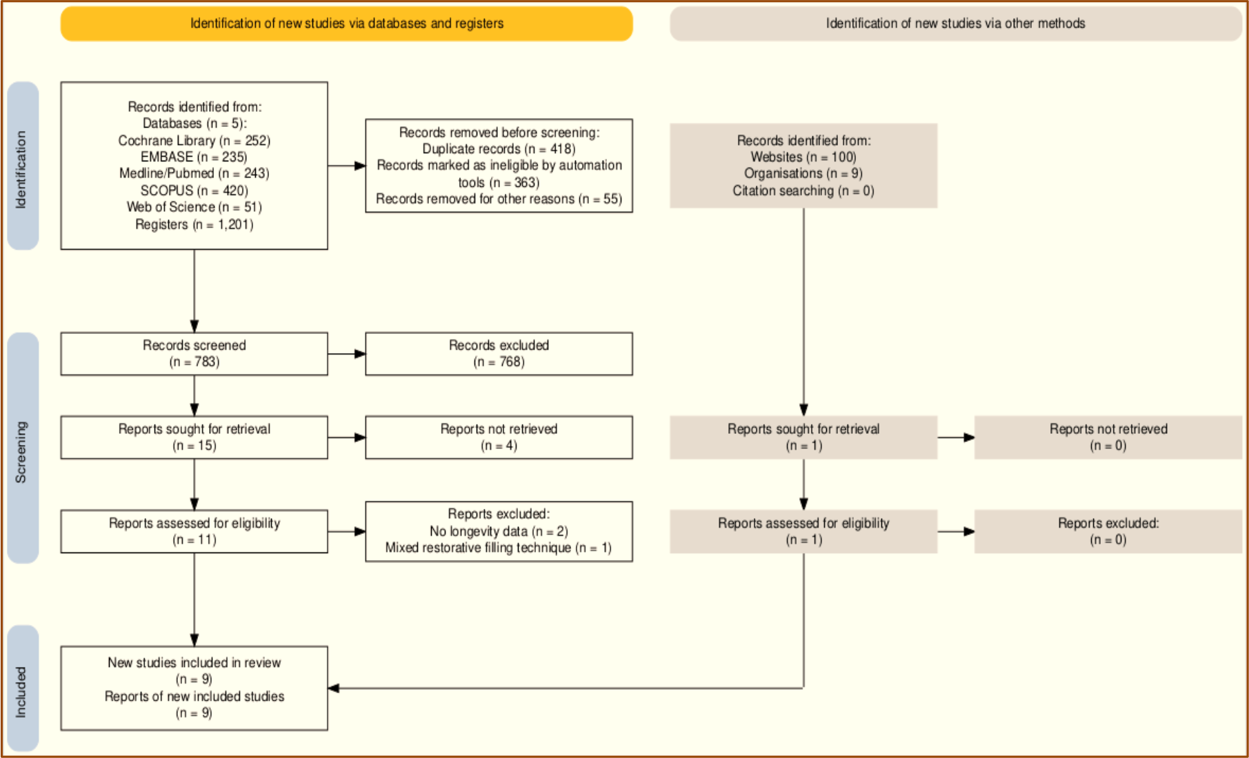
One thousand two hundred and one records were retrieved from the electronic databases. After removing duplicates, 783 records were screened. In phase I, the title and abstract of the records (783) were individually evaluated, with 15 records included. After full-text assessment, 8 studies were included from electronic databases, and 1 additional study was identified through grey literature, totaling 9 studies [1,2,4,7,8,10,19,20,21].
Figure 1 shows the detailed process for the search strategy. Meta-analysis was not feasible due to substantial heterogeneity among studies, including differences in study design (RCTs versus cohort studies), follow-up periods, outcome definitions, evaluation criteria, cavity designs, and adhesive protocols. Specifically, follow-up duration ranged from 3 months to approximately 6 years, with studies reporting outcomes either at a single end-point (e.g., 36, 48, or ~72 months) or at multiple time points (e.g., 6, 12, 18, and 24 months). Outcome definitions and evaluation criteria were not uniform (USPHS/modified USPHS, FDI, and Innes), and several studies included multiple intervention arms (e.g., different adhesive strategies, cavity designs, and substrate treatments), further limiting comparability. Importantly, overlap of identical follow-up time points across studies was sparse beyond 12 months, and denominators per evaluation period were not consistently reported, precluding reliable estimation of pooled proportions. Therefore, a descriptive synthesis was performed, emphasizing study-level differences in lesion severity, substrate condition at cavity margins (sound vs hypomineralized enamel), and adhesive protocols to aid interpretation of variability in reported longevity.
Flowchart of the search strategy process. The criteria used to assess clinical performance also varied among studies. Seven studies [1,2,4,7,8,20,21] employed the Ryge criteria according to the United States Public Health Service (USPHS or modified USPHS) [25], whereas one study [10] used the criteria proposed by Innes et al. [26], and another [19] adopted the World Dental Federation criteria. Clinical follow-up ranged from 12 months to 6 years, with most studies scheduling periodic evaluations every 3 months, 6 months, or annually [2,4,8,10,20,21]. Lygidakis et al. [2] and De Souza et al. [20] reported the provision of dietary and oral hygiene instructions every 6 months; in one study [2], fluoride varnish (5% NaF) was applied at every visit, whereas in another study [20], it was applied only when indicated. Differences in evaluation criteria, follow-up duration, and supportive preventive measures represent additional sources of variation that likely affected reported longevity rates across studies.
Study Characteristics
Table 2 shows the characteristics of the studies included in this systematic review. Among the 9 studies, 5 are observational cohort studies [1,2,7,10,19], and 4 are RCTs [4,8,20,21]. The studies were conducted in Brazil [8,20], Greece [1,2], Sweden [7], Germany [19], Colombia [10], Syria [21], and Turkey [4]. The mean age and number of participants were 8.6 and 45.25, respectively, with an average of 54.12 direct RC restorations per study. Only one study [2] did not explicitly report the MIH diagnostic criteria; however, its primary author is a leading researcher in the field and the author of the diagnostic criteria established and updated by the European Academy of Pediatric Dentistry (EAPD) [22-24]. All remaining studies adopted the EAPD criteria for MIH diagnosis, ensuring overall diagnostic consistency despite methodological heterogeneity.
The inclusion criteria differed considerably among studies, contributing to variability in clinical outcomes. Two studies restricted the number of restored surfaces [2,4]: Lygidakis et al. [2] included restorations with 2 or 3 surfaces, while Sönmez and Saat [4] limited restorations to 2 surfaces. Four studies included teeth with post-eruptive breakdown and/or previous unsuccessful atypical restorations, with or without associated carious lesions [8,10,20], whereas one study focused exclusively on MIH-affected teeth associated with carious lesions [4] and another included only teeth without carious involvement [19]. Two retrospective studies did not clearly report MIH severity [1,7]. Hakmi and Dashash [21] included only children with two hypomineralized permanent mandibular first molars, without previous dental treatment, and with carious or hypomineralized areas not exceeding two-thirds of the tooth. These differences in lesion severity, extension, and number of surfaces restored represent an important source of clinical heterogeneity and may partially explain the wide variation in longevity outcomes.
Regarding adhesive strategies, total etching was employed in most studies [2,4,8,19-21]. Two RCTs directly compared total etching (TEA) and self-etching (SEA) approaches in MIH-affected teeth [8,20], highlighting differences in marginal adaptation and retention over time. Sönmez and Saat [4] used selective enamel etching and specifically evaluated the effect of deproteinization of hypomineralized enamel with 5% NaOCl prior to universal adhesive application, demonstrating higher success when cavity margins were located in sound enamel. The same study also compared invasive cavity designs (margins in sound enamel) with non-invasive designs (margins in hypomineralized enamel), reinforcing the influence of substrate quality and cavity design on restoration survival.
The resin composites used included Filtek P60 (3M ESPE, New York, United States) [10], Tetric Evo Ceram (Ivoclar Vivadent AG, Schaan, Liechtenstein) [19], Brilliant/Synergy (Coltene) [2], Tetric N-Ceram Bulk Fill (Ivoclar Vivadent, Liechtenstein) [8], Grandio (VOCO, Cuxhaven, Germany) [4], and Filtek XT350 (3M ESPE, St. Paul, MN, USA) [20,21]. Most studies applied the incremental filling technique, while only one used a bulk-fill single-increment approach [8]. Two studies did not report the bonding strategy or resin composite used [1,7]. Variations in restorative materials and placement techniques further contribute to methodological heterogeneity and may influence outcomes, particularly in teeth with extensive defects or multiple restored surfaces.
Although one included study compared MIH-affected and non-affected teeth [1], no eligible studies were identified that included a parallel control group of non-affected teeth specifically designed to evaluate the longevity of direct resin composite restorations. Therefore, comparisons with sound enamel were limited and could not be systematically explored. Some information was not clearly addressed in the articles. After repeated unsuccessful attempts to contact the authors for further information, it was not possible to collect all data (Table 1).
Risk of Bias Among Studies
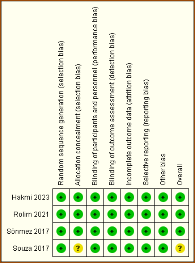
Among the cohort studies, three were judged at moderate risk of bias [1,2,7] and two at low risk of bias [10,19], according to the Newcastle-Ottawa Scale (Table 3). Regarding the randomized clinical trials, three were classified as having low risk of bias [4,8,21], and one raised some concerns due to issues related to allocation concealment [20], as assessed using the Cochrane Risk of Bias tool (ROB-2) (Figure 2). Overall, none of the included studies were judged to be at high risk of bias.
Risk of bias assessment of cohort studies included in the review, evaluated using the Newcastle-Ottawa Scale (NOS).

Risk of bias assessment of randomized clinical trials included in the review, evaluated using the Cochrane Risk of Bias tool (ROB-2).
Outcomes
Lygidakis et al. [2], in a prospective cohort study, evaluated 52 direct RC restorations over up to 48 months and reported a success rate of 100%. Of all the included studies, this [2] was the only one that primarily focused on evaluating the clinical performance of direct RC restorations placed in MIH-affected teeth. They showed the highest success rate and one of the longest clinical follow-up times. Despite the 100% success rate after 48 months, 10 restorations showed color mismatch, 3 showed altered surface, and 4 showed inadequate anatomic form.
The retrospective study by Mejarè et al. [7] evaluated 34 direct RC restorations for approximately 6 years. The success rate was 85.3%. Other restorative materials (GIC, stainless-steel crown, amalgam) were also included and 48% of the participants showed at least one molar with a clinically unacceptable restoration. The authors reported that tooth extraction followed by orthodontic treatment showed good or acceptable results in molars with severe defects of enamel (space closure was acceptable in 87% of the participants).
Kotsanos et al. [1] evaluated 59 RC restorations. The success rate was 74.6% after 48 months of follow-up. They evaluated different approaches in MIH-affected teeth (RC restorations, sealant, amalgam, and stainless-steel crown) compared with those performed in children without MIH-affected teeth. The odds ratio of children with MIH-affected teeth needing restorative reintervention in at least one restored molar was 3.70 times higher than that of children without MIH (95% confidence interval: 1.29 - 10.63).
In a retrospective cohort study, Linner et al. [19] evaluated 27 RC restorations, showing a success rate of 76.2% after 36 months of follow-up. Besides RC restorations, the study also included restorations placed with GIC, RC flow (non-invasive), and ceramic. The cumulative survival probability after 36 months was 7% for GIC, 29.9% for RC non-invasive (flow), 76.2% for RC conventional, and 100% for ceramic restorations. The RC conventional showed moderate to high survival rates. In contrast, non-invasive restorations were associated with lower survival rates.
De Farias et al. [10] evaluated 61 RC restorations after up to 24 months. The success rates were 59% and 49.2% after 12 and 24 months, respectively. They compared the survival of RC restorations with stainless-steel crowns. After 24 months, stainless-steel crowns showed a higher survival rate (94.4%) than RC restorations (49.2%). The authors report that tooth characteristics may have influenced this difference before restorative procedures.
The RCT by De Souza et al. [20] compared two bonding strategies on the survival rates of direct RC restorations in MIH-affected teeth. Forty-one teeth were randomly divided into two groups: TEA (total etching) and SEA (self-etching). Cumulative survival rates at 18 months were 68% for SEA and 54% for TEA (no significant difference between groups). The failures were attributed to marginal adaptation, loss of retention, and secondary caries.
Sönmez and Saat [4] evaluated the clinical effects of the deproteinization of the hypomineralised enamel and different cavity designs on the performance of RC restorations placed in MIH-affected molars. One hundred and twenty-six teeth were included, with 95 being MIH-affected. The teeth were randomized into four groups and followed-up for 24 months. In Group I, all hypomineralised tissue was removed until achieving sound enamel. In Group II, carious hypomineralised tissue until a reasonable resistance of the hypomineralized tissue. In Group III, cavities were designed as Group II; differently from this group, the deproteinization of the left hypomineralised tissue was performed prior to the placement of RC restorations. Group IV served as the control group, consisting of carious first molars without MIH. The success rate after 24 months was 81.25% (Group I), 58.06% (Group II), 78.12% (Group III), and 87.09% (Group IV). Group II showed the lowest success rate among all groups (p < 0.05). The deproteinization increases the retention rates, with the failures occurring predominantly when the cavity margins were in hypomineralised tissue. The failures were attributed to the anatomical form, secondary caries, adaptation, and marginal discoloration. Unexpectedly, no retention failures were recorded throughout the study.
Rolim et al. [8] clinically evaluated 64 RC restorations with 12 months of follow-up. The teeth were randomized into two groups: TEA (total etching) and SEA (self-etching). A universal adhesive system and bulk-fill resin composite were used. At the final evaluation, TEA and SEA showed success rates of 89.2% and 77.2%, respectively. Failures were attributed to loss of retention and marginal adaptation. Retention failures were most frequent after 12 months (7.6% for TEA and 18.2% for SEA). In the SEA group, 10% of the restorations failed after 6 months due to marginal adaptation, while no restorations failed for this reason in the TEA group. The authors concluded that a universal adhesive can be a viable option to restore MIH-affected teeth, with the total etching strategy showing higher survival rates.
Hakmi and Dashash [21] evaluated the longevity of 40 composite resin restorations, 20 performed using the direct technique (DCRR) and 20 using the indirect technique (ICRR) up to 12 months later. After 3 and 6 months of restoration, regardless of the technique, it was 100% successful. At 12 months, the composite resin survival rate was 90% with the indirect technique and 85% with the direct technique. The failures considered were: secondary caries (5% - ICRR and 10% - DCRR), change in anatomical shape (5% - ICRR and 15% - DCRR), surface texture (15% - ICRR and 40% - DCRR), discoloration marginal (20% - ICRR and 25% - DCRR), marginal maladaptation (20% for both) and postoperative sensitivity (5% - ICRR and 15% - DCRR).
Certainty of Evidence
According to the GRADE criteria, the cumulative certainty of the evidence was “very low” for the observational cohort studies and “moderate” for RCTs (Table 4). Downgrading was mainly driven by risk of bias (allocation concealment in RCTs and comparability issues in cohort studies), inconsistency due to substantial heterogeneity in follow-up periods and clinical protocols, and imprecision related to small sample sizes and wide confidence intervals. Indirectness and publication bias were not considered serious concerns.
Discussion
Resin composite is widely considered an appropriate direct restorative option for MIH-affected molars [1,5,7,12,19]. Nevertheless, restorations placed in MIH-affected teeth tend to require replacement more frequently than those in non-MIH teeth [1,2,14], which is consistent with the intrinsic fragility of hypomineralized substrates. Importantly, a previous systematic review on posterior composite restorations in non-MIH teeth reported annual failure rates of 1.8% after 5 years and 2.4% after 10 years of follow-up [27], suggesting that the substantially lower survival observed in several MIH studies likely reflects substrateand case-related challenges rather than composite performance alone. In the present review, variation in longevity across studies can be attributed to a combination of factors, particularly lesion severity, cavity design (margin location), adhesive approach, and follow-up/evaluation criteria.
Overall, higher success rates occurred when restorative protocols favored margins in sound enamel and when cases were carefully selected. Lygidakis et al. [2] reported 100% success for 52 RC restorations over 48 months, representing the highest longevity among the included studies and the only investigation primarily designed to assess direct RC performance in MIH-affected teeth. The favorable outcome may be partly explained by controlled clinical follow-up and cavity preparation strategies that favored margins in sound enamel. However, qualitative deterioration preceded overt failure, with color mismatch (10 restorations), altered surface texture (3), and inadequate anatomic form (4), indicating that esthetic and functional degradation may occur even when restorations remain “clinically successful” by conventional criteria. This observation is clinically relevant because it suggests that maintenance and possible repair strategies may be needed before complete failure is recorded.
Conversely, lower survival rates were commonly reported in studies involving more compromised substrates and/or protocols that retained hypomineralized enamel at cavity margins. De Farias et al. [10] reported success rates of 59% and 49.2% at 12 and 24 months, respectively, among 61 RC restorations, among the lowest values in this review. In the same study, stainless-steel crowns presented higher survival (94.4% at 24 months), supporting the interpretation that extensive structural compromise and unfavorable substrates can limit the predictability of adhesive restorations and may justify full-coverage approaches in selected cases. Similarly, Mejàre et al. [7] reported 85.3% success over approximately 6 years in 34 RC restorations, but 48% of participants had at least one clinically unacceptable molar restoration, and severe defects were associated with alternative treatment planning, including extraction followed by orthodontic management with acceptable outcomes in 87% of participants. Together, these findings reinforce that lesion severity and extent of breakdown substantially influence long-term prognosis and may necessitate treatment escalation.
The compromised MIH substrate appears to be a key determinant of restoration failure independent of the restorative material. Kotsanos et al. [1] reported 74.6% success after 48 months for 59 RC restorations and showed that children with MIH had a 3.70-fold higher likelihood of requiring restorative reintervention in at least one molar (95% confidence interval: 1.29-10.63) compared with children without MIH. This supports the concept that hypomineralized enamel is intrinsically more prone to marginal breakdown and degradation. Mechanistically, hypomineralized enamel has a higher organic content and lower mineral density, resulting in an altered bonding pattern relative to sound enamel, regardless of the adhesive system [30]. The transition between affected and apparently unaffected enamel may be diffuse, and even visually “sound” enamel can be structurally altered [7,29], which complicates margin placement and may explain why outcomes vary even among protocols that aim to bond to sound enamel.
Cavity design and margin location emerged as central drivers of clinical longevity. Sönmez and Saat [4] directly demonstrated that restorations placed in cavities with margins in sound enamel achieved higher success (81.25%) than those with margins in hypomineralized enamel (58.06%) after 24 months. In the same study, deproteinization with 5% NaOCl improved outcomes (78.12%), suggesting that substrate optimization can enhance retention and marginal integrity when complete removal of affected enamel is not feasible. These results align with the clinical dilemma between conservative approaches (removing enamel until “reasonable resistance” is obtained) and more extensive removal to reach a sound substrate [4,10,28]. While the conservative strategy preserves tooth structure, it may leave chippy enamel at margins and increase retreatment risk; conversely, extending preparation to sound enamel may improve bonding predictability at the cost of greater tissue removal and potential clinical difficulty in identifying the optimal boundary.
Adhesive strategy may further influence short-term performance, particularly regarding marginal stability and retention, but its effects are often intertwined with lesion severity and cavity design. De Souza et al. [20] compared total etching (TEA) and self-etching (SEA) strategies and found no statistically significant differences after 18 months (68% SEA vs 54% TEA), with failures mainly attributed to marginal adaptation, loss of retention, and secondary caries. Rolim et al. [8], using a universal adhesive and bulk-fill resin composite, reported higher success in the TEA group (89.2%) than in the SEA group (77.2%) after 12 months, with earlier failures more frequent in the SEA group. Taken together, these findings suggest that although total etching may offer more predictable marginal behavior in some settings, the compromised substrate remains a critical factor affecting longevity. This interpretation is also consistent with laboratory evidence indicating that phosphoric acid etching can increase mineral loss relative to self-etching systems [11,32], while cohesive failures within the altered enamel can compromise marginal integrity and lead to repair needs [30,31]. Therefore, no single adhesive approach can be universally recommended without considering substrate quality, margin location, and lesion extent.
Beyond adhesive strategy, restorative approach (direct vs indirect) and material selection may be relevant for specific clinical scenarios. Linner et al. [19] reported 76.2% success for conventional RC after 36 months, higher than non-invasive flowable RC (29.9%) and glass ionomer cement (7%), while ceramic restorations achieved 100% survival. These data suggest that both the mechanical behavior of the restorative option and the quality of the bonded substrate (influenced by the extent of preparation) contribute to performance in compromised enamel. Hakmi and Dashash [21] reported slightly higher survival for indirect composite restorations (90%) compared with direct restorations (85%) at 12 months, with failures related to surface texture changes, marginal discoloration/maladaptation, and secondary caries, suggesting that indirect approaches-potentially providing better control of contours and bonding conditions-may be advantageous in selected teeth with extensive hypomineralization.
Based on the available evidence, direct RC restorations appear most suitable for mild-to-moderate MIH lesions with limited surface involvement and when cavity margins can be placed in sound enamel [2,4]. When margins must remain in hypomineralized enamel or when lesions are severe with extensive breakdown, full-coverage approaches such as stainless-steel crowns may provide more predictable short-term survival [10] and may be preferable in selected cases. Substrate-oriented strategies, including careful removal of friable enamel and, when appropriate, deproteinization protocols (e.g., 5% NaOCl), may improve outcomes when complete removal of affected enamel is not feasible [4]. Follow-up and maintenance are essential, as qualitative deterioration (e.g., esthetic/functional shortcomings) may precede complete failure even in restorations classified as successful [2].
The strength of conclusions is limited by heterogeneity across studies, including differences in study design, inclusion/exclusion criteria, lesion severity profiles, cavity designs, adhesive protocols, evaluation criteria (USPHS, FDI, Innes), and follow-up intervals (1-6 years). This heterogeneity reduces comparability and precludes a meaningful meta-analysis, as pooling would likely yield misleading summary estimates. Additionally, the body of evidence includes retrospective cohorts, which are more susceptible to selection bias and uncontrolled confounding. Future research should prioritize well-designed multicenter randomized clinical trials with adequate sample sizes, standardized MIH diagnostic criteria and severity reporting, harmonized outcome measures and failure definitions, and protocols specifically tailored to hypomineralized enamel. Such steps would reduce heterogeneity, enable quantitative synthesis, and improve the certainty and applicability of evidence for clinical decision-making.
Finally, compared with broad MIH management reviews [5,6], the present review provides a more outcome-focused synthesis by concentrating specifically on the clinical longevity of direct composite restorations and identifying the key clinical determinants that likely explain variability in survival across studies. By integrating study-level findings with substrateand protocol-related interpretative factors, this review complements prior evidence and advances understanding of when direct composites are predictable, when alternative approaches may be preferable, and which methodological gaps should be addressed to strengthen future recommendations.
Conclusion
Longevity ranged from 49.2% to 100%, with very low to moderate certainty of evidence. Direct restorations may be suitable for mild to moderate MIH lesions, whereas severe cases require cautious indication and close follow-up. Failures were mainly related to loss of retention, marginal adaptation, secondary caries, and esthetic shortcomings. Methodological heterogeneity limits extrapolation and highlights the need for standardized, well-designed clinical trials.
-
Financial SupportNone.
Data Availability
The data used to support the findings of this study can be made available upon request to the corresponding author.
References
- [1] Kotsanos N, Kaklamanos EG, Arapostathis K. Treatment management of first permanent molars in children with Molar-Incisor Hypomineralisation. Eur J Paediatr Dent 2005; 6(4):179-184.
- [2] Lygidakis NA, Chaliasou A, Siounas G. Evaluation of composite restorations in hypomineralised permanent molars: A four year clinical study. Eur J Paediatr Dent 2003; 4(3):143-148.
-
[3] Bekes K, Melichar K, Stamm T, Elhennawy K. Dental students' knowledge, attitudes and beliefs regarding Molar Incisor Hypomineralization (MIH): A survey in Vienna, Austria. J Multidiscip Healthc 2021; 14:2881-2889. https://doi.org/10.2147/JMDH.S326846
» https://doi.org/10.2147/JMDH.S326846 -
[4] Sönmez H, Saat S. A clinical evaluation of deproteinization and different cavity designs on resin restoration performance in MIH-affected molars: Two-year results. J Clin Pediatr Dent 2017; 41(5):336-342. https://doi.org/10.17796/1053-4628-41.5.336
» https://doi.org/10.17796/1053-4628-41.5.336 -
[5] Elhennawy K, Schwendicke F. Managing molar-incisor hypomineralization: A systematic review. J Dent 2016; 55:16-24. https://doi.org/10.1016/j.jdent.2016.09.012
» https://doi.org/10.1016/j.jdent.2016.09.012 -
[6] Somani C, Taylor GD, Garot E, Rouas P, Lygidakis NA, Wong FSL. An update of treatment modalities in children and adolescents with teeth affected by molar incisor hypomineralisation (MIH): A systematic review. Eur Arch Paediatr Dent 2022; 23(1):39-64. https://doi.org/10.1007/s40368-021-00635-0
» https://doi.org/10.1007/s40368-021-00635-0 -
[7] Mejàre I, Bergman E, Grindefjord M. Hypomineralized molars and incisors of unknown origin: treatment outcome at age 18 years. Int J Paediatr Dent 2005; 15(1):20-28. https://doi.org/10.1111/j.1365-263X.2005.00599.x
» https://doi.org/10.1111/j.1365-263X.2005.00599.x -
[8] Rolim TZC, da Costa TRF, Wambier LM, Chibinski AC, Wambier DS, da Silva Assunção LR, et al. Adhesive restoration of molars affected by molar incisor hypomineralization: A randomized clinical trial. Clin Oral Investig 2021; 25(3):1513-1524. https://doi.org/10.1007/s00784-020-03459-2
» https://doi.org/10.1007/s00784-020-03459-2 -
[9] Americano GC, Jacobsen PE, Soviero VM, Haubek D. A systematic review on the association between molar incisor hypomineralization and dental caries. Int J Paediatr Dent 2017; 27(1):11-21. https://doi.org/10.1111/ipd.12233
» https://doi.org/10.1111/ipd.12233 -
[10] de Farias AL, Rojas-Gualdrón DF, Mejía JD, Bussaneli DG, Santos-Pinto L, Restrepo M. Survival of stainless-steel crowns and composite resin restorations in molars affected by molar-incisor hypomineralization (MIH). Int J Paediatr Dent 2022; 32(2):240-250. https://doi.org/10.1111/ipd.12849
» https://doi.org/10.1111/ipd.12849 - [11] Sapir S, Shapira J. Clinical solutions for developmental defects of enamel and dentin in children. Pediatr Dent 2007; 29(4):330-336.
-
[12] da Cunha Coelho ASE, Mata PCM, Lino CA, Macho VMP, Areias CMFGP, Norton APMAP, et al. Dental hypomineralization treatment: A systematic review. J Esthet Restor Dent 2019; 31(1):26-39. https://doi.org/10.1111/jerd.12420
» https://doi.org/10.1111/jerd.12420 - [13] Weerheijm KL. Molar incisor hypomineralisation (MIH). Eur J Paediatr Dent 2003; 4(3):114-120.
-
[14] Jälevik B, Klingberg G. Treatment outcomes and dental anxiety in 18-year-olds with MIH, comparisons with healthy controls - A longitudinal study. Int J Paediatr Dent 2012; 22(2):85-91. https://doi.org/10.1111/j.1365-263X.2011.01161.x
» https://doi.org/10.1111/j.1365-263X.2011.01161.x -
[15] Page MJ, McKenzie JE, Bossuyt PM, Boutron I, Hoffmann TC, Mulrow CD, et al. The PRISMA 2020 statement: An updated guideline for reporting systematic reviews. Syst Rev 2021; 10(1):89. https://doi.org/10.1186/s13643-021-01626-4
» https://doi.org/10.1186/s13643-021-01626-4 -
[16] Higgins JP, Altman DG, Gøtzsche PC, Jüni P, Moher D, Oxman AD, et al. The Cochrane Collaboration's tool for assessing risk of bias in randomised trials. BMJ 2011; 343:d5928. https://doi.org/10.1136/bmj.d5928
» https://doi.org/10.1136/bmj.d5928 -
[17] Lo CK, Mertz D, Loeb M. Newcastle-Ottawa Scale: Comparing reviewers' to authors' assessments. BMC Med Res Methodol 2014; 14:45. https://doi.org/10.1186/1471-2288-14-45
» https://doi.org/10.1186/1471-2288-14-45 -
[18] Guyatt GH, Oxman AD, Vist GE, Kunz R, Falck-Ytter Y, Alonso-Coello P, et al. GRADE: An emerging consensus on rating quality of evidence and strength of recommendations. BMJ 2008; 336(7650):924-926. https://doi.org/10.1136/bmj.39489.470347.AD
» https://doi.org/10.1136/bmj.39489.470347.AD -
[19] Linner T, Khazaei Y, Bücher K, Pfisterer J, Hickel R, Kühnisch J. Comparison of four different treatment strategies in teeth with molar-incisor hypomineralization-related enamel breakdown - A retrospective cohort study. Int J Paediatr Dent 2020; 30(5):597-606. https://doi.org/10.1111/ipd.12636
» https://doi.org/10.1111/ipd.12636 -
[20] de Souza JF, Fragelli CB, Jeremias F, Paschoal MAB, Santos-Pinto L, de Cássia Loiola Cordeiro R. Eighteen-month clinical performance of composite resin restorations with two different adhesive systems for molars affected by molar incisor hypomineralization. Clin Oral Investig 2017; 21(5):1725-1733. https://doi.org/10.1007/s00784-016-1968-z
» https://doi.org/10.1007/s00784-016-1968-z -
[21] Hakmi A, Dashash M. Direct or indirect composite for restoring permanent first molars affected by Molar Incisor Hypomineralisation (MIH): A randomized clinical controlled trial. BDJ Open 2023; 9(1):37. https://doi.org/10.1038/s41405-023-00165-5
» https://doi.org/10.1038/s41405-023-00165-5 - [22] Weerheijm KL, Duggal M, Mejàre I, Papagiannoulis L, Koch G, Martens LC, et al. Judgement criteria for molar incisor hypomineralisation (MIH) in epidemiologic studies: A summary of the European meeting on MIH held in Athens, 2003. Eur J Paediatr Dent 2003; 4(3):110-113.
-
[23] Lygidakis NA, Wong F, Jälevik B, Vierrou AM, Alaluusua S, Espelid I. Best Clinical Practice Guidance for clinicians dealing with children presenting with Molar-Incisor-Hypomineralisation (MIH): An EAPD Policy Document. Eur Arch Paediatr Dent 2010; 11(2):75-81. https://doi.org/10.1007/BF03262716
» https://doi.org/10.1007/BF03262716 -
[24] Lygidakis NA, Garot E, Somani C, Taylor GD, Rouas P, Wong FSL. Best clinical practice guidance for clinicians dealing with children presenting with molar-incisor-hypomineralisation (MIH): An updated European Academy of Paediatric Dentistry policy document. Eur Arch Paediatr Dent 2022; 23(1):3-21. https://doi.org/10.1007/s40368-021-00668-5
» https://doi.org/10.1007/s40368-021-00668-5 - [25] Ryge G. Clinical criteria. Int Dent J 1980; 30(4):347-358.
-
[26] Innes NP, Evans DJ, Stirrups DR. The Hall Technique; A randomized controlled clinical trial of a novel method of managing carious primary molars in general dental practice: Acceptability of the technique and outcomes at 23 months. BMC Oral Health 2007; 7:18. https://doi.org/10.1186/1472-6831-7-18
» https://doi.org/10.1186/1472-6831-7-18 -
[27] Opdam NJ, van de Sande FH, Bronkhorst E, Cenci MS, Bottenberg P, Pallesen U, et al. Longevity of posterior composite restorations: A systematic review and meta-analysis. J Dent Res 2014; 93(10):943-949. https://doi.org/10.1177/0022034514544217
» https://doi.org/10.1177/0022034514544217 - [28] William V, Burrow MF, Palamara JE, Messer LB. Microshear bond strength of resin composite to teeth affected by molar hypomineralization using 2 adhesive systems. Pediatr Dent 2006; 28(3):233-241.
-
[29] Bozal CB, Kaplan A, Ortolani A, Cortese SG, Biondi AM. Ultrastructure of the surface of dental enamel with molar incisor hypomineralization (MIH) with and without acid etching. Acta Odontol Latinoam 2015; 28(2):192-198. https://doi.org/10.1590/S1852-48342015000200016
» https://doi.org/10.1590/S1852-48342015000200016 -
[30] Lagarde M, Vennat E, Attal JP, Dursun E. Strategies to optimize bonding of adhesive materials to molar-incisor hypomineralization-affected enamel: A systematic review. Int J Paediatr Dent 2020; 30(4):405-420. https://doi.org/10.1111/ipd.12621
» https://doi.org/10.1111/ipd.12621 -
[31] Hosein I, Sherriff M, Ireland AJ. Enamel loss during bonding, debonding, and cleanup with use of a self-etching primer. Am J Orthod Dentofacial Orthop 2004; 126(6):717-724. https://doi.org/10.1016/j.ajodo.2003.10.032
» https://doi.org/10.1016/j.ajodo.2003.10.032 -
[32] Krämer N, Bui Khac NN, Lücker S, Stachniss V, Frankenberger R. Bonding strategies for MIH-affected enamel and dentin. Dent Mater 2018; 34(2):331-340. https://doi.org/10.1016/j.dental.2017.11.015
» https://doi.org/10.1016/j.dental.2017.11.015
Edited by
-
Academic Editor: Catarina Ribeiro Barros de Alencar