 Hydatidosis in Spix’s yellow-toothed cavy (Galea spixii) in Northeast Brazil
ABSTRACT:
Echinococcus spp. are zoonotic cestode parasites made up of eight identified species and one genotypic grouping (Echinococcus canadensis), which cause the echinococcosis-hydatidosis complex. The main intermediate hosts are cattle, sheep, goats, pigs, and wild animals such as rodents. The aim is to characterize the epidemiological and anatomopathological aspects of the first infection report with the cestode Echinococcus sp. in a prey (Galea spixii) in northeastern Brazil. An adult female Spix’s yellow-toothed cavy (Galea spixii) was preyed upon by a captive-bred bird (Parabuteo unicinctus) during falconry training in a closed forest located in the municipality of Livramento, Paraíba. The rodent was collected from predation, and during inspection, multiple cysts were observed covering organs in the abdominal and thoracic cavities. The rodent’s carcass was sent for necropsy. Macroscopically, it showed multiple traumas due to predation and an abdominal cavity with multiple hydatid cysts covering the omentum, liver, pancreas, thoracic cavity, and lungs. The cysts ranged from 2 to 10 mm in diameter, intact and occasionally collapsed, oval, well-delimited, randomly distributed, filled with translucent hydatid fluid, and visible white to yellowish structures. Organ fragments were fixed in 10% buffered formalin and then routinely processed. Histological sections of the liver, lung, pancreas, and omentum with cysts were selected and submitted to Masson’s trichrome (MT) and periodic acid-Schiff (PAS) for morphological characterization of the parasite and evaluation of the tissue architecture of the organs in wild rodents. Microscopically, viable and degenerated unilocular and multilocular cysts of varying sizes were observed in the liver, lung, pancreas, and omentum. The cysts were externally surrounded by a moderately thick adventitial layer of fibrous connective tissue and occasional neutrophils, lymphocytes, and macrophages, with parasite components in the center. The morphological characteristics of the parasite were compatible with a cestode belonging to the genus Echinococcus sp. On MT staining, areas of fibrosis were evident around the viable and degenerated cysts in the liver, pancreas, and lung, and the acellular and eosinophilic laminated layer was evident on PAS. Based on the epidemiological and anatomopathological findings associated with the morphological characteristics of the cestode Echinococcus sp., the diagnosis of hydatidosis in Spix’s yellow-toothed cavy was made. G. spixii should be considered intermediate hosts in the infection of Echinococcus sp. for domestic and wild canids and accidentally for humans.
INDEX TERMS:
Hydatid cyst; hydatidosis; Echinococcus sp.; wild rodent; zoonosis
RESUMO:
Echinococcus spp. são parasitos cestoides zoonóticos composto por oito espécies identificadas e um agrupamento genotípico (Echinococcus canadensis), causadores do complexo equinococose-hidatidose. Os principais hospedeiros intermediários são os bovinos, ovinos, caprinos, suínos e animais selvagens, como roedores. Objetiva-se caracterizar os aspectos epidemiológicos e anatomopatológicos do primeiro relato de infecção do cestódeo Echinococcus sp. em um preá (Galea spixii) no Nordeste do Brasil. Um preá (G. spixii), fêmea, adulta, foi predado por ave de rapina criada em cativeiro (Parabuteo unicinctus) em treinamento de falcoaria em mata fechada localizada no município de Livramento, Paraíba. O roedor foi recolhido da predação e durante inspeção foram observados múltiplos cistos recobrindo órgãos em cavidade abdominal e torácica. A carcaça do roedor foi encaminhada para necropsia. Macroscopicamente, apresentava múltiplos traumas por predação e cavidade abdominal com múltiplos cistos hidáticos recobrindo omento, fígado, pâncreas, cavidade torácica e pulmão. Os cistos variavam de 2 a 10 mm de diâmetro, intactos e ocasionalmente colapsados, ovoides, bem delimitados, distribuídos aleatoriamente, preenchidos com líquido hidático translúcido e estruturas brancacentas a amareladas visíveis. Fragmentos de órgãos foram fixados em formalina a 10% tamponada e, posteriormente, processados rotineiramente para confecção de lâminas histopatológicas. Secções histológicas de fígado, pulmão, pâncreas e omento com cistos foram selecionadas e submetidas ao tricrômio de Masson (TM) e ácido periódico de Schiff (PAS) para caracterização morfológica do parasito e avaliação da arquitetura tecidual dos órgãos. Microscopicamente, foram observados cistos de tamanhos variados, uniloculares e multiloculares, viáveis e degenerados no fígado, pulmão, pâncreas, omento e pulmão. Os cistos eram circundados externamente pela camada adventícia moderadamente espessa composta por tecido conjuntivo fibroso e ocasionais neutrófilos, linfócitos e macrófagos, com componentes do parasito ao centro. As características morfológicas do parasito foram compatíveis com cestódeo pertencente ao gênero Echinococcus sp. Na coloração de TM, as áreas de fibrose foram evidenciadas circundando os cistos viáveis e degenerados em fígado, pâncreas e pulmão, e a camada laminada acelular e eosinofílica evidenciada no PAS. Com base nos achados epidemiológicos e anatomopatológicos, associados às características morfológicas do cestódeo Echinococcus sp., foi realizado o diagnóstico de hidatidose em preá. G. spixii devem ser considerados hospedeiros intermediários na infecção de Echinococcus sp. para canídeos domésticos e silvestres, e acidentalmente para humanos.
TERMOS DE INDEXAÇÃO:
Cisto hidático; Echinococcus sp.; hidatidose; roedor silvestre; zoonose
Introduction
The echinococcosis-hydatidosis complex is a zoonotic disease caused by parasites of the genus Echinococcus with cosmopolitan distribution and greater occurrence in temperate zones (Soares 2023). Echinococcus sp. are cestode parasites composed of eight identified species and one genotypic grouping (Echinococcus canadensis), transmitted in indirect predator-prey cycles involving domestic and wild animals (Romig et al. 2017). In humans, Echinococcus granulosus s.s and Echinococcus multilocularis accidentally cause cystic and alveolar echinococcosis (Shi et al. 2019), which is a potential public health threat (Deplazes et al. 2017).
Domestic dogs and wild carnivores are definitive hosts of Echinococcus sp. infection in the adult stage, while intermediates are affected by the larval metacestode stage (Schantz et al. 2011) and are infected after ingesting parasite eggs released in the feces of carnivores (Nakao et al. 2013). Metacestodes reproduce asexually and develop the hydatid cyst, parasitizing visceral organs such as the liver and lungs in herbivores and omnivores (Thompson 2017). The main intermediate hosts are cattle, sheep, goats, and pigs (Schantz et al. 2011), and wild animals such as marsupials, hares, rabbits, monkeys, and rodents (Soares 2023).
Rodents are considered synanthropic animals susceptible to infection by pathogens and parasites with zoonotic risk (Studzinska et al. 2019). Hydatidosis in rodents is poorly reported, limited to isolated cases and studies of low occurrence in small and large rodents (Gardner et al. 2013, Umhang et al. 2013, Miller et al. 2016, Arrabal et al. 2017, Avcioglu et al. 2017, Studzinska et al. 2019). In Brazil, Echinococcus sp. infection has been reported in agouti (Dasyprocta leporina) (Zimmerman et al. 2009) and paca (Cuniculus paca) (Almeida et al. 2013, Bittencourt-Oliveira et al. 2018). There are no descriptions of Echinococcus sp. infection in Spix’s yellow-toothed cavy (Galea spixii), so this study aimed to characterize the anatomopathological aspects of Echinococcus sp. infection in G. spixii in Northeast Brazil.
Case Report
Ethical approval. The present study was conducted with tissues from the necropsied animal at the Animal Pathology Laboratory of the University Veterinary Hospital at the “Universidade Federal de Campina Grande” (HVU-UFCG), therefore, approval by the Animal Use Ethics Committee was not required.
An adult female Spix’s yellow-toothed cavy (Galea spixii Wagler, 1831) was preyed upon by a Harris’s hawk (Parabuteo unicinctus) during falconry training in a closed forest located in the municipality of Livramento, Paraíba (7° 22’ 26” South, 36° 56’ 45” West). The rodent was collected from predation, and during inspection, it had good body condition (score 3), and multiple cysts were observed covering organs in the abdominal and thoracic cavities. The rodent’s carcass was sent for necropsy.
Macroscopically, it showed multiple predation traumas with the absence of a right thoracic limb, subcutaneous hemorrhages, multiple rib fractures, and laceration of the intercostal muscles in the right lateral region. In the abdominal cavity, multiple hydatid cysts were observed covering the omentum (Fig. 1), the hepatic capsular surface (Fig. 2), and the pancreas. Cysts were also observed in the thoracic cavity and the left caudal lobe of the lung. The cysts ranged in diameter from 2 to 10 mm, were intact and occasionally collapsed, oval, well-defined, and randomly distributed, filled with translucent hydatid fluid and visible white to yellowish structures.
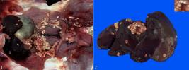
Hydatidosis in Galea spixii. (1) Abdominal cavity with multiple hydatid cysts, intact and occasionally collapsed, ovoid, well delimited, covering the omentum, liver and pancreas (arrows). (2) Liver with capsular surface and multiple randomly distributed hydatid cysts. Inset: cysts filled with translucent hydatid fluid and visible white to yellowish structures, ranging from 2 to 10 mm in diameter.
Fragments of organs from the abdominal and thoracic cavities, central nervous system, and skin were fixed in 10% buffered formalin and then routinely processed for histopathological evaluation using hematoxylin and eosin (HE). Histological sections of the liver, lung, pancreas, and omentum with cysts were subjected to histochemical techniques using Masson’s trichrome (MT) and periodic acid-Schiff (PAS) for morphological characterization of the parasite and evaluation of the tissue architecture of the organs.
Histological sections of the liver, lung, pancreas, and omentum showed cysts of varying sizes, unilocular and multilocular, viable and degenerated. The cysts were externally surrounded by a moderately thick adventitial layer composed of fibrous connective tissue and occasional neutrophils, lymphocytes, and macrophages, with parasite components in the center. From the outermost portion towards the inside of the viable cysts, there was an acellular and eosinophilic laminated layer, evident in PAS, followed by a thin, eosinophilic, and poorly cellular germ layer (Fig. 3 and 4). In the center was hydatid sand made up of small and large protoscolices surrounded by a prolific capsule attached to the germ layer and membrane remnants. The protoscolices had small and large hooks, calcareous corpuscles, an integument, and no gastrointestinal tract (Fig. 5 and 6). The morphological characteristics of the parasite were compatible with a cestode belonging to the genus Echinococcus sp.

Hydatidosis in Galea spixii. (3) Omentum. Hydatid cyst surrounded by adventitious layer (AL), followed by laminated layer (LL), germinal layer (GL) and 20 specimens of protoscolices (P). HE, obj. 10x. (4) Omentum. Acellular and strongly eosinophilic laminated layer (LL). PAS, obj. 40x. (5) Omentum. Small proboscis composed of small hook (GP), large hook (GG), calcareous corpuscles (CC), tegument (T) and absence of gastrointestinal tract. HE, obj. 40x. (6) Omentum. Hydatid cyst with large protoscolice composed of small hook (GP), large hook (GG), calcareous corpuscles (CC), tegument (T), proligerous capsule (PC) and absence of gastrointestinal tract. HE, obj. 10x. (7) Liver. Hydatid cyst containing protoescolice (P) surrounded by proligerous capsule (PC) and delimited by fibrosis (F), adjacent to hepatic parenchyma (HP) and Glisson’s capsule (G). HE, obj. 10x. (8) Lung. A degenerated cyst (DC) in the center surrounded by fibrous connective tissue strongly stained in blue (FC), partially replacing the lung parenchyma. MT, obj. 10x.
In the degenerated cysts, there were remnants of eosinophilic amorphous membrane in the center surrounded by calcareous corpuscles, necrosis and absence of protoscolices. The viable and degenerated cysts in the liver and pancreas were limited to a connective tissue capsule with preserved parenchyma and slight centrilobular congestion (Fig. 7). The lung parenchyma showed degenerated cysts associated with edema and hemorrhage in alveolar spaces and bronchioles, congestion of alveolar septa, and macrophages with hemosiderin. MT staining showed areas of fibrosis surrounding the viable and degenerated cysts in the liver, pancreas, and lung (Fig. 8).
Discussion
Based on the anatomopathological findings observed in the multiple organs associated with the morphological characteristics of the cestode Echinococcus sp. (Gardiner & Poynton 1999, Panziera et al. 2020), the diagnosis of hydatidosis was made in the Spix’s yellow-toothed cavy (Galea spixii). Wild rodents infected with Echinococcus sp. have been detected in Brazil in agouti (Dasyprocta leporina) associated with Echinococcus oligarthrus infection (Zimmerman et al. 2009), and paca (Cuniculus paca) with Echinococcus vogeli (Almeida et al. 2013, Bittencourt-Oliveira et al. 2018). Although this study did not determine the species of the genus Echinococcus sp., two species of E. oligarthrus and E. vogeli are commonly associated with infection in rodents and wild felids in South America and Asia (Gardner et al. 2013, Romig et al. 2017). Additionally, Echinococcus multilocularis has been identified in other rodent species in Europe (Umhang et al. 2013, Miller et al. 2016, Avcioglu et al. 2017, Studzinska et al. 2019) and should be considered as a differential in wild rodent infections.
Animals affected by hydatid cysts usually show no clinical signs, and depending on the location and size of the cysts, they can cause necrosis and atrophy of the affected tissues, calcification, and secondary bacterial infections (Brunetti et al. 2010). In a study described by Thiele et al. (2023), a muskrat (Ondatra zibethicus) showed abdominal distension and multiple generalized cysts in the liver and abdominal cavity associated with apathy, which contributed to capture by the predator. Organs affected by hydatid cysts in intermediate hosts can cause functional alterations and general weakness of the organism, which can consequently favor predation (Arrabal et al. 2017). Although a bird of prey preyed on the rodent, it is not possible to confirm that the hydatid cysts contributed to the predation.
The macroscopic lesions identified in G. spixii were similar to those observed in other rodents diagnosed with hydatidosis (Miller et al. 2016, Avcioglu et al. 2017, Studzinska et al. 2019). Histopathological examination is an essential tool for identifying parasitic lesions in tissues (Bittencourt-Oliveira et al. 2018) and is fundamental for the differential diagnosis of abscesses, calcifications, and neoplasms (Brunetti et al. 2010).
The findings of inflammation and fibrosis were mild to moderate. They differed from those of other authors (Almeida et al. 2013, Bittencourt-Oliveira et al. 2018, El Saftawy et al. 2021), possibly related to the initial phase of the parasitosis or an efficient immune response in the individual. Hydatid cysts induce an inflammatory response in the host through secretory proteins and the concentration of calcium, phosphorus, and magnesium released from the calcareous corpuscles present in the parasite and through enzymes released from tissue cells undergoing apoptosis, degeneration, and necrosis (Meng et al. 2012, Profumo et al. 2014). Fibrosis occurs secondary to the interaction of the cyst and vascular alterations with the tissue (Simonetto et al. 2015), best evidenced by Masson’s trichrome staining (El Saftawy et al. 2021).
Spix’s yellow-toothed cavy is a species of rodent widely distributed in subtropical and temperate regions of South America (Guenther & Trillmich 2022), with a high prevalence in the Sertão region of Northeast Brazil (Queiroz et al. 2020). The hydatidosis diagnosed in this prey demonstrates the species’ participation as an intermediate host in the echinococcosis cycle for domestic and wild canids in the region.
Wild rodents play an essential role in transmitting and signaling potentially zoonotic parasites (Duscher et al. 2015), as observed in this study. It is suggested that the consumption of meat from wild animals in Northeast Brazil (Oliveira et al. 2017) may be a factor that contributes to the transmission of this parasitic disease in humans. The echinococcosis-hydatidosis complex in humans has been reported in all regions of Brazil (Lise et al. 2022). Monitoring and diagnosing emerging diseases in wild animals are means of epidemiological surveillance that contribute to understanding the prevalence of diseases and identifying reservoirs and infectious agents (Sanches & Lavín 2014).
New species of Echinococcus could be identified during epidemiological investigations in wild animals (Thompson 2017). Epidemiological studies aimed at the wild cycle with the detection of Echinococcus sp. are believed to have contributed to a better understanding of the parasite and the adoption of control measures for public health.
Conclusion
We conclude that hydatidosis occurs in Galea spixii in Northeast Brazil, and that this seems to be the first description of this species. Spix’s yellow-toothed cavy should be considered as intermediate hosts in the infection of Echinococcus sp. for domestic and wild canids and accidentally for humans.
Acknowledgments
To the “Programa de Pós-Graduação em Ciência e Saúde Animal” (PPGCSA-UFCG). To the “Coordenação de Aperfeiçoamento de Pessoal de Nível Superior” (CAPES) for the scholarship of the first author.
References
-
Almeida F, Caldas R, Corrêa C, Rodrigues-Silva R, Siqueira N, Machado-Silva JR. Co-infections of the cestode Echinococcus vogeli and the nematode Calodium hepaticum in the hystricomorphic rodent Agouti paca from a forest reserve in Acre, Brazil. J Helminthol 2013; https://doi.org/10.1017/S0022149X12000661
» https://doi.org/10.1017/S0022149X12000661 -
Arrabal JP, Avila HG, Rivero MR, Camicia F, Salas MM, Costa SA, Nocera CG, Rosenzvit MC, Kamenetzky L. Echinococcus oligarthrus in the subtropical region of Argentina: First integration of morphological and molecular analyses determines two distinct populations. Vet Parasitol 2017; https://doi.org/10.1016/j.vetpar.2017.03.019
» https://doi.org/10.1016/j.vetpar.2017.03.019 -
Avcioglu H, Guven E, Balkaya I, Kirman R, Bia MM, Gulbeyen H, Kurt A, Yaya S, Demirtas S. First detection of Echinococcus multilocularis in rodent intermediate hosts in Turkey. Parasitol 2017; https://doi.org/10.1017/S0031182017001226
» https://doi.org/10.1017/S0031182017001226 -
Bittencourt-Oliveira FB, Teixeira P, Alencar A, Menezes R, Corrêa C, Neves L, Almeida F, Daipert-Garcia D, Machado-Silva JR, Rodrigues-Silva R. First parasitological, histopathological and molecular characterization of Echinococcus vogeli Rausch and Bernstein, 1972 from Cuniculus paca Linnaeus, 1766 in the Cerrado biome (Mato Grosso do Sul, Brazil). Vet Parasitol 2018; https://doi.org/10.1016/j.vetpar.2017.12.003
» https://doi.org/10.1016/j.vetpar.2017.12.003 -
Brunetti E, Kern P, Vuitton DA, Writing Panel for the WHO-IWGE. Expert consensus for the diagnosis and treatment of cystic and alveolar echinococcosis in humans. Acta Trop 2010; https://doi.org/10.1016/j.actatropica.2009.11.001
» https://doi.org/10.1016/j.actatropica.2009.11.001 -
Deplazes P, Rinaldi L, Alvarez Rojas CA, Torgerson PR, Harandi MF, Romig T, Antolova D, Schurer JM, Lahmar S, Gringoli G, Magambo J, Thompson RCA, Jenkins EJ. Global distribution of alveolar and cystic echinococcosis. Adv Parasitol 2017; https://doi.org/10.1016/bs.apar.2016.11.001
» https://doi.org/10.1016/bs.apar.2016.11.001 -
Duscher GG, Leschnik M, Fuehrer H-P, Joachim A. Wildlife reservoirs for vector-borne canine, feline and zoonotic infections in Austria. Int J Parasitol Parasites Wildl 2015; https://doi.org/10.1016/j.ijppaw.2014.12.001
» https://doi.org/10.1016/j.ijppaw.2014.12.001 -
El Saftawy EA, Abdelmoktader A, Sabry MM, Alghandour SM. Histological and immunological insights to hydatid disease in camels. Vet Parasitol Reg Stud Rep 2021; https://doi.org/10.1016/j.vprsr.2021.100635
» https://doi.org/10.1016/j.vprsr.2021.100635 - Gardiner CH, Poynton SL. An Atlas of Metazoan Parasites in Animal Tissues. Washington: Armed Forces Institute of Pathology, American Registry of Pathology; 1999.
-
Gardner SL, Dursahinhan AT, Rácz GR, Batsaikhan N, Ganzorig S, Tinnin DS, Damidinbazar D, Wood C, Peterson AT, Alandia E, Mollericona JL, Salazar-Bravo J. Sylvatic species of Echinococcus from rodent intermediate hosts in Asia and South America. Museum of Texas Tech University 2013; https://digitalcommons.unl.edu/parasitologyfacpubs/759
» https://digitalcommons.unl.edu/parasitologyfacpubs/759 -
Guenther A, Trillmich F. Photoperiod influences the development and the expression of personality traits and social behaviour in wild cavies (Cavia aperea). Ethol 2022; https://doi.org/10.1111/eth.13343
» https://doi.org/10.1111/eth.13343 -
Lise MLZ, Widdicombe J, Lise CRZ, Oliveira SV, Caldas EP, Entezami M, Prada JM, Ghiotti N, Silva RRE, Vizcaychipi KA, Vilas VDR. Records of human deaths from echinococcosis in Brazil, 1995-2016. Vet Sci 2022; https://doi.org/10.3390/vetsci9080436
» https://doi.org/10.3390/vetsci9080436 -
Meng F, Wang K, Aoyama T, Grivennikov SI, Paik Y, Scholten D, Cong M, Iwaisako K, Liu X, Zhang M, Österreicher CH, Stickel F, Ley K, Brenner DA, Kisseleva T. Interleukin-17 signaling in inflammatory, Kupffer cells, and hepatic stellate cells exacerbates liver fibrosis in mice. Gastroenterol 2012; https://doi.org/10.1053/j.gastro.2012.05.049
» https://doi.org/10.1053/j.gastro.2012.05.049 -
Miller AL, Olsson GE, Walburg MR, Sollenberg S, Skarin M, Ley C, Wahlström H, Höglund J. First identification of Echinococcus multilocularis in rodent intermediate hosts in Sweden. Int J Parasitol Parasites Wildl 2016; https://doi.org/10.1016/j.ijppaw.2016.03.001
» https://doi.org/10.1016/j.ijppaw.2016.03.001 -
Nakao M, Lavikainen A, Yanagida T, Ito A. Phylogenetic systematics of the genus Echinococcus (Cestoda: Taeniidae). Int J Parasitol 2013; https://doi.org/10.1016/j.ijpara.2013.06.002
» https://doi.org/10.1016/j.ijpara.2013.06.002 -
Oliveira GB, Oliveira REM, Bezerra FVF, Câmara FV, Araújo Júnior HN, Oliveira MF. Origin and distribution of the celiac artery in yellow-toothed cavies (Galea spixii Wagler, 1831). Ciênc Anim Bras 2017; https://doi.org/10.1590/1089-6891v18e-32918
» https://doi.org/10.1590/1089-6891v18e-32918 -
Panziera W, Bianchi MV, Vielmo A, Bianchi RM, Pavarini SP, Sonne L, Soares JF, Driemeier D. Atypical parasitic lesions in slaughtered cattle in Southern Brazil. Braz J Vet Parasitol 2020; https://doi.org/10.1590/S1984-29612020043
» https://doi.org/10.1590/S1984-29612020043 -
Profumo E, Ludovisi A, Buttari B, Angeles M, Morales G, Riganò R. Parasite-mediated immune modulation during the development of human cystic echinococcosis. In: Jirillo E, Magrone T, Miragliotta G. Immune Response to Parasitic Infections: immunity to helminths and novel therapeutic approaches. 2014; https://doi.org/10.2174/9781608059850114020007
» https://doi.org/10.2174/9781608059850114020007 -
Queiroz JPAF, Souza-Junior JBF, Oliveira VRM, Costa LLM, Oliveira MF. How does Spix’s yellow-toothed cavy (Galea spixii Wagler, 1831) face the thermal challenges of the Brazilian tropical dry forest? J Therm Biol 2020; https://doi.org/10.1016/j.jtherbio.2020.102525
» https://doi.org/10.1016/j.jtherbio.2020.102525 -
Romig T, Deplazes P, Jenkins D, Giraudoux P, Massolo A, Craig PS, Wassermann M, Takahashi K, de la Rue M. Ecology and life cycle patterns of Echinococcus species. Adv Parasitol 2017; https://doi.org/10.1016/bs.apar.2016.11.002
» https://doi.org/10.1016/bs.apar.2016.11.002 - Sanches IM, Lavín S. Methods of epidemiological surveillance in wildlife. In: Cubas ZS, Silva JCR, Catão-Dias JL. Tratado de Animais Selvagens: medicina veterinária. Vol.1. 2a ed. São Paulo: Roca; 2014.
-
Schantz PM, Kern P, Brunetti E. Echinococcosis, p.824-238. In: Guerrant RL, Walker DH, Weller PF. Tropical Infectious Diseases: principles, pathogens and practice. 2011; https://doi.org/10.1016/B978-0-7020-3935-5.00120-8
» https://doi.org/10.1016/B978-0-7020-3935-5.00120-8 -
Shi Y, Wan X, Wang Z, Li J, Jiang Z, Yang Y. First description of Echinococcus ortleppi infection in China. Parasit Vectors 2019; https://doi.org/10.1186/s13071-019-3653-y
» https://doi.org/10.1186/s13071-019-3653-y -
Simonetto DA, Yang H-Y, Yin M, Assunção TM, Kwon JH, Hilscher M, Pan S, Yang L, Bi Y, Beyder A, Cao S, Simari RD, Ehman R, Kamath PS, Shah VH. Chronic passive venous congestion drives hepatic fibrogenesis via sinusoidal thrombosis and mechanical forces. Hepatol 2015; https://doi.org/10.1002/hep.27387
» https://doi.org/10.1002/hep.27387 - Soares MP. Hidatidose, p.692-698. In: Riet-Correa F, Schild AL, Lemos RAA, Borges JRJ, Mendonça FS, Machado M. Doenças de Ruminantes e Equídeos. Vol.1. 4a ed. São Paulo: Medvep; 2023.
-
Studzinska MB, Kutrzepa MD, Karamon J, Cencek T, Tomczuk K. Echinococcus multilocuraris - first recorded case of Norway rat (Rattus norvegicus) in Poland. Ann Agri Environ Med 2019; https://doi.org/10.26444/aaem/113470
» https://doi.org/10.26444/aaem/113470 -
Thiele T, Morf N, Grimm F, Kipar A, Hetzel U. Muskrat (Ondatra zibethicus) with alveolar echinococcosis bitten to death by a dog - a challenge for the forensic pathologist as an expert witness. J Comp Pathol 2023; https://doi.org/10.1016/j.jcpa.2022.11.005
» https://doi.org/10.1016/j.jcpa.2022.11.005 -
Thompson RCA. Biology and systematics of Echinococcus Adv Parasitol 2017; https://doi.org/10.1016/bs.apar.2016.07.001
» https://doi.org/10.1016/bs.apar.2016.07.001 -
Umhang G, Richomme C, Boucher J-M, Guedon G, Boué F. Nutrias and muskrats as bioindicators for the presence of Echinococcus multilocularis in new endemic areas. Vet Parasitol 2013; https://doi.org/10.1016/j.vetpar.2013.05.003
» https://doi.org/10.1016/j.vetpar.2013.05.003 -
Zimmerman DM, Douglas M, Reavill DR, Greiner EC. Echinococcus oligarthrus cystic hydatidosis in Brazilian agouti (Dasyprocta leporina). J Zoo Wildl Med 2009; https://doi.org/10.1638/2009-0004.1
» https://doi.org/10.1638/2009-0004.1
-
Funding.- The authors are grateful to “Coordenação de Aperfeiçoamento de Pessoal de Nível Superior” (CAPES), Brazil, for financial support for this study (financial code 001), and “Conselho Nacional de Desenvolvimento Científico e Tecnológico” (CNPq) for the Productivity Scholarship (CNPq/process number 315813/2023-7).
-
Data Availability Statement
The authors declare that all information used in this manuscript is available. Any data related to the case may be requested from the corresponding author, in accordance with institutional and ethical guidelines.
The authors declare that all information used in this manuscript is available. Any data related to the case may be requested from the corresponding author, in accordance with institutional and ethical guidelines.


