 Proposal of a putative necropsy technique for post mortem examination in Giant River Prawn (Macrobrachium rosenbergii)
ABSTRACT:
Crustacean production is a vital aquaculture activity in Brazil and worldwide, contributing to food security for various population groups. Quickly and accurately diagnosing emerging and re-emerging diseases poses a significant challenge to shrimp production. Even though several techniques can render a definitive diagnosis, the initial step involves identifying macroscopic lesions and collecting samples for testing. Standardizing a necropsy technique for shrimp is essential. This study aims to establish a necropsy protocol that can be applied orderly and systematically. Eight adult giant river prawn (Macrobrachium rosenbergii) specimens were used: six females and two males, with an average weight of 41 g ± 15 g, an average total length of 14.38 cm ± 1.64 cm, and an age of 6 months. The animals were euthanized using eugenol (100 mg/L) and potassium chloride (100 mg/100 g of body weight) and then subjected to hemolymph collection by ventral hemolymphatic sinus puncture. The specimens were initially examined externally and subsequently sectioned into cephalothorax and abdomen, each undergoing a dorsal incision along the midline to expose anatomical structures such as the intestine, heart, gonads, stomach, hepatopancreas, and nervous tissues. Out of the eight specimens evaluated, three exhibited alterations: absence of appendages, areas of erosion and melanization, and reduced exoskeleton hardness. The technique described by the authors enabled a thorough evaluation of these animals’ main organs and can be adapted to other shrimp species, considering anatomical variations and lesion sites.
INDEX TERMS:
Necropsy technique; prawn farming; macroscopy; invertebrates
RESUMO:
A produção de crustáceos é uma importante atividade aquícola no Brasil e no mundo, contribuindo para manutenção da segurança alimentar de diversos grupos populacionais. É um desafio na produção de camarões diagnosticar com celeridade e precisão doenças emergentes e reemergentes. O diagnóstico das enfermidades pode ser realizado pelo emprego de diversas técnicas; contudo, como passo inicial tem-se a identificação de lesões macroscópicas e a coleta de amostras para testagem. A padronização de uma técnica de necropsia para camarões é necessária. O objetivo deste estudo foi estabelecer um roteiro ordenado e sistemático de necropsia. Oito espécimes adultos de camarão-da-Malásia (Macrobrachium rosenbergii) foram utilizados, seis fêmeas e dois machos, com peso médio de 41 g (± 15 g), comprimento total médio de 14,38 cm (± 1,64 cm) e seis meses de idade. Antes da eutanásia, foram submetidos a coleta de hemolinfa pela punção do seio hemolinfático ventral. Os animais foram eutanasiados com emprego de eugenol (100 mg/L) e cloreto de potássio (100 mg/100 g de peso vivo). Os espécimes foram avaliados externamente e em segundo momento foram seccionados em cefalotórax e abdômen, cada qual submetida a incisão dorsal em linha média e exposição dos componentes anatômicos, como intestino, coração, gônadas, estômago, hepatopâncreas e tecidos nervosos. Três dos oito avaliados apresentaram alterações, sendo elas: ausência de anexos, áreas de erosões, melanização, e redução da dureza no exoesqueleto. A técnica descrita pelos autores possibilitou a avaliação dos principais órgãos desses animais e pode ser empregada nas demais espécies de camarões, respeitando variações anatômicas e sítios lesivos.
TERMOS DE INDEXAÇÃO:
Técnica de necropsia; carcinicultura; macroscopia; invertebrados
Introduction
Crustaceans constitute a significant population of fisheries and aquaculture exploitation worldwide and play fundamental ecological roles (Ross et al. 2019, Smolowitz 2021, Berzins et al. 2022). Given this substantial relevance to global food security, combined with previous documented events of mass mortality within the group, it is essential to identify, understand, and mitigate the diseases that affect these organisms (Ross et al. 2019, Berzins et al. 2022, Santos 2023). In addition to creating food risks, such threats generate economic losses in fishing activities, as they reduce the number of specimens captured and increase production costs (Ross et al. 2019).
Worldwide, prawn farming’s production in 2024 was estimated at more than five million tons (FAO 2024, Guerrelhas & Ferreira 2024). In Brazil, prawn farming is one of the most important and expressive activities in aquaculture, and despite periods of decline in its recent history, it has shown notable recovery (Rocha & Fernandes 2022). Aspects such as developing new methods and technologies have been on the agenda in national prawn farming to tackle the most diverse diseases (Rocha & Fernandes 2022, Andrade et al. 2023).
In the Brazilian scenario, emerging and re-emerging diseases, such as white spot syndrome virus (WSSV) and white tail disease (WTD) (Andrade et al. 2023), pose actual obstacles to prawn production. Worldwide, the need to monitor, mitigate, diagnose, respond to, and overcome these diseases has already been established to enhance the sustainability and profitability of the production chain (Andrade et al. 2023). It is essential to prepare laboratories and train professionals who will conduct diagnostic activities.
Disease diagnosis involves techniques such as isolation, molecular tests, fresh tissue analysis, and histopathological evaluation (Andrade et al. 2023, Santos 2023). The initial step is the identification of macroscopic lesions by necroscopic examination and sample collection for diagnostic tests. Proper handling and collection of samples are essential for disease diagnosis (Berzins et al. 2022). For invertebrates, a quick and precise collection is recommended, with the evaluation of more than one specimen, given that tissue autolysis rates are higher than in vertebrates (Garcês & Pires 2020, Berzins et al. 2022). It is of utmost importance to propose work guidelines outlining a primary technique for evaluation, necropsy, and collection of material for shrimp, as there are no publications in this regard. This study aimed to establish a necropsy script that can be used orderly and systematically.
Materials and Methods
Ethical approval. Studies involving invertebrates do not require ethical approval. All procedures were performed in accordance with international guidelines for the protection of animals used in scientific research, including those of the U.S. Environmental Protection Agency, the Animal Behavior Society, and the European Parliament and the Council of the European Union.
The experiment was conducted at the “Laboratório de Patologia Animal” (Animal Pathology Laboratory) at the “Universidade Federal do Paraná” (UFPR) in the city of Palotina, state of Paraná, in partnership with the “Laboratório de Carcinicultura” (Carciniculture Laboratory) of the same institution.
Animals and clinical history. Six female and two male adult giant river prawns (Macrobrachium rosenbergii De Man, 1879) were used. The animals had an average weight of 41 g ± 15 g, an average total length of 14.38 cm ± 1.64 cm and were six months old. The prawns used in this study came from the reproductive maintenance system of the UFPR prawn farming laboratory.
Animal capture and transport. The animals were kept in maintenance tanks at the UFPR-Palotina Sector prawn farming laboratory. The crustaceans were captured and transferred to plastic containers filled with water from the breeding tank. These containers were sent to the “Laboratório de Patologia Animal”; the water volume was established based on a ratio of 2 liters for 2.5 cm of the animal. The travel time between the collection and the necropsy sites was 15 minutes.
Anesthesia and euthanasia. The researchers previously anesthetized the animals by immersion in a special container with eugenol (100 mg/L) and water (Murray 2022). They remained in the anesthetic solution for a minimum of 15 minutes and were euthanized by potassium chloride (KCl) injection into the ventral hemolymphatic sinus. The recommended dose was 100 mg of KCl for every 100 g of live weight (Murray 2022). Before KCl administration, hemolymph was collected from the same site.
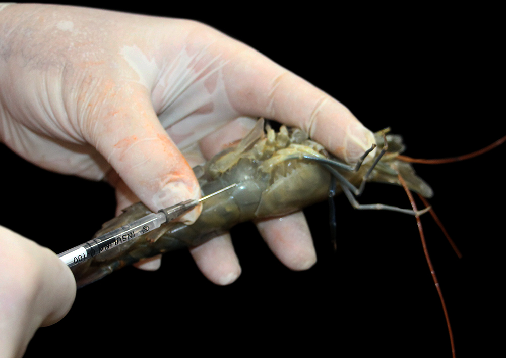
Hemolymph collection. In this study, hemolymph samples were collected by puncturing the ventral hemolymphatic sinus. The procedures were performed with the animals properly restrained and anesthetized (Fig. 1). Samples were collected immediately before euthanasia with 13 x 0.45 mm needles and sterile 1 mL syringes.
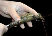
Anesthesiated giant river prawn positioning (Macrobrachium rosenbergii De Man, 1879) for hemolymph collection from the ventral hemolymphatic sinus. The absence of coloration in the hemolymph already present in the syringe is noticeable.
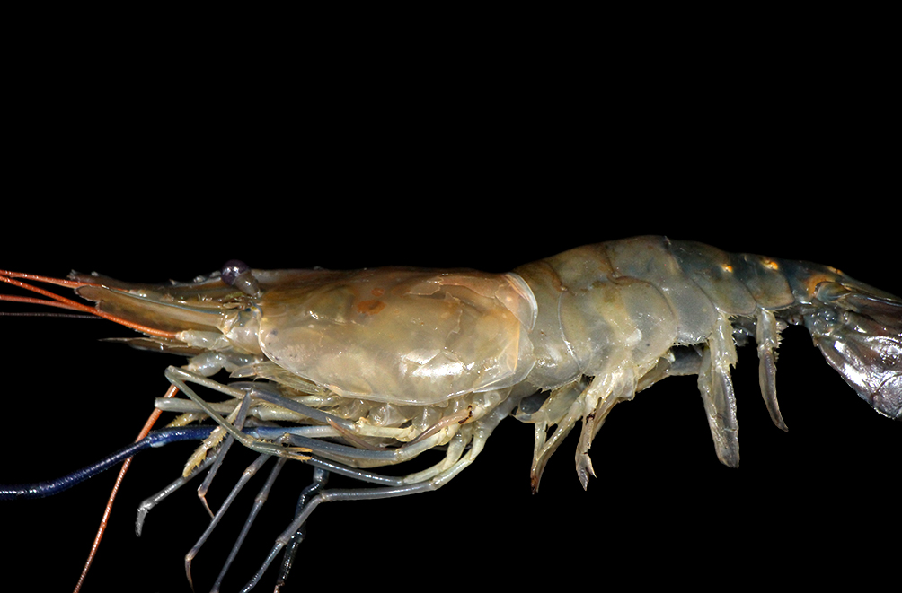
External examination. The prawns were first evaluated inside the transport container, observing their ability to move, respond to stimuli, and position in the water column. Once euthanized, the total body length was measured (rostrum to the base of the telson). The animals were meticulously evaluated for erosions and stains (color changes) on the exoskeleton, darkening of the gills, and total or partial absence of appendages (antennae, pereiopods, pleopods, and eyes, for example) (Fig. 2). Each change was described, when present, following the standardized description with the organ, lesion, distribution, intensity, and time.
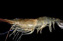
External evaluation of a specimen of giant river prawn (Macrobrachium rosenbergii De Man, 1879). The cephalothorax contains a focally extensive area of exoskeleton erosion, in addition to multifocal blackened areas in the cephalothorax region, abdomen, and limbs.
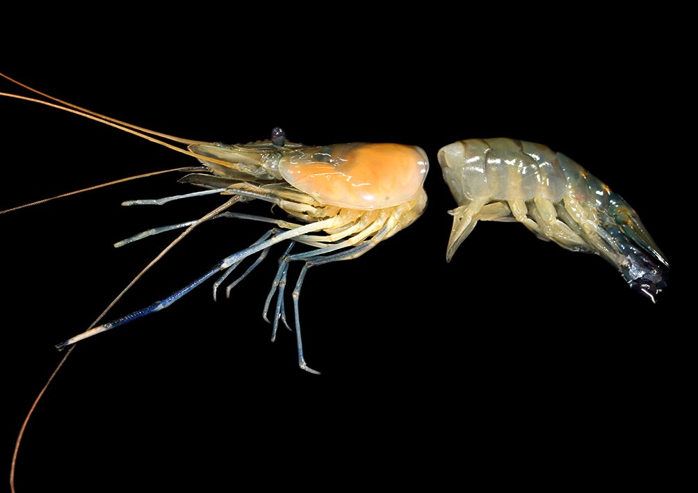
Dissection.Post mortem examination employed scalpels, anatomical tweezers, and scissors. The first step involved separating the cephalothorax from the abdomen by making an incision and transecting the muscles connecting these structures, using the intersegmental membranes of the region as a guide. This procedure resulted in two fragments: the cephalothorax, which contains most organs, and the abdomen and tail region, which comprises the intestine, muscles, and ventral nerve cord (Fig. 3).
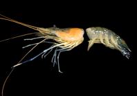
Section of a specimen of giant river prawn (Macrobrachium rosenbergii De Man, 1879) in the intersegmental membrane region between the cephalothorax and the first abdominal somite.
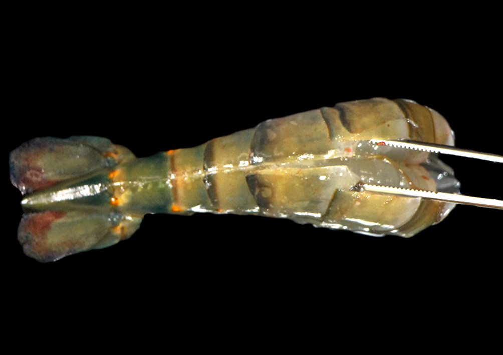
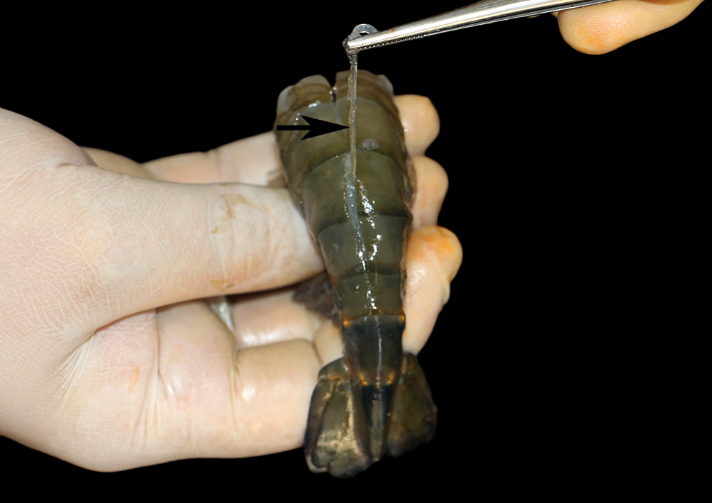
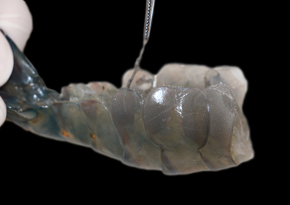
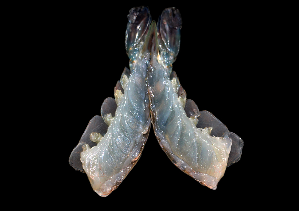
The tail region was then dorsally incised to expose the caudal segment of the digestive system. In cases where the exoskeleton and its connection to the musculature are not the objective of the analysis, this structure can be removed; otherwise, the incision will also include the exoskeleton (Fig. 4). The intestine was clamped at the most cranial portion and pulled caudally through the incision, culminating in its removal for sampling (Fig. 5). A midline incision of the caudal fragment was also made on the ventral side, giving access to the ventral nerve cord. Similarly to the intestine, the nervous tissue was identified, isolated, and removed (Fig. 6 and 7). In the abdominal fragment, it was possible to evaluate the muscle groups after removing the previously mentioned structures (Fig. 8).
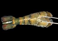
Section of the caudal portion of a specimen of giant river prawn (Macrobrachium rosenbergii De Man, 1879). A dorsal incision is observed in the midline, exposing the bowel and muscle segments.
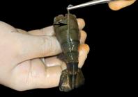
Section of the caudal portion of a specimen of giant river prawn (Macrobrachium rosenbergii De Man, 1879). The removal of the bowel’s caudal portion (arrow) is observed by craniocaudal displacement of the cranial end of the organ.
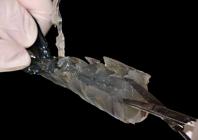
Section of the caudal portion of a specimen of giant river prawn (Macrobrachium rosenbergii De Man, 1879). A ventral incision is observed in the midline, exposing the ventral nerve cord.
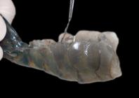
Section of the caudal portion of a specimen of giant river prawn (Macrobrachium rosenbergii De Man, 1879). Craniocaudal traction of the ventral nerve cord is observed.
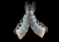
Section of the caudal portion of a specimen of giant river prawn (Macrobrachium rosenbergii De Man, 1879). The abdominal muscle groups can be observed after the separation of the caudal portion into left and right antimeres.
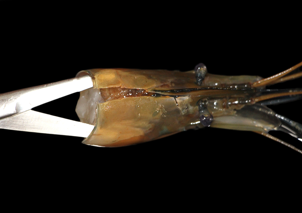
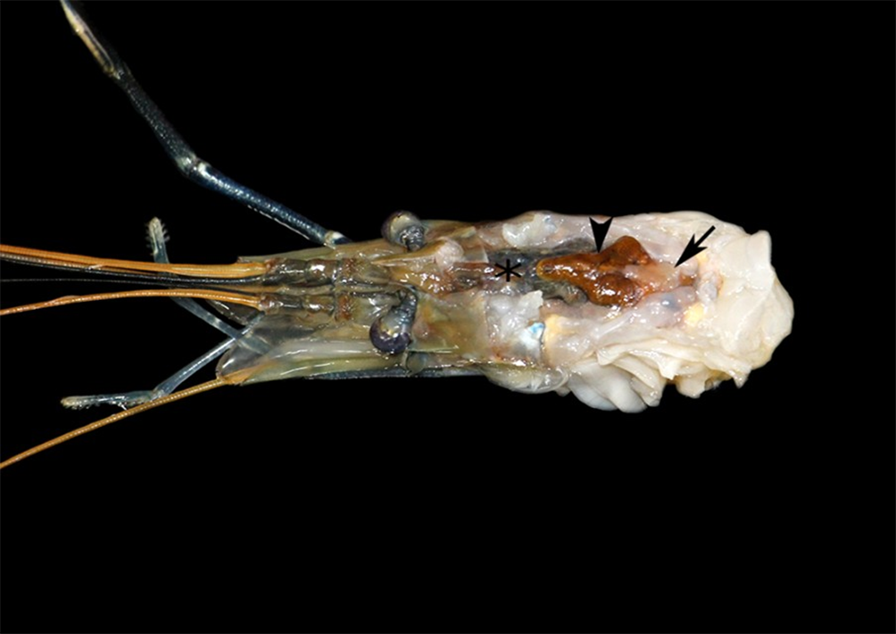
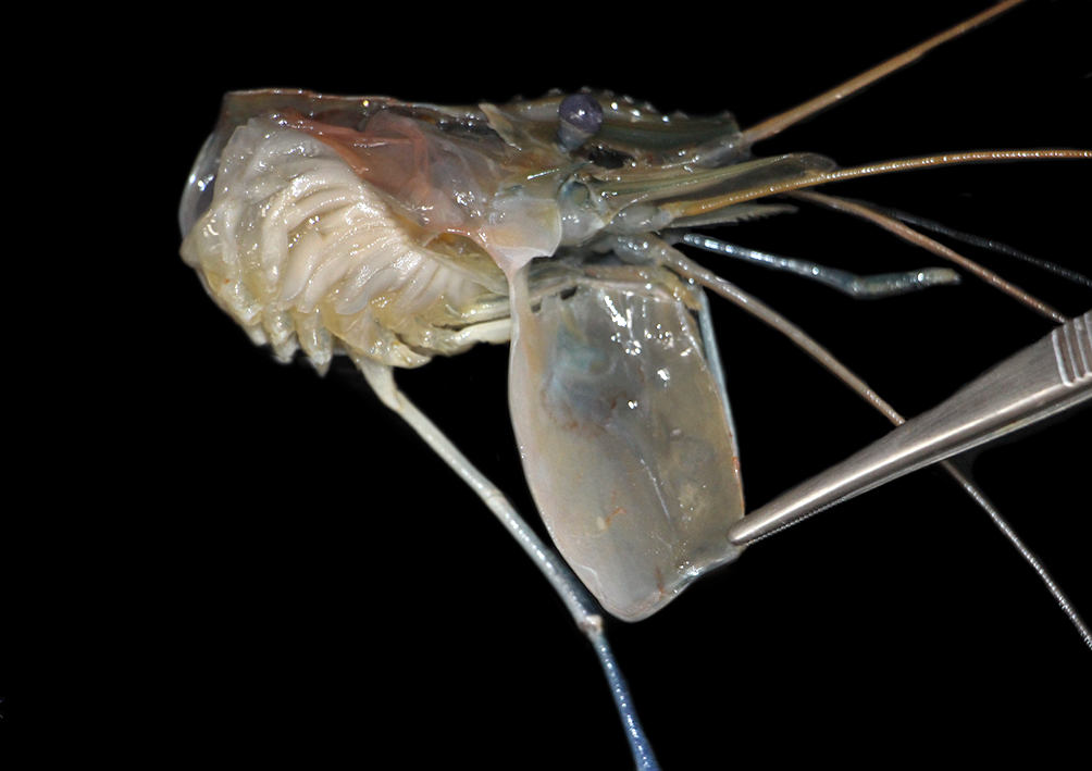
The cranial fragment was opened dorsally with a midline incision made in the caudo-rostral direction, carefully to avoid cutting into adjacent structures (Fig. 9). After the incision, the two exoskeleton fragments were pulled laterally, exposing the muscular and connective tissues of the cephalothorax. The tissues were removed using scissors and anatomical forceps, revealing the heart, hepatopancreas, stomach, and gonads (Fig. 10). In the lateral view of the cephalothorax, now devoid of the exoskeleton, the gills became evident (Fig. 11).

Section of the cranial portion (cephalothorax) of a specimen of giant river prawn (Macrobrachium rosenbergii De Man, 1879). A dorsal incision is observed in the midline.
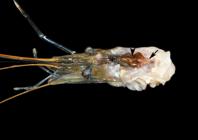
Section of the cranial portion of a specimen of giant river prawn (Macrobrachium rosenbergii De Man, 1879). Arrow = cardiac tissue, arrowhead = gonadal tissue, asterisk = stomach.
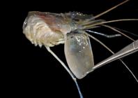
Section of the cranial portion (cephalothorax) of a specimen of giant river prawn (Macrobrachium rosenbergii De Man, 1879). The displacement of the carapace in a cranial and lateral direction can be observed, with exposure of the gill chamber.
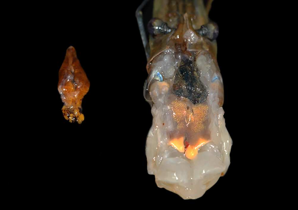
The heart was the first organ removed, considering its dorsal position, followed by the gonads, which became more visible after the removal of the cardiac tissue (Fig. 12). The stomach and the cranial portion of the intestine were then removed, with sectioning of the esophageal tissue (Fig. 13). Following this, the hepatopancreas was removed.

Section of the cranial portion (cephalothorax) of a specimen of giant river prawn (Macrobrachium rosenbergii De Man, 1879). Cephalothorax containing stomach (darker tissue) and hepatopancreas can be seen, with gonadal tissue removed from the piece.

Section of the cranial portion (cephalothorax) of a specimen of giant river prawn (Macrobrachium rosenbergii De Man, 1879). Cephalothorax containing hepatopancreas can be seen, and the stomach is detached from the piece.
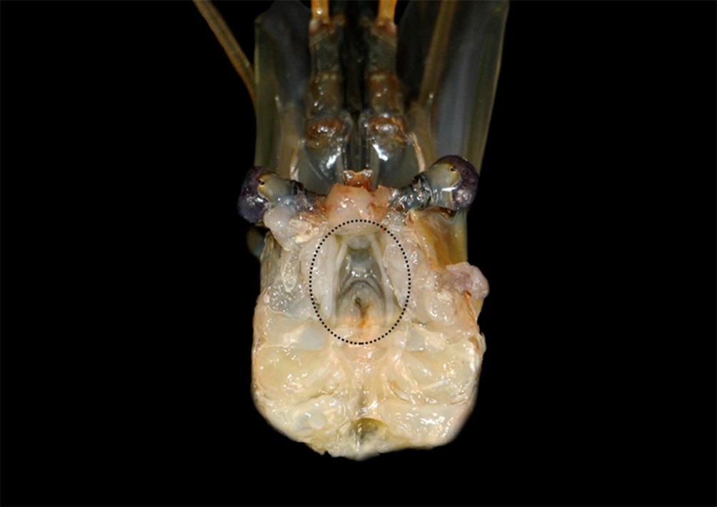
During its removal, the antennal glands, also known as green glands, were located at the base of the antenna. Furthermore, it was possible to identify the supraesophageal ganglion (Fig. 14).
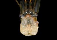
Section of the cranial portion (cephalothorax) of a specimen of giant river prawn (Macrobrachium rosenbergii De Man, 1879). Cephalothorax containing ganglion tissue is observed after the removal of the hepatopancreas (circled area).
Once separated, the organs were evaluated. Structures such as the stomach and intestine were opened when possible, and their contents were examined macroscopically.
Results
The proposed post mortem examination technique allowed for a standardized and sequential examination of the specimens studied. Changes in technique may ensue depending on the examiner’s objective and hypothesis. Through the method described here, however, it was possible to identify, remove, and evaluate the organs of the shrimp submitted to the examination. Furthermore, although basic, the standardized technique enabled rapid and straightforward opening and examination of the animals.
Absence of the second right pereiopod was observed in one of the specimens, in addition to light multifocal areas of erosions and blackening (melanization of the exoskeleton) concentrated in the uropods. In another, mild multifocal melanization areas were observed in the left chela, along with the absence of its last segment. Finally, a reduction in exoskeleton hardness was identified when compared to the other sampled animals. Multiple areas of erosion/ulceration and melanization throughout the exoskeleton were also observed.
No alterations were found in the internal evaluation, except for the absence of intestinal and stomach contents in the animal that presented exoskeleton hardness alterations. No alterations were found in the gills or the macroscopic evaluation of hemolymph.
Discussion
Obtaining the animals’ prior history before the necropsy examination is important, but its absence does not hinder the procedure. Owners or production units must provide as much information as possible to help create differential lists and feasible causes for the animals’ afflictions (Cuéllar-Anjel 2008). Conditions such as stress, development stage, and molting stage, for example, influence the appearance of diseases in crustaceans (Alcivar-Warren et al. 2023). Keepers must also provide appropriate samples of food, water, and other inputs (Berzins et al. 2022). Information on the animals’ origin, the location of production units, the form of production, and environmental parameters must be collected (Cuéllar-Anjel 2008, Berzins et al. 2022). The animals in this study came from the UFPR prawn farming laboratory and had no history of disease. They were euthanized for the experiment.
Environmental water samples must be sent separately from those used to transport animals, always in clean bottles, and analyzed immediately. When immediate analysis is impossible, the sample can be kept refrigerated and tested within 24 hours of collection (Berzins et al. 2022). Key parameters for evaluating water quality in production units include oxygen concentration and temperature (more assertive when measured in loco), in addition to pH, nitrogen compounds (ammonia, nitrite, nitrate), salinity (especially for marine or estuarine species), and alkalinity (freshwater species) (Berzins et al. 2022, Alcivar-Warren et al. 2023). Due to the objective of the present study, water samples were not collected. However, the prawn farming laboratory routinely assesses the water quality of the tanks housing the animals and determines whether all quality parameters are within the recommended range for the species at the time of capture. Environmental characteristics such as acidification, high temperatures, and poor water quality are predisposing factors for diseases and weakening of the exoskeleton in prawns (Alcivar-Warren et al. 2023).
Capturing specimens in tanks or production units usually does not present challenges. However, handling must be gentle and careful, using capture tools that cause the least harm to the animals. Some authors recommend talc-free latex procedure gloves during handling (Berzins et al. 2022). This study employed hand nets, and the animals were transferred to water without long periods of manual restraint, minimizing distress and reducing possible physical damage.
The specimens used in this study were healthy, without any clinical manifestations. However, animals captured in field conditions for diagnostic purposes should preferably be moribund. In some viral diseases diagnosed through hemolymph samples, capture must include clinically healthy individuals that cohabit with sick animals since, in the latter, hemocytes tend to be absent due to cell consumption resulting from the infection (Cuéllar-Anjel 2008, Alcivar-Warren et al. 2023). Juvenile and subadult specimens are the preferred sampling groups in health surveillance cases (Alcivar-Warren et al. 2023). The choice of specimens with a lower development index is recommended (Cuéllar-Anjel 2008). This study chose adult animals to enable better visualization of their anatomical structures, thus facilitating organ identification and execution of the technique.
Animal transport was carried out in suitable-sized and shaped containers. Prawns must be transported and sampled quickly to avoid distress (Cuéllar-Anjel 2008, Alcivar-Warren et al. 2023). Under usual conditions, water from the source tank is adequate for transport, and 2 liters of water for 2.5 cm of animal is recommended (Berzins et al. 2022). If the interval between transfer and examination is too long, the water must be oxygenated and its temperature kept between 25 and 27 °C (Cuéllar-Anjel 2008). In this study, the transfer time from the animal holding unit to the necropsy site was 15 minutes, reducing the need to aerate and cool the transport water.
Assessing invertebrates with signs of stress or disease can be difficult. Conditions such as anorexia, lethargy, erosions, darkening, or spots on the exoskeleton, as well as the absence of appendages, are commonly observed in sick shrimp (Garcês & Pires 2020, Berzins et al. 2022, Alcivar-Warren et al. 2023). External evaluation is an important step and must be carried out meticulously. At this stage, external lesions related to the animal’s cause of death can be observed (Garcês & Pires 2020). In the external evaluation, traumatic lesions, parasitic structures, pustules, blisters, and malformations are assessed (Garcês & Pires 2020). Exoskeleton melanization lesions, such as erosion, ulceration, and absence (total or partial) of attached structures, were observed during the evaluation.
Several prawn-affecting diseases cause color changes, such as white spot disease, named after spots on the exoskeleton caused by calcium deposition. Many conditions cause redness and correlate to the expansion of chromatophores with carotenoid pigments as part of the necrosis process (Alcivar-Warren et al. 2023). Sometimes, due to the translucent characteristic of some shrimps’ exoskeletons, it is possible to observe changes in internal structures, which can be expressed as color changes at first (Alcivar-Warren et al. 2023). The stomach and intestine filling, the musculature coloration, and the appearance of the hepatopancreas can be observed externally (Alcivar-Warren et al. 2023). Color changes of physiological origin, recorded in some crustaceans, require attention; they should be assessed according to the evaluated animals’ species and life stages (Alcivar-Warren et al. 2023).
The presence of parasites in the gill chamber is a recurrent condition, indicating the need for a thorough analysis of this structure during the external evaluation of individuals (Alcivar-Warren et al. 2023, Santos 2023). In crustaceans, the assessment of gill coloration is crucial, as gill darkening can indicate fungal infections and other conditions (Garcês & Pires 2020). A complementary fresh tissue analysis of the gills is recommended. It must be performed with a slide, a coverslip, and a regular optical microscope that allows the visualization of sediments, ciliated protozoa, and oligochaetes that can interfere with the animal’s health (Santos 2023). Macroscopic parasitic structures in the gills and color changes of these structures were not observed in this study.
In the external evaluation, the rigidity of the exoskeleton, which can be assessed with comparative analyses between healthy and sick animals, requires attention. This assessment must consider the molting stage of these animals, as thinner, softer exoskeletons are expected during the post-molt period (Alcivar-Warren et al. 2023). Due to the physiological fragility of the exoskeleton at this stage, animals are more prone to trauma and disease (Alcivar-Warren et al. 2023). Only one of the specimens sampled in the study showed changes in the hardness of the exoskeleton; the individuals presented multiple areas of erosion and melanization, which may indicate inflammation, necrosis, and immune response to a possible pathogen.
The assessment of the animal’s body length and antennae length is called morphometry. This procedure measures live weight and is valuable in studies evaluating growth factors, maturity, and body score (Atherley et al. 2020). In this study, the animals were weighed and their body lengths measured. In field situations, these measurements can be compared with those of healthy animals of the same age or with other groups to detect developmental delays.
The sites for hemolymph collection are diverse and vary among different invertebrate species; most of these animals, including crustaceans, have an open circulatory system (Berzins et al. 2022). Preferred collection sites include the haemocoelomic spaces and the heart. There are cases of limb amputation in which the hemolymph is collected at the wound site (Berzins et al. 2022). In this study, the collection was from the ventral hemolymphatic sinus puncture, which proved less traumatic and easier to perform than limb amputation. Animals were anesthetized for hemolymph collection, as recommended by Berzins et al. (2022). The collected volume can prepare smears, assess coagulation time, perform molecular biology techniques, cultivate and isolate infectious agents, and measure toxins (Berzins et al. 2022). The hemolymph must be stored in a sterile bottle and kept at 4 °C in anticoagulant solutions such as modified Alsever (Santos 2023) or sodium citrate (Barretto 2009) when intended for cell counts and deposited in dry tubes or slides when intended for coagulation time tests (Santos 2023).
Macroscopic evaluation of hemolymph is important primary data; although not diagnostic, it sometimes indicates some conditions in crustaceans (Garcês & Pires 2020, Berzins et al. 2022). Normally, hemolymph is colorless or bluish, the latter resulting from the reaction between hemocyanin and oxygen (Berzins et al. 2022, Alcivar-Warren et al. 2023). The hemolymph of animals infected by Aerococcus viridans var. homari tends to be reddish, as in animals positive for Paramoeba, for example (Garcês & Pires 2020, Berzins et al. 2022, Alcivar-Warren et al. 2023). In the specimens evaluated, the hemolymph did not show any color changes. The study did not include microscopic evaluation of the hemolymph, but the results of this analysis have been significant for assessing the health conditions of shrimp.
The practice of euthanasia must minimize tissue artifacts or changes associated with acute stress and is essential for animal welfare (Atherley et al. 2020, Murray 2022). Euthanasia techniques must take into account the animals’ anatomical characteristics. In arthropods, the presence of a decentralized nervous system (NS) must be accounted for to validate the techniques as humane (Murray 2022). Although commonly used in crustaceans, euthanasia techniques that use relaxing compounds followed by freezing are contraindicated, especially in conditions where the tissues will undergo histopathological evaluations (Murray 2022). This technique was promptly excluded from this study since obtaining a diagnosis involves tissue evaluation for numerous diseases in shrimp farming.
According to Murray (2022), euthanasia in aquatic invertebrates is recommended in two stages. In the first, the anesthesia is followed by euthanasia methods, with the use of chemical substances or physical procedures, such as sectioning of the nervous tissue. The interval between one stage and the next is 15 minutes. The person responsible for euthanasia must pay attention to the animals’ breathing and responses to stimuli. Several components can be used in the first stage of invertebrate euthanasia, such as magnesium chloride, eugenol, tricaine methanesulfonate (MS-222), and ethanol; however, not all are recommended for crustaceans. Studies on crustaceans’ euthanasia focus mainly on lobsters. In these species, compounds such as MS-222 and eugenol (75-100 mg/L) are used in the first stage. In the second, potassium chloride (KCl) (100 mg/100 g of live weight) is administered into the hemolymph and supraesophageal ganglion. In this protocol, death occurs due to cardiac arrest since KCl promotes depolarization of neurons and blocks nerve signals. According to Murray (2022), this protocol has shown worthy results in other species of crustaceans, so it was the author’s chosen method.
Materials intended for post mortem examinations of small vertebrate specimens can be used and adapted for shrimp necropsy (Garcês & Pires 2020, Berzins et al. 2022). Understanding anatomical structures is essential for necropsy examination, since a critical part of this evaluation is distinguishing the abnormal from the normal (Berzins et al. 2022). Basic texts on the anatomy and anatomical variations found in these species should be consulted; in cases where information is lacking, maintaining serial photographic records can be of great value (Berzins et al. 2022). Furthermore, these animals’ routine necropsy allows professionals to increase their knowledge of the tissues and, consequently, helps in distinguishing lesions from non-lesions (Garcês & Pires 2020).
After opening the exoskeleton and exposing the organs, the tissue consistency and color should be analyzed and checked for cystic areas or neoformations (Garcês & Pires 2020). In the evaluated specimens, no alterations suggestive of disease were observed. The authors did not expect lesions, as the animals used were healthy and had no history of disease. The anatomical organization observed in the examinations followed that described by Atherley et al. (2020) and Smolowitz (2021) for crustaceans. The chosen technique was based on that described for larger decapods, such as lobsters (Atherley et al. 2020).
When collected, organ fragments should be promptly fixed. Fixation solutions for invertebrate histological samples are diverse, such as 10% buffered formalin, Bouin’s fixative, Davidson’s fixative, Dietrich’s fixative, and Helly’s fixative (Berzins et al. 2022, Alcivar-Warren et al. 2023). Choosing one fixation solution over another should consider the research objective and the species. Phenolic compounds and strong acids or alkalis should be avoided due to the possibility of damage to the assessed tissue (Berzins et al. 2022). As with obtaining and processing vertebrate samples, freezing samples for histological processing should be avoided due to the large amount of tissue artifacts generated by this practice (Berzins et al. 2022). Fixative solutions were not evaluated, and no microscopic evaluation was conducted in this study. Davidson’s solution is commonly used as an alternative to formalin, usually employed in samples of mollusks, crustaceans, and echinoderms (Berzins et al. 2022, Alcivar-Warren et al. 2023).
Although necropsy is a fundamental tool for diagnosing diseases in aquaculture species, there is no standardized, widely recognized technique specifically described for shrimp. This gap contrasts with the existence of documented protocols for other decapod crustaceans, such as lobsters, in which necropsy methods are well established and applied for anatomical, pathological, and health assessments (Atherley et al. 2020). The lack of a validated procedure specific to shrimp hinders the uniformity of diagnostic practices and the reproducibility of research results, highlighting the need to adapt and develop protocols that consider the particular anatomical and physiological characteristics of the species.
Conclusion
The post mortem examination technique used on the Malaysian shrimp (Macrobrachium rosenbergii) specimens was effective and enabled the opening and evaluation of the animals’ anatomical components. As such, an adaptation for other species of this group is suggested, considering the development phases, size, and possible anatomical variations of different species. Macroscopic evaluation standardization is vital, as it provides fundamental diagnostic practices to mitigate economic and environmental losses.
Acknowledgments
The author(s) would like to thank the “Centro de Assessoria de Publicação Acadêmica” (Academic Publishing Advisory Center - CAPA; http://www.capa.ufpr.br) of the “Universidade Federal do Paraná” (UFPR) for assistance with English language translation and developmental editing.
References
-
Alcivar-Warren A, Bateman K, Clinton M, Foyle L, Lewbart G, Loh R, Tepper J. Aquatic Invertebrates, p.3-80. In: Urdes L, Walster C, Tepper J. Pathology and Epidemiology of Aquatic Animal Diseases for Practitioners. 2023; https://doi.org/10.1002/9781119839729.ch1
» https://doi.org/10.1002/9781119839729.ch1 - Andrade TP, Cruz-Flores R, Mai HN, Dhar AK. Doenças emergentes que afetam a carcinicultura marinha brasileira e mundial. Rev Assoc Bras Criadores Camarão 2023;1(3):98-99.
-
Atherley NAM, Freeman MA, Dennis MM. Post-mortem examination of the Caribbean spiny lobster (Panulirus argus, Latreille 1804) and pathology in a fishery of the Lesser Antilles. J Invertebrate Pathol 2020; https://doi.org/10.1016/j.jip.2020.107453
» https://doi.org/10.1016/j.jip.2020.107453 - Barretto ACG. Contagem total de hemócitos de camarões marinhos Litopenaeus vannamei (Boone, 1931). Dissertação de Mestrado, Universidade Federal Rural de Pernambuco, Recife, 2009. 52p.
-
Berzins IK, Smolowitz R, Lewbart GA. Diagnostic techniques and sample handling, p. 615-630. In: Lewbart GA. Invertebrate Medicine. 2022; https://doi.org/10.1002/9781119569831.ch25
» https://doi.org/10.1002/9781119569831.ch25 - Cuéllar-Anjel J. Métodos de diagnóstico de enfermedades en camarones marinos de cultivo, p.2-54. In: Morales VQ, Cuéllar-Anjel J. Guía técnica: patología e inmunología de camarones penaidos. Panamá: Programa CYTED Red II-D Vannamei, Rep. de Panamá; 2008. 270p.
-
FAO. The State of World Fisheries and Aquaculture 2024: blue transformation in action. 2024; https://doi.org/10.4060/cd0683en
» https://doi.org/10.4060/cd0683en -
Garcês A, Pires I. Necropsy Techniques for Examining Wildlife Samples. 2020; https://doi.org/10.2174/97898114683391200101
» https://doi.org/10.2174/97898114683391200101 - Guerrelhas ACB, Ferreira MC. 2º Evento Shrimp Summit 2024 na Índia: estimular as pessoas a comerem mais camarão e acima de tudo “fazê-las se apaixonar por camarão”. Rev Assoc Bras Criadores Camarão 2024;26(2):10-11.
-
Murray MJ. Euthanasia, p.645-650. In: Lewbart GA. Invertebrate Medicine. 2022; https://doi.org/10.1002/9781119569831.ch27
» https://doi.org/10.1002/9781119569831.ch27 - Rocha IP, Fernandes BRS. Censo da carcinicultura dos Estados do Ceará, Piauí e Rio Grande do Norte. Natal: Associação Brasileira de Criadores de Camarão (ABCC), Deza´s; 2022. 205p.
-
Ross EP, Behringer DC, Muñoz A, Díaz D, Bojko J. A histological atlas for the Palinuridae (Crustacea: Decapoda: Achelata): a guide to parasite discovery and spotting the abnormal in spiny lobsters. J Invertebrate Pathol 2019; https://doi.org/10.1016/j.jip.2019.03.001
» https://doi.org/10.1016/j.jip.2019.03.001 - Santos LC. Protocolo de análises como indicador preditivo do estado de saúde de Macrobrachium rosenbergii Dissertação de Mestrado, Universidade Federal do Paraná, Palotina, 2023. 59p.
-
Smolowitz R. Arthropod, p.277-299. In: Ladouceur EE. Invertebrate histology. 2021; https://doi.org/10.1002/9781119507697.ch11
» https://doi.org/10.1002/9781119507697.ch11
-
Data availability statement
The data presented in this study are available on request from the corresponding author.
The data presented in this study are available on request from the corresponding author.