 Apocrine adenocarcinoma in Oryctolagus cuniculus (domestic rabbit) with osteolysis: case report
ABSTRACT:
Apocrine adenocarcinoma is a malignant tumor arising from the apocrine gland. It’s well-reported in dogs, uncommon in cats, and rarely described in other species. A 4-year-old female domestic rabbit with lameness in the left pelvic limb, which progressed to loss of function over seven months, was treated in a medical routine. Upon palpation of the affected region, the rabbit presented alertness without exhibiting painful sensitivity. Radiographic imaging revealed pronounced alterations in the bone trabeculae of the left femur, accompanied by areas of corticomedullary osteolysis. A biopsy from the left femur was performed for histopathological and immunohistochemical analysis. The morphological diagnosis comprises apocrine adenocarcinoma. This report aims to present what seems to be the first worldwide case of apocrine adenocarcinoma invading muscle tissue and causing bone lysis in the joint region in an Oryctolagus cuniculus.
INDEX TERMS:
apocrine adenocarcinoma; Oryctolagus cuniculus; rabbit; apocrine gland; osteolysis; histopathology; immunohistochemistry
RESUMO:
O adenocarcinoma apócrino é um tumor maligno que surge da glândula apócrina. Este é bem relatado em cães, incomum em gatos e raramente descrito em outras espécies. Uma coelha doméstica, fêmea, de quatro anos de idade, com claudicação em membro pélvico esquerdo, que evoluiu para impotência funcional em sete meses, foi tratada em rotina médica. Esta apresentou estado de alerta sem sensibilidade dolorosa à palpação da região afetada. A imagem radiográfica revelou alterações pronunciadas em trabéculas ósseas do fêmur esquerdo, acompanhadas de áreas de osteólise corticomedular. Foi realizada biópsia do fêmur esquerdo para análise histopatológica e imunohistoquímica. O diagnóstico morfológico compreende o que parece ser o primeiro caso mundial de adenocarcinoma apócrino invadindo o tecido muscular e causando lise óssea em região articular de um Oryctolagus cuniculus.
TERMOS DE INDEXAÇÃO:
adenocarcinoma apócrino; Oryctolagus cuniculus; coelho; glândula apócrina; osteólise; histopatologia; imuno-histoquímica
Introduction
Apocrine adenocarcinoma is a malignant tumor originating from the apocrine gland present in the dermis and hypodermis. It is usually solitary, and the clinical appearance may vary depending on the histologic subtype (Gross et al. 2008). Sweat gland adenocarcinoma encompasses various subtypes, including cystadenocarcinoma, simple and complex (mixed) adenocarcinoma, ductal carcinoma, solid-cystic ductal carcinoma, and clear cell ductal carcinoma. Among these, simple apocrine adenocarcinoma is the most prevalent (Miwa et al. 2006, Gross et al. 2008).
This entity is common in dogs and rare cats, with only one case reported in a rabbit, but there is no report of this tumor causing osteolysis. Therefore, immunohistochemistry is essential to know the origin and type of tumor (Bahrami et al. 2008). The documentation of a case of apocrine adenocarcinoma in a domestic rabbit is crucial due to the scarcity of available literature on this condition, with only one previous report existing to date. Given the rarity of this neoplasm in rabbits, additional case studies are essential for expanding the veterinary understanding of its clinical and histopathological presentations, progression, and potential treatment options.
This report aims to contribute valuable insights into the diagnosis and management of apocrine adenocarcinoma in rabbits, thereby enriching the limited knowledge base and guiding future research and clinical practices in veterinary oncology.
Case Description
Ethical approval. No approval of research ethics committees was required to accomplish the goals of this study because experimental work was conducted with samples from the cadaver of a domestic rabbit (Oryctolagus cuniculus) donated for academic purposes by the owner to the “Universidade de Santo Amaro”.
A 4-year-old female domestic rabbit (O. cuniculus) presented left pelvic limb lameness. Upon gross evaluation, the rabbit exhibited alertness and activity, showing no signs of pain or sensitivity upon palpation of the bilateral coxal region. Radiographic examination revealed osteolysis and irregular bone proliferation in the caudal portion of the left acetabulum, indicative of a bone injury. Additionally, a well-defined radiolucent area with regular margins was observed in the proximal diaphysis of the tibia, suggesting the presence of a bone cyst. The prescribed treatment regimen included gabapentin (10 mg/kg, orally, t.i.d.), dipyrone (40 mg/kg, orally, t.i.d.), and prednisolone (0.5 mg/kg, orally, b.i.d.). A fine needle aspiration cytology was performed to investigate the suspected bone cyst further, indicating the presence of a heterophilic inflammatory process.
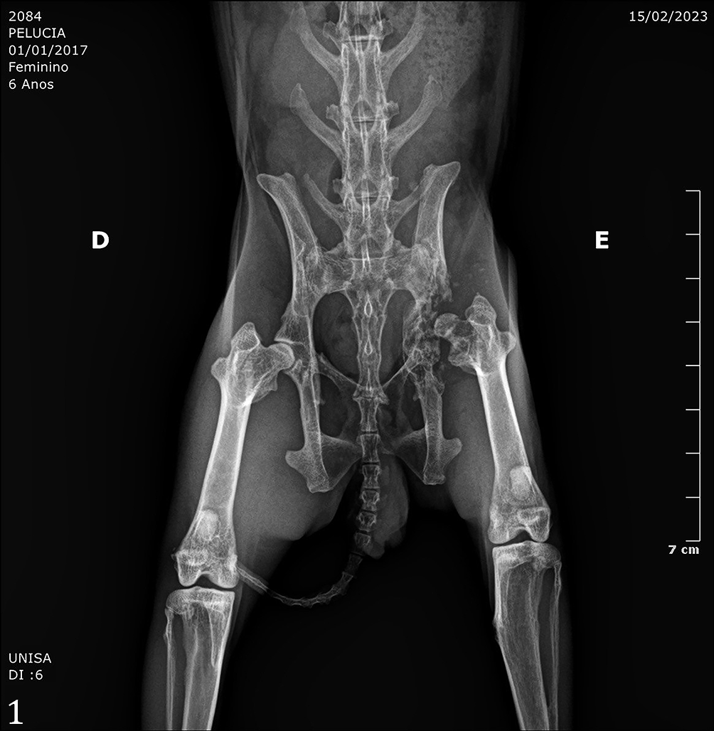
After two months, the rabbit exhibited prostration and experienced pain upon palpation of the left hip joint region. The treatment plan was modified, replaced with prednisolone (0.5 mg/kg, orally, b.i.d.) with tramadol (12 mg/kg, orally, t.i.d.) and metoclopramide (0.5 mg/kg, orally, b.i.d.). Subsequent radiographic evaluation revealed significant alterations in the bone, characterized by irregular proliferation and areas of osteolysis in the left acetabulum and pubis, indicative of aggressive bone injury (Fig. 1). Additionally, the caudal margin of the acetabulum exhibited loss of definition. In contrast, there was irregular bone proliferation adjacent to the cranial margin of the left acetabulum.
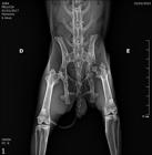
Radiographic evaluation of an aggressive bone injury chacracterized by alterations in the bone trabeculae, composed by irregular proliferation and areas of osteolysis. Osteolysis, left acetabulum and pubis, rabbit. São Paulo, 2024.
After seven months, the rabbit’s condition worsened, displaying increased prostration, loss of limb function, and muscle atrophy. Two bone formations encompassing the head and neck of the left femur region were collected for histopathological analysis. Fragments of the limb and femur bone measured 7.2 x 3.9 x 2.9 cm and 6.5 x 6.4 x 1.9 cm; the bigger one contained bone, muscular, adipose tissue and inner dermis, while the smaller one contained epidermis and superficial dermis. Both were irregular in shape and varied in consistency from hard to soft. They exhibited heterogeneous colors, ranging from blackened and brownish to whitish, presenting a multinodular appearance, interspersing the dermis, muscle and bone tissue.
Immunohistochemical analysis was conducted using cytokeratin (CK) 5/6, CK7, AE1/AE3, and vimentin, such as anti-CK5/6 antibody (D5/16 B4, Agilent Santa Clara/CA), anti-CK7 antibody (OV-TL 12/30, Agilent Santa Clara/CA), anti-Cytokeratin (AE1/AE3, Agilent Santa Clara/CA) and anti-vimentin (V9, Agilent Santa Clara/CA). Proper positive and negative external controls were included in the analysis. The internal positive controls were considered from the tissue near the neoplasm. The clinical therapies were ineffective with the progressive worsening of the case, and euthanasia was performed, but the owner did not authorize a post mortem examination.
Results
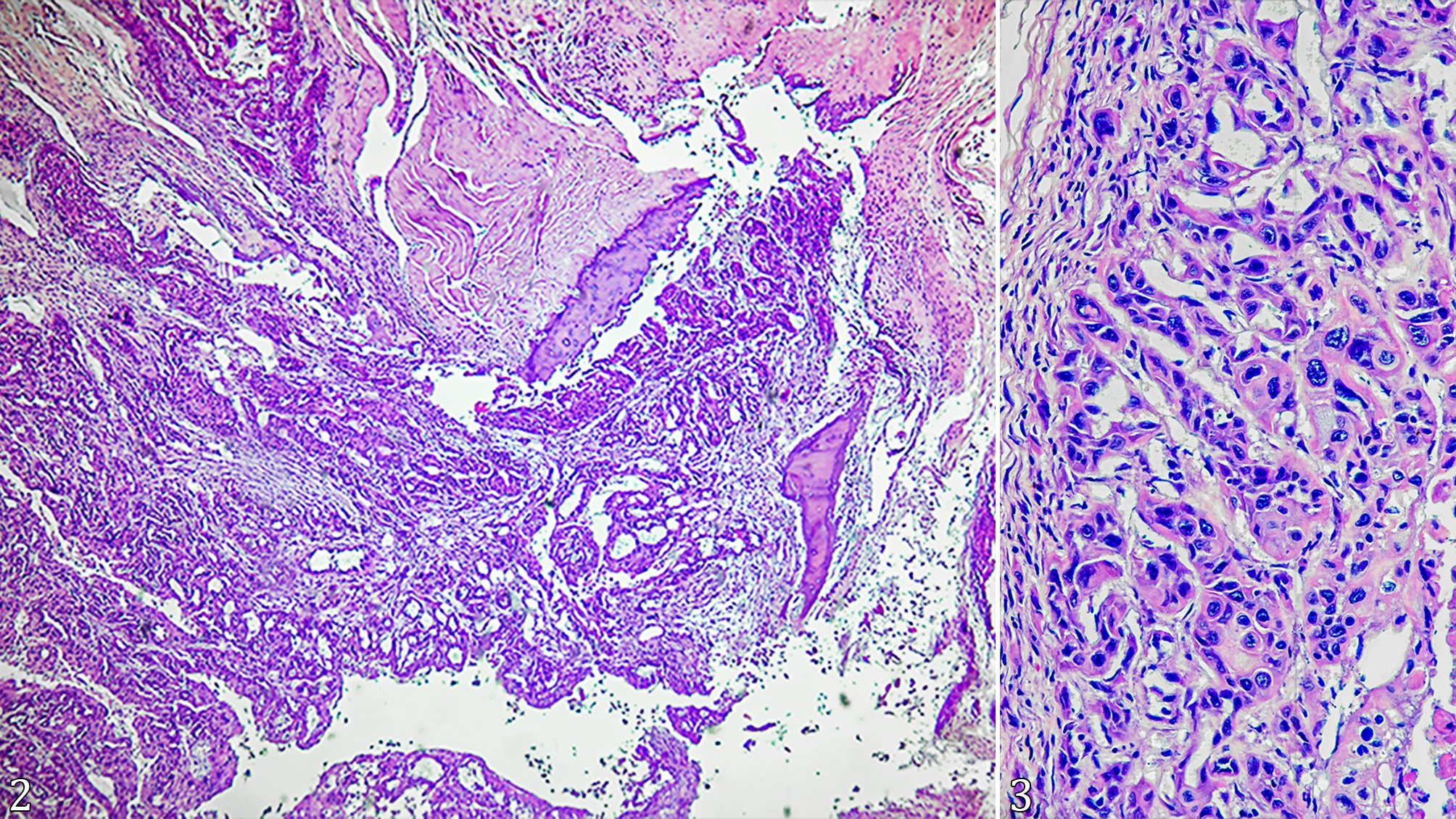
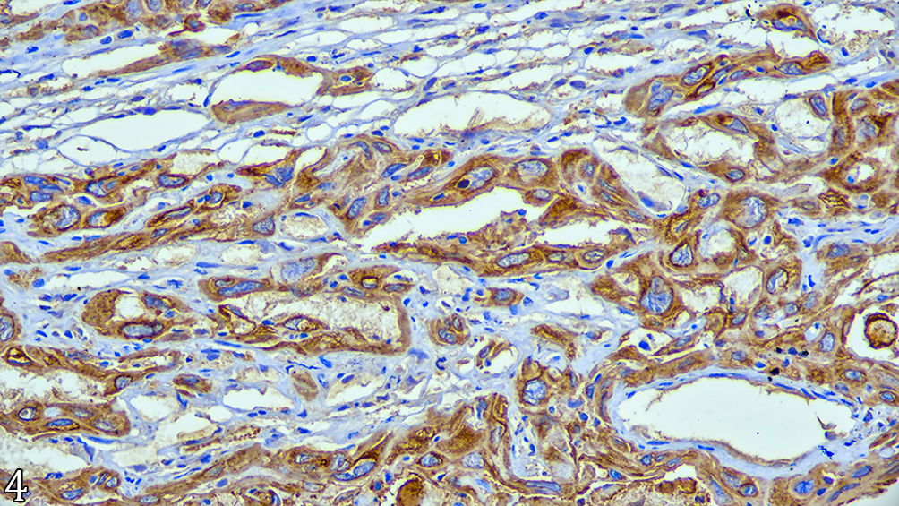
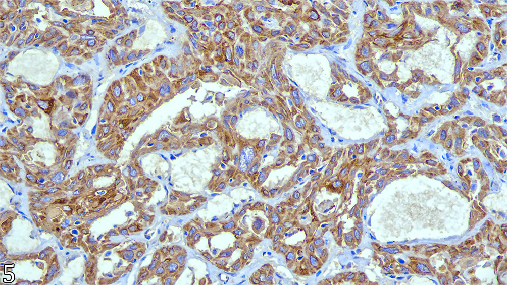
Microscopic examination of the neoplasm revealed a moderate cellular infiltrate within the dermis, subcutaneous, skeletal striated muscle and bone. The tumor appeared poorly delimited, non-encapsulated, and irregular. The cells exhibited an irregular tubular arrangement, and the tubular lumen was rarely filled with eosinophilic fibrillar material and cellular debris, indicating secretory coil differentiation. The stroma surrounding the cells displayed a moderate fibro-collagenous composition. The neoplastic cells were small, polyhedral to round epithelial cells with indistinct and eosinophilic cytoplasm. They had large, rounded polyhedral central nuclei with loose chromatin and one to four distinct, small, eosinophilic nucleoli. Marked anisocytosis, anisokaryosis, and cellular pleomorphism were observed. The neoplasm exhibited low mitotic activity, with nine mitotic figures observed in 2.37 mm². Atypical mitoses were also present. Additionally, a slight presence of binucleated, multinucleated, and karyomegalic cells, multifocal necrosis, a small number of apoptotic cells, and multifocal emperipolesis was also observed. There was the presence of multifocal areas of bone lysis (Fig. 2 and 3). On immunohistochemistry analysis, the controls were strongly positive for AE1/AE3, for CK5/6, for CK7 and Vimetin, all of them with cytoplasmatic immunolabeling. In the case of the tumor, it revealed diffuse cytoplasmatic immunolabeling with strong intensity for AE1/AE3. In contrast, Vimentin immunolabeling was negative (Vimentin -). CK5/6 exhibited strong and diffuse cytoplasmatic immunolabeling with perinuclear enhancement in all epithelial cells (CK5/6 +) (Fig. 4). Strong and diffuse cytoplasmatic immunolabeling for CK7 was also observed (CK7 +) (Fig. 5).
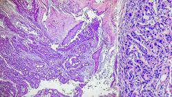
Apocrine adenocarcinoma in acetabulum of Oryctolagus cuniculus envolving muscular and bone tissues. (2) The tumor is poorly delimited, nonencapsulated, irregular and exhibited an irregular tubular arrangement. HE, obj. 4x. (3) Small epithelial cells with indistinct and eosinophilic cytoplasm, with large, polyhedral central nuclei with 1 to 4 nucleoli. Marked anisocytosis, anisokaryosis, and cellular pleomorphism, with presence of binucleated, multinucleated and karyomegalic cells. HE, obj. 40x.
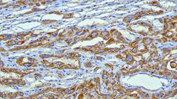
Immunohistochemical labelling of anti-CK5/6 antibody (D5/16 B4) exhibited strong and diffuse cytoplasmic staining with perinuclear enhancement. IHC, obj. 40x.
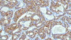
Immunohistochemical of anti-CK7 antibody (OV-TL 12/30) labelling strong and diffuse cytoplasmic staining. IHC, obj. 40x.
Discussion
Apocrine adenocarcinomas have been extensively studied in dogs, the most common species, and, to a lesser extent, in cats (Withrow et al. 2013). However, there is one case report of a male rabbit with a possible sweat gland-derived apocrine adenocarcinoma located on the left caudal abdominal wall (Miwa et al. 2006), but without the involvement of deeper tissues like in this report. Across all these species, this neoplasm exhibits similar clinical and pathological features. The characterization of apocrine differentiation relies on several factors, including its typical anatomical location, association with follicular tumors, and decapitation secretion, which has traditionally been considered a key feature of apocrine glandular differentiation (secretory coil). In addition, the apocrine adenocarcinoma has a slow-growing pattern, and metastasis is uncommon. Therefore, the use of immunohistochemistry analysis is indispensable for accurate tumor diagnosis and determination of its origin site because the animal presented bone and joint infiltration with bone lysis as observed in the radiographic evaluation (Leong & Wright 1987, Bellizzi 2020, Meuten 2020).
This neoplasm, common in dogs and rare in cats, exhibits histological similarities to their benign counterparts, although they tend to be larger, less circumscribed, and demonstrate cytologic features of malignancy. They can be classified as poor or well-differentiated. Poorly differentiated variants are characterized by infiltrative neoplasms with ill-defined borders, composed of large cuboidal to polygonal cells arranged in glandular and solid aggregates in varying proportions (Gross et al. 2008, Mauldin & Peters-Kennedy 2016). Decapitation secretion and luminal accumulation of secretory material are frequently observed (Robson et al. 2008). The cells exhibit amphophilic cytoplasm, large vesicular nuclei with pleomorphism, possible loss of nuclear polarity, prominent nucleoli, and variable mitotic activity. Atypical mitotic figures may be present. The formation is composed of a moderate to abundant stroma of reactive collagenous tissue. Squamous metaplasia and caseous necrosis can also occur in small areas (Gross et al. 2008, Mauldin & Peters-Kennedy 2016). Due to the lack of published studies on this neoplasm in domestic rabbits, the authors used the same description for dogs and cats, similar to the previously published work (Miwa et al. 2006).
The differential diagnoses include apocrine adenoma, mammary carcinoma, and other poorly differentiated carcinomas, both primary and metastatic (Gross et al. 2008, Robson et al. 2008, Mauldin & Peters-Kennedy 2016). In this case, physical examination and radiography are insufficient to completely rule out the possibility of metastasis, and benign neoplasms were also ruled out due to the infiltrative pattern of the tumor itself. The histologic distinction is often challenging, particularly in poorly differentiated neoplasms, thus highlighting the essential role of immunohistochemistry.
AE1/AE3, an anti-pan-cytokeratin antibody, is suggested as the primary antibody to define epithelial origin. These cytokeratins are commonly expressed in epithelial tissues and recognize a broad range of cytokeratins involved in epithelial cell differentiation (Painter et al. 2010, Toniti et al. 2010). Cytokeratin subtypes (CK1 through CK20) play a role in distinguishing different epithelial cell lineages. For instance, CK7 is a low molecular weight cytokeratin predominantly found in various ductal and glandular epithelia (Qureshi et al. 2004, Hrudka et al. 2021), whereas CK5/6 is a high molecular weight cytokeratin expressed in neoplasms originating from stratified epithelia (Chu & Weiss 2002, Gurda et al. 2015).
In this case report, the positive immunolabeling of AE1/AE3 and negative immunolabeling of vimentin confirm epithelial proliferation. Strong and diffuse positive immunolabeling of CK5/6 and CK7 validates the differentiation of apocrine and sebaceous secretory epithelial cells (Yamamoto & Yasuda 1999, Qureshi et al. 2004).
Conclusion
The morphological diagnosis corresponds to apocrine adenocarcinoma, consistent with the descriptions found in the literature. The conclusive evidence provided by immunohistochemistry strengthens this diagnosis. This study seems to be the first documented case of apocrine adenocarcinoma in Oryctolagus cuniculus worldwide with muscle and bone tissue invasion in the joint region, supported by compelling evidence through immunohistochemistry analysis.
Acknowledgments
This study was supported by the Veterinary Hospital of the “Universidade de Santo Amaro” (UNISA), São Paulo/SP.
References
-
Bahrami A, Truong LD, Ro JY. Undifferentiated tumor: true identity by immunohistochemistry. Arch Pathol Lab Med 2008; https://doi.org/10.5858/2008-132-326-UTTIBI
» https://doi.org/10.5858/2008-132-326-UTTIBI -
Bellizzi AM. An algorithmic immunohistochemical approach to define tumor type and assign site of origin. Adv Anat Pathol 2020; https://doi.org/10.1097/PAP.0000000000000256
» https://doi.org/10.1097/PAP.0000000000000256 - Chu PG, Weiss LM. Expression of cytokeratin 5/6 in epithelial neoplasms: an immunohistochemical study of 509 cases. Modern Pathol 2002;15(1):6-10. PMid:11796835
- Gross TL, Ihrke PJ, Walder EJ, Affolter VK. Skin diseases of the dog and cat: clinical and histopathologic diagnosis. New Jersey: John Wiley & Sons; 2008.
-
Gurda GT, Zhang L, Wang Y, Chen L, Geddes S, Cho WC, Askin F, Gabrielson E, Li QK. Utility of five commonly used immunohistochemical markers TTF-1, Napsin A, CK7, CK5/6 and P63 in primary and metastatic adenocarcinoma and squamous cell carcinoma of the lung: a retrospective study of 246 fine needle aspiration cases. Clin Transl Med 2015; https://doi.org/10.1186/s40169-015-0057-2
» https://doi.org/10.1186/s40169-015-0057-2 -
Hrudka J, Fišerová H, Jelínková K, Matěj R, Waldauf P. Cytokeratin 7 expression as a predictor of an unfavorable prognosis in colorectal carcinoma. Sci Rep 2021; https://doi.org/10.1038/s41598-021-97480-4
» https://doi.org/10.1038/s41598-021-97480-4 -
Leong AS-Y, Wright J. The contribution of immunohistochemical staining in tumour diagnosis. Histopathology 1987; https://doi.org/10.1111/j.1365-2559.1987.tb01874.x
» https://doi.org/10.1111/j.1365-2559.1987.tb01874.x -
Mauldin EA, Peters-Kennedy J. Integumentary system. In: Maxie MG. Jubb, Kennedy & Palmer’s Pathology of Domestic Animals. 2016; https://doi.org/10.1016/B978-0-7020-5317-7.00006-0
» https://doi.org/10.1016/B978-0-7020-5317-7.00006-0 - Meuten DJ. Tumors in domestic animals. 5th ed. Iowa: Wiley-Blackwell; 2020.
-
Miwa Y, Mochiduki M, Nakayama H, Shibuya N, Ogawa H, Sasaki N. Apocrine adenocarcinoma of possible sweat gland origin in a male rabbit. J Small Anim Pract 2006; https://doi.org/10.1111/j.1748-5827.2006.00113.x
» https://doi.org/10.1111/j.1748-5827.2006.00113.x -
Painter JT, Clayton NP, Herbert RA. Useful immunohistochemical markers of tumor differentiation. Toxicol Pathol 2010; https://doi.org/10.1177/0192623309356449
» https://doi.org/10.1177/0192623309356449 -
Qureshi HS, Ormsby AH, Lee MW, Zarbo RJ, Ma CK. The diagnostic utility of p63, CK5/6, CK 7, and CK 20 in distinguishing primary cutaneous adnexal neoplasms from metastatic carcinomas. J Cutan Pathol 2004; https://doi.org/10.1111/j.0303-6987.2004.00147.x
» https://doi.org/10.1111/j.0303-6987.2004.00147.x -
Robson A, Lazar AJF, Nagi JB, Hanby A, Grayson W, Feinmesser M, Granter SR, Seed P, Warneke CL, McKee PH, Calonje E. Primary cutaneous apocrine carcinoma: a clinico-pathologic analysis of 24 cases. Am J Surg Pathol 2008; https://doi.org/10.1097/PAS.0b013e3181590ba4
» https://doi.org/10.1097/PAS.0b013e3181590ba4 - Toniti P, Sirivisoot S, Jandee P, Srimontri P, Puchadapirom P, Doungchawee G, Kasorndorkbua C. AE1/AE3, vimentin and p63 immunolocalization in canine mammary gland tumours: Roles in differentiation between luminal epithelial and myoepithelial lineages. Asian Pac J Cancer Prev 2010;11(1):227-230. PMid:20593961
-
Withrow SJ, Vail DM, Page RL. Withrow and macewen’s small animal clinical oncology. 2013; https://doi.org/10.1016/C2009-0-53135-2
» https://doi.org/10.1016/C2009-0-53135-2 -
Yamamoto O, Yasuda H. An immunohistochemical study of the apocrine type of cutaneous mixed tumors with special reference to their follicular and sebaceous differentiation. J Cutan Pathol 1999; https://doi.org/10.1111/j.1600-0560.1999.tb01836.x
» https://doi.org/10.1111/j.1600-0560.1999.tb01836.x
-
Data availability statement
The data supporting the findings of this case report are available from the corresponding author upon reasonable request. Given the routine nature of the pathology findings, no publicly accessible dataset has been generated. Any additional pathological details, images, or histological data related to the case can be provided upon request, in accordance with institutional and ethical guidelines.
The data supporting the findings of this case report are available from the corresponding author upon reasonable request. Given the routine nature of the pathology findings, no publicly accessible dataset has been generated. Any additional pathological details, images, or histological data related to the case can be provided upon request, in accordance with institutional and ethical guidelines.