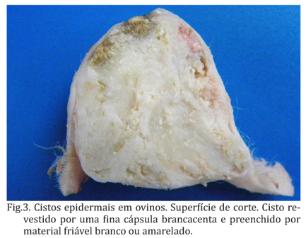
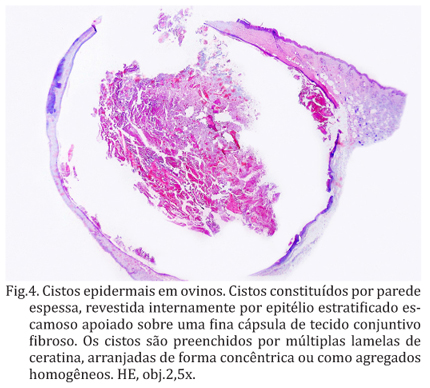
The clinic and pathological aspects of four cases of epidermal cysts in sheep are described. The affected two to 12-year-old sheep were from farms in the Rio Grande do Sul state, Brazil. Three sheep showed multiple nodules, scattered randomly throughout the body, while one sheep had a single nodule on the cervical region. The period between the emergence of the first nodule to appearance of multiple nodules was approximately eight months in a case, one year in two cases, and unknown in the remaining case. These four were the only affected sheep in their respective flocks. The cutaneous nodules were round to oval, raised, fluctuant, non-pruritic and covered by wooless skin. These nodules had 1-7cm in diameter and, occasionally, revealed a small 1-2mm central pore communicating the interior of the nodule with external surface. On cut surface, the nodules were cystic, demarcated by 0.5-0.8cm in thickness white wall, and filled by abundant white to yellow, friable material. Histologically, the cyst wall was lined by a rim of tissue containing all layers of a stratified squamous epithelium. This epithelium was anchored on and supported by a thin capsule of fibrous connective tissue. The cysts were filled with homogenous aggregates or concentrically arranged streams of keratin. Based on the location and on the gross and histological findings these nodules were diagnosed as infundibular epidermal cysts.
Epidermal cysts; skin; dermatology; sheep diseases


