 Outbreak of swinepox in subsistence pig herds in Santa Catarina state, Brazil
ABSTRACT:
Swinepox (SWPV), caused by Suipoxvirus, primarily affects pigs and typically presents progressive skin lesions. This study aims to describe and characterize an outbreak of SWPV on four subsistence-farming properties in the state of Santa Catarina, South region of Brazil. Clinical, histopathological, molecular, and immunohistochemical analyses were performed on skin samples from one pig per property. The outbreak involved four subsistence pig farms, affecting the herds with proliferative, crusted skin lesions. Skin samples from four pigs, one from each property, were collected and routinely processed for histopathological analysis. Immunohistochemistry (IHC) was performed using the peroxidase method with a polyclonal vaccinia virus antibody, and the Max Polymer Detection System was utilized. Frozen skin fragments underwent polymerase chain reaction (PCR) for Suipoxvirus, targeting the FP-DNApol/RP-DNApol sequences, amplifying 543 base pairs. Sequencing and phylogenetic analysis were performed using the Sanger method. The farm facilities were located near water reservoirs, where constant flies and mosquitoes plagued. The number of affected pigs was 14, 9, 21 and 2, respectively, in the four farms, resulting in 98% morbidity; however, no pig died. Macroscopically, erythematous and crusted lesions with a crateriform appearance were observed on the dorsal, ventral and limb regions, as well as the face, ears, and snout, associated with intense pruritus. Histopathology revealed proliferative pustular ulcerative dermatitis with eosinophilic intracytoplasmic inclusion bodies. The IHC showed positivity in the cytoplasm of epithelial cells for poxvirus 1/4, and the PCR was positive for SWPV in 3/4 cases. The phylogenetic analysis revealed 75% similarity with previously described Brazilian strains. After three weeks, the affected animals experienced spontaneous recovery from the disease. This study emphasizes the significance of disease monitoring in subsistence pig farming, particularly for differential diagnoses, as the observed lesions are suggestive of other viral agents, such as foot-and-mouth disease.
INDEX TERMS:
Swine farming; viral dermatitis; pathology; virus
RESUMO:
A varíola suína (SWPV), causada pelo Suipoxvirus, afeta principalmente os suínos e geralmente se manifesta por lesões cutâneas progressivas. Este estudo tem como objetivo descrever e caracterizar um surto de SWPV em quatro propriedades de subsistência no estado de Santa Catarina, Sul do Brasil. Foram realizadas análises clínicas, histopatológicas, moleculares e imuno-histoquímicas em amostras de pele de um suíno por propriedade. O surto envolveu quatro criações de subsistência, cujos rebanhos apresentaram lesões cutâneas proliferativas e crostosas. Amostras de pele de quatro suínos, sendo um de cada propriedade foram coletadas e processadas rotineiramente para análise histopatológica. A imuno-histoquímica (IHQ) foi conduzida pelo método da peroxidase, utilizando anticorpo policlonal para o vírus vaccinia com o sistema Max Polymer Detection. Fragmentos de pele congelados foram submetidos à reação em cadeia da polimerase (PCR) para Suipoxvirus, com alvo nas sequências FP-DNApol/RP-DNApol, amplificando 543 pares de bases. O sequenciamento e a análise filogenética foram realizados pelo método de Sanger. As instalações das propriedades estavam próximas a reservatórios de água, com presença constante de moscas e mosquitos. O número de suínos afetados foi de 14, 9, 21 e 2, respectivamente, nas quatro propriedades, atingindo até 98% de morbidade; entretanto, nenhum animal morreu. Macroscopicamente, observaram-se lesões eritematosas e crostosas, de aspecto crateriforme, localizadas em dorso, abdômen, membros, face, orelhas e focinho, associadas a intenso prurido. A histopatologia revelou dermatite pustular proliferativa e ulcerativa, com inclusões intracitoplasmáticas eosinofílicas. A IHQ mostrou marcação positiva no citoplasma das células epiteliais para poxvírus em 1/4 dos casos, e a PCR foi positiva para SWPV em 3/4. A análise filogenética revelou 75% de similaridade com cepas brasileiras previamente descritas. Após três semanas, os animais afetados apresentaram recuperação espontânea da doença. Este estudo ressalta a importância da vigilância sanitária na suinocultura de subsistência, sobretudo para diagnósticos diferenciais, uma vez que as lesões observadas podem ser sugestivas de outros agentes virais, como o vírus da febre aftosa.
TERMOS DE INDEXAÇÃO:
Produção suína; dermatite viral; patologia; vírus
Introduction
The Brazilian swine industry stands out globally, ranking fourth in pork production and exportation (USDA 2019). Skin diseases in pigs can negatively affect productivity, resulting in treatment costs, reduced growth rates, and, in extreme cases, animal mortality (Turton 2001, Bender et al. 2011, Pereira et al. 2020). A wide range of diseases can affect the skin of pigs, many of which are of infectious origin (bacterial, viral, fungal, and parasitic) or non-infectious (environmental, nutritional, hereditary, and neoplastic) in nature, which can be limited to the skin or present as cutaneous manifestations of a systemic condition (Torrison & Cameron 2019).
Viral diseases that affect pigs’ skin can cause characteristic lesions, impact animal welfare and performance, and in some cases, lead to high morbidity and mortality rates (Pereira et al. 2020). Swinepox is a contagious disease that can affect both young and adult pigs, caused by a virus belonging to the Suipoxvirus genus of the Poxviridae family (Olinda et al. 2016). It is a double-stranded DNA enveloped virus that replicates in the cytoplasm of the cell, where the characteristic cytoplasmic inclusions of the virus are formed (Bersano et al. 2003). This disease is characterized by high morbidity and low mortality, with pigs serving as the natural hosts. It manifests as progressive skin lesions that begin as erythematous pinpoint areas, evolve into papules and pustules, and eventually form crusts (Afonso et al. 2002, Olinda et al. 2016).
The virus infects pigs through direct contact between sick and susceptible animals or indirectly, with the participation of mechanical vectors such as lice, flies, and mosquitoes (Canal 2007). Transmission is associated with biosafety failures, such as poor hygienic conditions and the presence of flies and lice, which act as vectors, facilitating the viral entry through pruritus-induced skin lesions (Bersano et al. 2003, Olinda et al. 2016).
In Brazil, the first recorded case of swinepox occurred in 1976 in the State of São Paulo (SP), affecting 17 animals on a subsistence farm (Bersano et al. 2003). Outbreaks have also been reported in Holambra/SP (Medaglia et al. 2011), as well as in other states such as Tocantins (Bersano et al. 2003) and in the Northeast region of Brazil (Olinda et al. 2016). The objective of this study is to report a swinepox outbreak in subsistence pigs in the state of Santa Catarina (SC), South region of Brazil, and describe the epidemiological, clinical-pathological, molecular, and immunohistochemical findings.
Materials and Methods
Ethical approval. Approval from the Ethics Committee on the Use of Animals (CEUA) was not required for this study, as the analyses were performed on samples from pigs submitted for post-mortem and histopathological examination at the “Laboratório de Patologia Animal” (LAPA), “Universidade do Estado de Santa Catarina” (UDESC).
In 2021, four subsistence pig farms in the county of Pouso Redondo/SC were affected by proliferative crusted skin lesions. Skin biopsies were performed on all farms, with one pig sampled from each of them. The samples were fixed in 10% buffered formalin, routinely processed for histopathology, stained with hematoxylin and eosin (HE), and evaluated under optical microscopy. Additionally, immunohistochemistry (IHC) was performed. Skin fragments were also collected and frozen for polymerase chain reaction (PCR) analysis.
Immunohistochemistry (IHC) technique. For the IHC technique, endogenous peroxidase activity was blocked by immersing the slides in a hydrogen peroxide solution diluted in methanol at a 1:10 ratio for 20 minutes. Antigen retrieval was performed using Tris-EDTA buffer (pH 9.0) at 96 °C for 40 minutes (Dakocytomaker). Blocking of nonspecific reactions was performed with Protein Block from the Novolink Kit (LEICA® REF RE7280-K) for 15 minutes. The primary antibody used was the Vaccinia Virus Polyclonal Antibody (Invitrogen® REF PA1-7258), diluted 1:500 in PBS, and added to the histological sections for incubation at room temperature for 12 hours (overnight). The detection system used was the NovolinkTM Max Polymer Detection System (LEICA® REF RE7280-K). The slides were developed with the Romulin AEC Chromogen Kit (BIOCARE Medical® REF RAEC810L) chromogen and counterstained with Meyer’s hematoxylin for microscopic evaluation. A skin fragment from a pig previously positive for poxvirus was used as a positive control, and a universal negative control reagent (EasyPath, SP, Brazil) was also applied.
PCR identification. Skin fragments from four pigs, one from each affected farm, were subjected to PCR for Suipoxvirus detection. DNA was extracted from approximately 3 g of skin, which was individually macerated, and then manually extracted using phenol and chloroform (Withoeft et al. 2024). PCR was performed targeting the DNA polymerase gene, amplified with the primer sequences FP-DNApol (5’-ATACAGAGCTAGTAC/ideoxyl/TTAATAAAAG-3’) and RP-DNApol (5’-CTATTTTTAAATCCCATTAAACC-3’) (Olinda et al. 2016). The reaction was performed in a pool with a final volume of 150 μL, containing 6X buffer (5 μL), 7.5 μL dNTPs (1.25 μL), 22.5 μL MgCl2 (3.75 μL), 1.25 μL FP-DNApol primer, 1.25 μL RP-DNApol primer, 7.5 μL of each primer, and 1.8 μL Taq DNA (0.3 μL). The reaction conditions were as follows: initial denaturation at 94 °C for 1 minute, followed by 39 cycles at 94 °C for 30 seconds, 45 °C for 30 seconds, and 72 °C for 30 seconds, and a final extension at 72 °C for 10 minutes. The amplified samples were subjected to agarose gel electrophoresis (1.5%) for visualization. The positive control was a skin sample known to be positive for poxvirus by IHC, and ultrapure autoclaved water was used as the negative control.
DNA polymerase gene sequencing. The sequencing of the 543 bp fragment of the DNA polymerase gene was performed by PCR amplification and manual purification of the amplified products using sodium acetate (3 M) and 100% ethanol precipitation, followed by overnight storage at -20 °C. The samples were then centrifuged at 14,000 rpm for 15 minutes at 4 °C. The precipitate was resuspended in 70% ethanol, centrifuged again, and then suspended in 20 μL of ultrapure autoclaved H2O. Sequencing was performed using the BigDyeTM Terminator version 3.1 Cycle Sequencing Kit according to the manufacturer’s instructions (Applied Biosystems®, Inc., Foster City/CA, USA). Each sample was sequenced three times in forward and reverse directions using the Sanger method, on an automatic sequencer (ABI 3130 Genetic Analyzer).
The sequences obtained were evaluated for quality and edited in BioEdit Sequence Alignment Software (Hall 1999). The edited and concatenated sequences were analyzed in BLAST to assess similarity to poxvirus sequences deposited in GenBank. The study sequences and selected GenBank sequences were aligned using Clustal W (Thompson et al. 1994) in MEGA 11 (Tamura et al. 2021). Phylogenetic analysis was conducted using the Maximum Likelihood method with 1000 bootstrap replications and the Tamura 3-parameter statistical model. The phylogenetic tree was constructed in MEGA 11 (Tamura et al. 2021).
Results
The four subsistence pig farms featured facilities constructed of bricks and wood or entirely wooden pens. These were situated near water sources, including ponds and streams, with some pens located directly adjacent to the ponds. The farms also had a constant presence of flies and mosquitoes, with no screen protection in place. In all properties, pigs were raised in collective pens without biosafety measures. Farms 1, 2, 3, and 4 had the following numbers of affected animals: 14, 10, 21, and 2, respectively, totaling 47 pigs affected by the virus. Younger pigs were more affected, particularly those with a higher degree of genetic purity. The morbidity reached 100%, but no animals died.
The pigs did not have a defined breed, were of both sexes, and ranged from piglets 30 days old to sows and boars (Table 1). The cases occurred during the summer and in February. The four farms were in the same county and close to one another, which may have facilitated the virus’s spread. A case was reported on one of the affected farms in May of the same year; however, the lesions also healed spontaneously.
Number of animals by age group affected by swinepox in subsistence pig farms in the Santa Catarina state, South region of Brazil
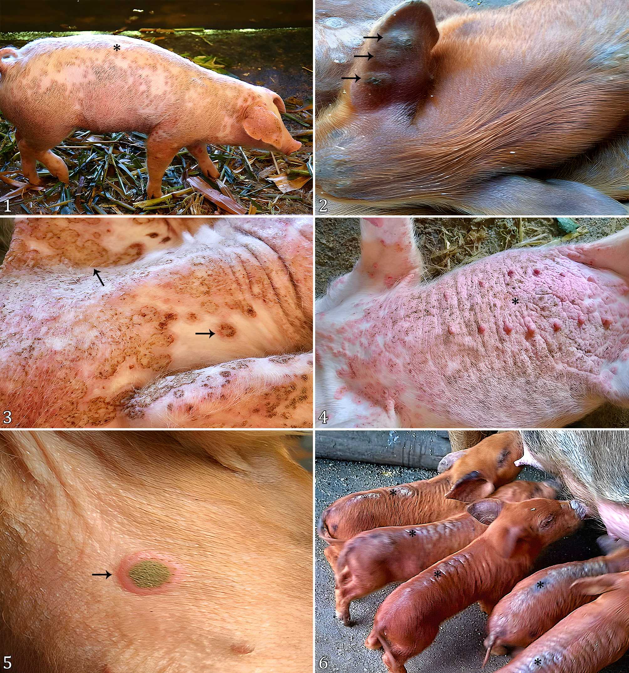
The cutaneous lesions were erythematous and crusty with a crateriform appearance, sometimes disseminated throughout the entire body of the pig (Fig. 1), or localized on the face, ears (Fig. 2), snout, pelvic limbs, thoracic limbs (Fig. 3), and ventral regions (Fig. 4). In some cases, umbilicated pustule lesions were present in both ventral (Fig. 5) and dorsal region (Fig. 6), associated with intense pruritus.
Subsistence pigs with macroscopic skin lesions caused by swinepox in the municipality of Pouso Redondo, Santa Catarina. (1) Multifocal to coalescent erythematous crusty lesions diffusely distributed on the pig’s skin (asterisk). (2) Multifocal, moderate erythematous crusty lesions with a crateriform appearance on the pig’s ear (arrows). (3) Focally extensive erythematous crusty lesions with a crateriform appearance on the neck and thoracic limbs (arrows). (4) Diffuse erythematous crusty lesions on the ventral region of the thorax and abdomen (asterisk). (5) Umbilicated pustule on the abdominal skin of a piglet (arrow). (6) Erythematous crusty lesions with a crateriform appearance on the dorsal region of piglets (asterisks).
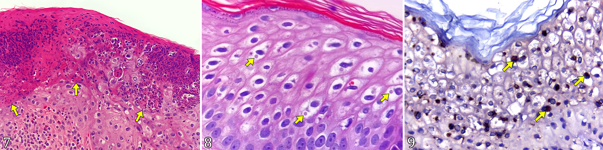
Histologically, in two of the four skin samples, only serocellular crusts were observed. In comparison, in the other two samples, acanthosis with ballooning degeneration of the epithelium and pronounced multifocal spongiosis (Fig. 7) were seen, accompanied by eosinophilic intracytoplasmic inclusions in the epidermis (Fig. 8). Ulcerated areas with serocellular crust deposition were observed, accompanied by a multifocal, moderate infiltrate of both intact and degenerating intraepithelial neutrophils. In the superficial dermis, a moderate, multifocal infiltrate was present, consisting of neutrophils, macrophages, lymphocytes, plasma cells, and eosinophils.
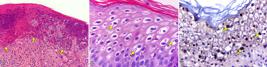
Subsistence pigs with histological and immunohistochemical alterations caused by swinepox. (7) Proliferative pustular dermatitis with focal accentuation (arrow). HE, obj. 10x. (8) Moderate multifocal eosinophilic intracytoplasmic inclusion bodies (arrows). HE, obj. 40x. (9) Skin with moderate immunostain in the intracytoplasmic inclusion bodies (polymer linked to endogenous peroxidase, Romulin AEC Chromogen Kit (BIOCARE Medical® REF RAEC810L) (arrows). IHC, obj. 10x.
In the IHC evaluation for poxvirus, positive immunolabeling (Table 2) was observed in the cytoplasm of the epidermal epithelial cells, showing moderate diffuse immunostaining (Fig. 9 and 10).
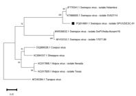
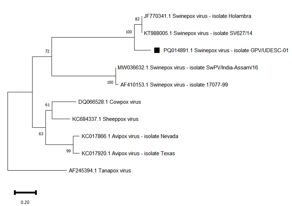
Phylogenetic tree based on the nucleotide sequences of the DNA polymerase gene from different genera of the Poxviridae family. The outgroup was composed of the respective gene from Tanapoxvirus. The sample positive for swinepox in the present study is identified by the symbol (■).
In the PCR for Suipoxvirus, three out of the four skin biopsies were positive. The genetic sequence was then aligned with other known poxvirus sequences from GenBank. Based on the DNA polymerase sequence, a phylogenetic tree was constructed as shown in Figure 3.
The phylogenetic tree (Fig. 3), based on the DNA polymerase sequence, revealed that the sequence detected in this study (PQ014891) clustered with swinepox virus sequences detected in the Northeast (KT988005) and Southeast of Brazil (JF770341), with similarity percentages of 91.4% in both. The same level of similarity was observed with two other sequences, one originating from the United States (AF410153) and the other from India (MW036632).
Discussion
The SWPV can cause disease in up to 100% of young pig herds (Barlow & Grist 2000). In outbreaks reported in the states of São Paulo and Tocantins, morbidity reached 100% of the animals on the affected farms, with a mortality rate of 3% (Bersano et al. 2003). In the Holambra/SP outbreak, 24% (850/3,460) of the pigs were affected by the virus, but no deaths were recorded (Medaglia et al. 2011). In the present study, 46 of 47 pigs were affected, with only one adult pig not presenting skin lesions; 85% of the affected pigs were young. An outbreak involving five subsistence farms in the state of Rio Grande do Norte was reported, with three of these farms experiencing 100% morbidity in young pigs, while the remaining two farms had morbidity rates of 65% and 33%. Mortality rates of 60% and 55% in young pigs were reported in two farms, whereas no mortality was observed in young or adult pigs from the other farms (Olinda et al. 2016).
Swinepox is self-limiting due to the immune responses in the affected pigs (Medaglia et al. 2011, Olinda et al. 2016). After infection, the animals develop an adaptive immune response, including the production of specific antibodies against the virus (Bonilla & Oettgen 2010). In outbreaks in the Northeast and Holambra/SP, the animals showed improvement between 15 and 25 days after the clinical signs appeared (Olinda et al. 2016).
Macroscopically, the lesions consisted of red papules and pustules covered by scabs, affecting the ears, periocular region, abdomen, and thoracic and pelvic limbs, as well as the dorsal and lateral regions of the flank (Barlow & Grist 2000, Radostits et al. 2002, Olinda et al. 2016). Other studies have reported proliferative lesions with ulcerated or scabbed aspects throughout the bodies of the animals (Borst et al. 1990, Bersano et al. 2003, Medaglia et al. 2011). In a report involving a young wild boar, the lesions described were multifocal to coalescing papules, ranging from 0.5 to 0.8 cm in diameter, located on the skin of the snout, neck, chest, abdomen, and limbs, without involving the interdigital space (Guardone et al. 2023). In the present study, the lesions were similar to those previously described.
Histopathological lesions revealed proliferative and ulcerative vesiculo-pustular dermatitis with ballooning degeneration of epithelial cells, parakeratotic hyperkeratosis, and acanthosis, as well as eosinophilic intracytoplasmic inclusion bodies in keratinocytes, in a subsistence pig outbreak (Olinda et al. 2016). Another report primarily described epidermal hyperplasia and degeneration of keratinocytes in the stratum spinosum, with rare visualization of eosinophilic intracytoplasmic inclusion bodies (Guardone et al. 2023), which were similar to the lesions observed in the present study. In contrast, Avipoxvirus is characterized by the presence of large eosinophilic intracytoplasmic inclusion bodies (Schoemaker et al. 1998). The formation of intracytoplasmic inclusion bodies differs between Suipoxvirus and Avipoxvirus due to their distinct viral replication strategies and interactions with host cells. Avipoxvirus forms large structures, known as Bollinger bodies, during intense replication in the epithelial cells of birds’ skin and mucous membranes. At the same time, Suipoxvirus does not induce the prominent formation of these large intracytoplasmic bodies during its replication (Olson et al. 2009).
The use of PCR is an important tool for confirming the diagnosis of Suipoxvirus (Massung & Moyer 1991), as performed in the present study. This technique has been increasingly used in studies to diagnose this disease, as it is a faster, more sensitive, and specific method (Souza et al. 2019). In reports of swinepox in wild boars in Italy and Germany, molecular tests were used to diagnose the virus (Kaiser et al. 2021, Guardone et al. 2023). During an outbreak of swinepox in India, Suipoxvirus was detected by PCR, and all skin samples tested positive (Riyesh et al. 2016). In Brazil, studies also utilized PCR to confirm the virus; during the outbreak in the Northeast region, PCR was performed on four skin samples, all of which were positive (Olinda et al. 2016). In a study conducted in Holambra, seven scabs from animals were collected, all of which were positive (Medaglia et al. 2011).
The phylogenetic analysis of the sequence obtained in this study is closely related to isolates detected in skin fragments of pigs from the Southeast region, as well as in cutaneous crusts from pigs in the Northeast, indicating considerable genetic proximity and a common origin, as well as shared circulation of the virus across different Brazilian regions (Medaglia et al. 2011, Olinda et al. 2016). Regarding isolates of foreign origin, the analysis indicates an older temporal divergence, demonstrating that similar clustering among isolates from different regions can occur and suggesting that the virus exhibits genetic diversity based on geographic location (Koltsov et al. 2023).
An important distinction for Suipoxvirus includes infection by the vaccinia virus (VACV), a member of the Orthopoxvirus genus in the Poxviridae family, which has a worldwide distribution and infects humans, cattle, horses, pigs, rodents, rabbits, and other mammals (Trindade et al. 2004, Chapman et al. 2010, Essbauer et al. 2010, Medeiros-Silva et al. 2010, Caetano et al. 2016, Peres et al. 2016). Although swinepox is not considered a zoonosis, it is important to highlight the significant similarity between the P42 protein of the SWPV envelope and the P37 protein of VACV, both of which are implicated in the transmission process of the disease (Bersano et al. 2003). Both SWPV and the VACV cause skin lesions in proliferative forms, with ulcerated or crusted aspects (Medeiros-Silva et al. 2010, Koltsov et al. 2023), making it essential to perform molecular tests like PCR to confirm the diagnosis. In this study, PCR analysis of skin samples confirmed the presence of the SWPV genome in the pig DNA. Therefore, distinguishing between infections caused by SWPV and VACV during outbreaks of pustular disease in pigs is crucial for determining the potential spread of VACV among swine populations and verifying the presence of SWPV in Brazil (Medaglia et al. 2011).
The negative PCR result for the sample from Farm 1 does not rule out Suipoxvirus involvement, as the skin crust used may contain a viral load insufficient to detect the agent (Koltsov et al. 2023). In the same sample, positive immunolabeling was observed using a polyclonal anti-vaccinia antibody. However, it is unlikely that the lesions are associated with VACV, which primarily affects humans and cattle (Medeiros-Silva et al. 2010, Peres et al. 2016). The antibody used in IHC recognizes epitopes broadly conserved among members of the Poxviridae family, showing cross-reactivity with different genera (Zachary 2021). Thus, the observed labeling strongly suggests a poxvirus infection, with Suipoxvirus as the most probable agent in pigs, given its genus-specificity and the morphological pattern of the lesions (Olinda et al. 2016, Kaiser et al. 2021).
A wide variety of skin lesions can affect pigs (Pereira et al. 2020). Exudative epidermitis is an important differential diagnosis for SWPV and, in addition to causing intense pruritus, is characterized by generalized, reddened skin lesions that are sometimes covered with crusts and have purulent exudate accumulation. The causative agent, Staphylococcus hyicus, is a bacterium typically present in the skin microbiota and requires predisposing factors for disease development (Wegener et al. 1993). Erysipelas is another disease caused by bacteria of the genus Erysipelothrix sp., which is zoonotic and characterized by reddish skin lesions in a diamond-shaped pattern (Bender et al. 2011). In a study involving 8,071 pigs sent to slaughterhouses, 172 had lesions suggestive of Erysipelothrix, and upon histopathological and immunohistochemical analysis, 43 cases were confirmed as positive (Pereira et al. 2022). Porcine Circovirus type 2 (PCV2) can cause porcine nephropathy and dermatitis syndrome, characterized by large macules and plaques, round to irregular in shape, primarily red in the skin of the perineal area, limbs, ears, abdomen, and ventral thorax (França et al. 2005).
IHC has been used in cases of poxvirus in other animal species and humans, as it is a rapid and specific tool (Molina-Ruiz et al. 2015). Monkeypox virus primarily affects monkeys, but it is a zoonotic viral disease, and important diagnostic tests include IHC (Bayer-Garner 2005). Another zoonosis in which IHC was used is the Orf virus, a member of the Parapoxvirus genus, which causes the disease known as contagious ecthyma (Molina-Ruiz et al. 2015). In a study conducted in the United States, IHC was used to diagnose Flowpox virus, with positive staining in the cytoplasm (Tadese et al. 2007). There are also cases of IHC being used for sheeppox (Capripoxvirus) in lambs (Gulbahar et al. 2000). In this study, IHC was performed to aid in the final diagnosis of Suipoxvirus, highlighting the importance of further studies with IHC as a diagnostic method in pigs.
Suipoxvirus should be included in the differential diagnosis for skin diseases, such as dermatitis in pigs with lice and fly infestations (Zanella et al. 2016). Infection with Suipoxvirus has been associated with contact between infected animals and ectoparasites, such as lice or flies, which mechanically transmit the virus (Medaglia et al. 2011, Roehe et al. 2012), facilitating the spread of the disease (Olinda et al. 2016). Therefore, disease control primarily relies on preventing the introduction of virus-carrying animals and on maintaining stringent hygienic and sanitary measures, including the effective ectoparasite control management (Guardone et al. 2023).
Suipoxvirus is species-specific, meaning it is recognized for its host specificity. Its spread is limited to pigs (Bersano et al. 2003, Olinda et al. 2016). Some poxviruses are zoonotic and are often responsible for skin lesions. The main culprits are from the Orthopoxvirus and Parapoxvirus genera, which include diseases such as cowpox, monkeypox, and contagious ecthyma, all of which can affect humans (Haig et al. 2002, Batista et al. 2009, Bohelay & Duong 2017).
Conclusion
This study seems to be the first to report a swinepox outbreak caused by Suipoxvirus in subsistence pig farming in the state of Santa Catarina. The diagnosis of swinepox was determined through epidemiological data and clinical signs supported by histopathological examination, polymerase chain reaction (PCR), and immunohistochemistry tests. It is important to have a definitive diagnosis, as it is a differential for zoonotic diseases and mandatory reporting.
Acknowledgments
This study was supported by the “Laboratório de Patologia Animal”, “Centro de Ciências Agroveterinárias”, “Universidade do Estado de Santa Catarina” (UDESC), Lages/SC, and “Coordenação de Aperfeiçoamento de Pessoal de Nível Superior” (CAPES) for the postgraduate scholarship for this paper.
References
-
Afonso CL, Tulman ER, Lu Z, Zsak L, Osorio FA, Balinsky C, Kutish GF, Rock DL. The genome of swinepox virus. J Virol 2002; https://doi.org/10.1128/jvi.76.2.783-790.2002
» https://doi.org/10.1128/jvi.76.2.783-790.2002 -
Barlow AM, Grist C. An outbreak of congenital pig pox. Pig J 2000; https://www.cabidigitallibrary.org/doi/full/10.5555/20003014263
» https://www.cabidigitallibrary.org/doi/full/10.5555/20003014263 - Batista VH, Scremin J, Aguiar LM, Schatzmayr HG. Vulvar infection and possible human-to-human transmission of bovine poxvirus disease. Virus Rev Res 2009;14(1):7.
-
Bayer-Garner IB. Monkeypox virus: histologic, immunohistochemical and electron-microscopic findings. J Cutan Pathol 2005; https://doi.org/10.1111/j.0303-6987.2005.00254.x
» https://doi.org/10.1111/j.0303-6987.2005.00254.x -
Bender JS, Irwin CK, Shen H-G, Schwartz KJ, Opriessnig T. Erysipelothrix spp. genotypes, serotypes, and surface protective antigen types associated with abattoir condemnations. J Vet Diagn Invest 2011; https://doi.org/10.1177/104063871102300126
» https://doi.org/10.1177/104063871102300126 -
Bersano JG, Catroxo MHB, Villalobos EMC, Leme MCM, Martins AMCRPF, Peixoto ZMP, Portugal MASC, Monteiro RM, Ogata RA, Curi NA. Varíola suína: estudo sobre a ocorrência de surtos nos estados de São Paulo e Tocantins, Brasil. Arq Inst Biol 2003; https://doi.org/10.1590/1808-1657v70p2692003
» https://doi.org/10.1590/1808-1657v70p2692003 -
Bohelay G, Duong T-A. Infecciones por poxvirus en humanos. EMC Dermatol 2017; https://doi.org/10.1016/S1761-2896(17)85923-9
» https://doi.org/10.1016/S1761-2896(17)85923-9 -
Bonilla FA, Oettgen HC. Adaptive immunity. J Allergy Clin Immunol 2010; https://doi.org/10.1016/j.jaci.2009.09.017
» https://doi.org/10.1016/j.jaci.2009.09.017 - Borst GH, Kimman TG, Gielkens AL, van der Kamp JS. Four sporadic cases of congenital swinepox. Vet Rec 1990;127(3):61-63. PMid:2169139.
-
Caetano AL, Araujo JM, Silva MO, Leal AA. Estomatite papular em bovinos no município de São Miguel do Araguaia, estado de Goiás, Brasil. Rev Med Vet Zoot CRMV-SP 2016; https://www.revistamvez-crmvsp.com.br/index.php/recmvz/article/view/31930
» https://www.revistamvez-crmvsp.com.br/index.php/recmvz/article/view/31930 - Canal CW. Poxviridae, p. 491-509. In: Flores EF. Virologia Veterinária. Santa Maria: Ed. UFSM; 2007.
-
Chapman JL, Nichols DK, Martinez MJ, Raymond JW. Animal models of orthopoxvirus infection. Vet Pathol 2010; https://doi.org/10.1177/0300985810378649
» https://doi.org/10.1177/0300985810378649 -
Essbauer S, Pfeffer M, Meyer H. Zoonotic poxviruses. Vet Microbiol 2010; https://doi.org/10.1016/j.vetmic.2009.08.026
» https://doi.org/10.1016/j.vetmic.2009.08.026 -
França TN, Ribeiro CT, Cunha BM, Peixoto PV. Circovirose suína. Pesq Vet Bras 2005; https://doi.org/10.1590/S0100-736X2005000200001
» https://doi.org/10.1590/S0100-736X2005000200001 -
Guardone L, Varello K, Listorti V, Peletto S, Wolfsgruber L, Zoccola R, Montemurro V, Messana E, Bozzetta E, Acutis P, Masoero L, Razzuoli E. First report of swinepox in a wild boar in Italy: pathologic and molecular findings. Pathogens 2023; https://doi.org/10.3390/pathogens12030472
» https://doi.org/10.3390/pathogens12030472 -
Gulbahar MY, Cabalar M, Gul Y, Icen H. Immunohistochemical detection of antigen in lamb tissues naturally infected with sheeppox virus. J Vet Med 2000; https://doi.org/10.1046/j.1439-0450.2000.00321.x
» https://doi.org/10.1046/j.1439-0450.2000.00321.x -
Haig DM, McInnes CJ. Immunity and counter-immunity during infection with the parapoxvirus orf virus. Virus Res 2002; https://doi.org/10.1016/S0168-1702(02)00117-X
» https://doi.org/10.1016/S0168-1702(02)00117-X -
Hall TA. BioEdit: a user-friendly biological sequence alignment editor and analysis program for Windows 95/98/NT. Nucleic Acids Symp Ser 1999; https://shre.ink/q8hV
» https://shre.ink/q8hV -
Kaiser FK, Wiedemann A, Kühl B, Menke L, Beineke A, Baumgärtner W, Wohlsein P, Rigbers K, Becher P, Peters M, Osterhau ADME, Ludlow M. Swinepox virus strains isolated from domestic pigs and wild boar in Germany display altered coding capacity in the terminal genome region encoding for species-specific genes. Viruses 2021; https://doi.org/10.3390/v13102038
» https://doi.org/10.3390/v13102038 -
Koltsov A, Sukher M, Kholod N, Namsrayn S, Tsybanov S, Koltsova G. Isolation and Characterization of swinepox virus from outbreak in Russia. Animals 2023; https://doi.org/10.3390/ani13111786
» https://doi.org/10.3390/ani13111786 -
Massung RF, Moyer RW. The molecular biology of swinepox virus I. A characterization of the viral DNA. Virology 1991; https://doi.org/10.1016/0042-6822(91)90039-E
» https://doi.org/10.1016/0042-6822(91)90039-E -
Medaglia MLG, Pereira AC, Freitas TRP, Damaso CR. Swinepox virus outbreak, Brazil, 2011. Emerg Infect Dis 2011; https://doi.org/10.3201/eid1710.110549
» https://doi.org/10.3201/eid1710.110549 -
Medeiros-Silva DC, Moreira-Silva EAS, Gomes JAS, Fonseca FG, Correa-Oliveira R. Clinical signs, diagnosis, and case reports of Vaccinia virus infections. Braz J Infect Dis 2010; https://doi.org/10.1016/S1413-8670(10)70025-8
» https://doi.org/10.1016/S1413-8670(10)70025-8 -
Molina-Ruiz AM, Santonja C, Rütten A, Cerroni L, Kutzner H, Requena L. Immunohistochemistry in the diagnosis of cutaneous viral infections- part II: cutaneous viral infections by parvoviruses, poxviruses, paramyxoviridae, picornaviridae, retroviruses and filoviruses. Am J Dermatopathol 2015; https://doi.org/10.1097/dad.0000000000000200
» https://doi.org/10.1097/dad.0000000000000200 -
Olinda RG, Maia LA, Cargnelutti JF, Gois RCS, Batista JS, Dantas AFM, Flores EF, Riet-Correa F. Swinepox dermatitis in backyard pigs in Northeastern Brazil. Pesq Vet Bras 2016; https://doi.org/10.1590/S0100-736X2016000600002
» https://doi.org/10.1590/S0100-736X2016000600002 -
Olson VA, Regnery RL, Damon IK. Poxviruses. In: Specter S. Clinical Virology Manual. 2009; https://doi.org/10.1128/9781555815974.ch29
» https://doi.org/10.1128/9781555815974.ch29 -
Pereira PR, Bianchi RM, Hammerschmitt ME, Cruz RAS, Hesse KL, Sonne L, Pavarini SP, Driemeier D. Primary skin diseases and cutaneous manifestations of systemic diseases in swine. Pesq Vet Bras 2020; https://doi.org/10.1590/1678-5150-PVB-6704
» https://doi.org/10.1590/1678-5150-PVB-6704 -
Pereira PR, De Lorenzo C, Lopes BC, Sonne L, Pavarini SP, Driemeier D. Anatomopathological aspects and the use of immunohistochemistry in slaughter pigs with cutaneous lesions of erysipelas. Pesq Vet Bras 2022; https://doi.org/10.1590/1678-5150-PVB-6997
» https://doi.org/10.1590/1678-5150-PVB-6997 -
Peres MG, Barros CB, Appolinário CM, Antunes JMAP, Mioni MSR, Bacchiega TS, Allendorf SD, Vicente AF, Fonseca CR, Megid J. Dogs and opossums positive for vaccinia virus during outbreak affecting cattle and humans, São Paulo State, Brazil. Emerg Infect Dis 2016; https://doi.org/10.3201/eid2202.140747
» https://doi.org/10.3201/eid2202.140747 - Radostits OM, Gay CC, Blood DC, Hinchcliff KW. Clínica Veterinária: um tratado de doenças dos bovinos, ovinos, suínos, caprinos e eqüinos. 9th ed. Rio de Janeiro: Guanabara Koogan; 2002.
-
Riyesh T, Barua S, Kumar N, Jindal N, Bera BC, Narang G., Mahajan NK, Arora D, Anand T, Vaid RK, Yadav M, Chandel SS, Malik P, Tripathi BN, Singh RK. Isolation and genetic characterization of swinepox virus from pigs in India. Comp Immunol Microbiol Infect Dis 2016; https://doi.org/10.1016/j.cimid.2016.04.001
» https://doi.org/10.1016/j.cimid.2016.04.001 - Roehe P, Brito WD. Varíola, p. 409-411. In: Sobestiansky J, Barcellos D. Doenças dos Suínos. 2ª ed. Goiânia: Canône; 2012.
-
Schoemaker NJ, Dorrestein GM, Lumeij JT. An avipoxvirus infection in a goshawk (Accipiter gentilis). Avian Pathol 1998; https://doi.org/10.1080/03079459808419282
» https://doi.org/10.1080/03079459808419282 -
Souza FA, Santos Júnior EM, Laguardia-Nascimento M, Freitas TRP, Damaso CR, Rivetti Júnior AV, Camargos MF, Fonseca Júnior AA. Validation of a real-time PCR assay for detection of swinepox virus. Arch Virol 2019; https://doi.org/10.1007/s00705-019-04403-w
» https://doi.org/10.1007/s00705-019-04403-w -
Tadese T, Potter AE, Fitzgerald S, Reed WM. Concurrent infection in chickens with fowlpox virus and infectious laryngotracheitis virus as detected by immunohistochemistry and a multiplex polymerase chain reaction technique. Avian Dis 2007; https://doi.org/10.1637/0005-2086(2007)51[719:CIICWF]2.0.CO;2
» https://doi.org/10.1637/0005-2086(2007)51[719:CIICWF]2.0.CO;2 -
Tamura K, Stecher G, Kumar S. MEGA11: molecular evolutionary genetics analysis version 11. Mol Biol Evol 2021; https://doi.org/10.1093/molbev/msab120
» https://doi.org/10.1093/molbev/msab120 -
Thompson JD, Higgins DG, Gibson TJ. Clustal W: improving the sensitivity of progressive multiple sequence alignment through sequence weighting, position-specific gap penalties and weight matrix choice. Nucleic Acids Res 1994; https://doi.org/10.1093/nar/22.22.4673
» https://doi.org/10.1093/nar/22.22.4673 -
Torrison J, Cameron R. Integumentary system: skin, hoof, and claw. In: Zimmerman JJ, Karriker LA, Ramirez A, Schwartz KJ, Stevenson GW, Zhang J. Diseases of Swine. 2019; https://doi.org/10.1002/9781119350927.ch17
» https://doi.org/10.1002/9781119350927.ch17 -
Trindade GS, Fonseca FG, Marques JT, Diniz S, Leite JA, De Bodt S, Van der Peer Y, Bonjardim CA, Ferreira PCP, Kroon EG. Belo Horizonte virus: a vaccinia-like virus lacking the A-type inclusion body gene isolated from infected mice. J Gen Virol 2004; https://doi.org/10.1099/vir.0.79840-0
» https://doi.org/10.1099/vir.0.79840-0 - Turton J. Skin conditions in pigs. Department of Agriculture in cooperation with the ARC-Onderstepoort Veterinary Institute. IOP Publishing Physics Web; 2001.
-
USDA. This report contains assessments of commodity and trade issues made by USDA staff and not necessarily statements of official U.S. government policy. GAIN Report Number: GM19015. Berlin: Global Agricultural Information Network (GAIN), Foreign Agricultural Service, United States Department of Agriculture; 2019. Accessed Jun 12, 2024. https://apps.fas.usda.gov/newgainapi/api/Report/DownloadReportByFileName?fileName=Trade+Show+Overview+Austria_Berlin_Germany_4-17-2019.pdf
» https://apps.fas.usda.gov/newgainapi/api/Report/DownloadReportByFileName?fileName=Trade+Show+Overview+Austria_Berlin_Germany_4-17-2019.pdf -
Wegener HC, Andresen LO, Bille-Hansen V. Staphylococcus hyicus virulence in relation to exudative epidermitis in pigs. Can J Vet Res 1993; https://pmc.ncbi.nlm.nih.gov/articles/PMC1263605/pdf/cjvetres00034-0055.pdf
» https://pmc.ncbi.nlm.nih.gov/articles/PMC1263605/pdf/cjvetres00034-0055.pdf -
Withoeft JA, Marian L, Costa LS, Fernandes FD, Vogel FSF, Neves GB, Miletti LC, Casagrande RA. Sheep abortions associated with Neospora caninum and Toxoplasma gondii infections in multiple flocks from Southern Brazil. Vet Res Comm 2024; https://doi.org/10.1007/s11259-024-10390-4
» https://doi.org/10.1007/s11259-024-10390-4 - Zachary JF. Pathologic Basis of Veterinary Disease. 7th ed. St. Louis: Elsevier; 2021.
-
Zanella JRC, Morés N, Barcellos DESN. Main endemic health threats in the swine production chain in Brazil. Pesq Agropec Bras 2016; https://doi.org/10.1590/S0100-204X2016000500004
» https://doi.org/10.1590/S0100-204X2016000500004
-
Data availability statement
The authors declare that all data used are available in this article.
Edited by
-
Editor-in-Chief
Fabiano José Ferreira de Sant’Ana
The authors declare that all data used are available in this article.