 Expanding the known geographic range of Leishmania (Mundinia) enriettii: clinical and molecular characterization in naturally infected guinea pigs from southern Brazil
ABSTRACT:
Three domestic guinea pigs (Cavia porcellus) from Ampére, Paraná State, Brazil, presenting crusted nodular lesions on the pinnae, snout, and digits of all limbs were examined at the University Veterinary Hospital of the Federal University of the Southern Border in February 2023. Cytological evaluation by fine-needle aspiration revealed Leishmania amastigotes within macrophages. Two animals were euthanized for further histopathological, immunohistochemical, parasitological, and molecular investigations. Skin samples exhibited a diffuse mixed inflammatory infiltrate, predominantly histiocytic, with macrophages densely parasitized by amastigotes. Immunohistochemistry (IHC) using an anti-mTXNPx monoclonal antibody confirmed the abundant presence of amastigotes in the skin, providing key diagnostic support. Liver sections showed mild lymphocytic infiltrates, with specific IHC labeling in pericentral hepatocytes indicating antigenic presence despite the absence of visible parasites. Kidneys, lungs, and spleen exhibited mild architectural changes with no detectable amastigotes on microscopy. Promastigote forms were successfully isolated in NNN/LIT medium from one liver sample. Molecular analysis using hsp70-targeted PCR and sequencing confirmed the presence of Leishmania DNA in the skin, spleen, and liver, identifying the species as Leishmania (Mundinia) enriettii. The absence of microscopically visible amastigotes in tissues, despite molecular and immunohistochemical evidence, underscores the value of a multimodal diagnostic approach. This study expands the known geographic distribution of L. enriettii in Paraná State and reinforces the guinea pig’s role as the only confirmed reservoir of this parasite in Brazil.
INDEX TERMS:
Leishmania enriettii; guinea pig; Cavia porcellus; infection; Brazil
RESUMO:
Três porquinhos-da-índia domésticos (Cavia porcellus), provenientes de Ampére, no estado do Paraná, Brasil, apresentando lesões nodulares crostosas no pavilhão auricular, focinho e dígitos de todos os membros, foram examinados na Unidade Hospitalar Veterinária da Universidade Federal da Fronteira Sul em fevereiro de 2023. A avaliação citológica por punção aspirativa com agulha fina revelou formas amastigotas de Leishmania no interior de macrófagos. Dois animais foram eutanasiados para realização de exames histopatológicos, imuno-histoquímicos, parasitológicos e moleculares. As amostras de pele apresentaram infiltrado inflamatório misto difuso, predominantemente histiocítico, com macrófagos densamente parasitados por amastigotas. A imuno-histoquímica (IHQ), utilizando anticorpo monoclonal anti-mTXNPx, confirmou a presença abundante de amastigotas na pele, contribuindo significativamente para o diagnóstico. As seções hepáticas revelaram infiltrado linfocitário discreto, com marcação IHQ específica em hepatócitos pericentrais, indicando presença antigênica mesmo na ausência de parasitos visíveis. Rins, pulmões e baço apresentaram alterações arquiteturais leves, sem detecção de amastigotas à microscopia. Formas promastigotas foram isoladas com sucesso em meio bifásico NNN/LIT a partir de uma amostra de fígado. A análise molecular por PCR direcionada ao gene hsp70, seguida de sequenciamento, confirmou a presença de DNA de Leishmania em amostras de pele, baço e fígado, com identificação da espécie como Leishmania (Mundinia) enriettii. A ausência de amastigotas nos tecidos viscerais, apesar da imuno-histoquímica e positividade molecular, reforça o valor de uma abordagem diagnóstica multimodal. Este estudo amplia a distribuição geográfica conhecida de L. enriettii no estado do Paraná e reforça o papel do porquinho-da-índia como único reservatório confirmado desse parasito no Brasil.
TERMOS DE INDEXAÇÃO:
Leishmania enriettii; porquinho-da-índia; Cavia porcellus; infecção; Brasil
Introduction
Parasites of the genus Leishmania are known to infect a wide range of vertebrate hosts, causing leishmaniasis, a group of diseases with a variable clinical spectrum that affects millions of people in tropical and subtropical regions (Bruschi & Gradoni 2018). However, several species within the genus are not pathogenic to humans. One such example is Leishmania (Mundinia) enriettii, which has the guinea pig (Cavia porcellus) as its primary vertebrate host (Muniz & Medina 1948).
The discovery of L. enriettii marked the beginning of a significant trajectory for this protozoan. Currently, it serves as an important model organism in experimental research, particularly in studies of immunology, intracellular survival, cutaneous leishmaniasis dissemination, and chemotherapy (Lainson 1997, Paranaíba et al. 2017). Despite its extensive use in laboratory models, the transmission ecology of L. enriettii remains poorly understood, with both its natural vectors and reservoir hosts still unconfirmed. Although Pintomyia monticola has been suggested as a potential vector (Luz et al. 1967), guinea pigs have been proposed as natural reservoirs in Brazil (Luz et al. 1967, Machado et al. 1994, Thomaz-Soccol et al. 1996, Soccol et al. 2021). Despite their Andean origin, guinea pigs are widely kept as exotic companion animals in Brazil, primarily in domestic and peri-urban environments, where their role as potential sentinels for zoonotic pathogens is increasingly recognized.
Reports of guinea pigs naturally infected with L. enriettii in Brazil appear to be increasing. However, the geographic distribution seems limited to the southeastern and southern regions, particularly the states of São Paulo (Machado et al. 1994), Paraná (Luz et al. 1967, Thomaz-Soccol et al. 1996, Soccol et al. 2021), and Rio Grande do Sul (Rosa et al. 2020, Ribeiro et al. 2023). Most of the available information on the clinical manifestations of leishmaniasis in guinea pigs, however, originates from experimental studies (Muniz & Medina 1948, Neal & Miles 1977, Alves-Sobrinho et al. 2022). Nevertheless, nodular lesions, mainly on the ears and nose, have been documented in naturally infected animals (Thomaz-Soccol et al. 1996, Soccol et al. 2021, Rosa et al. 2020).
Although leishmaniasis caused by L. enriettii was first described nearly 80 years ago (Muniz & Medina 1948), its characteristic nodular lesions are likely underdiagnosed, often being mistaken for neoplasms or scabies (Soccol et al. 2021). This diagnostic challenge highlights the need for a detailed characterization of the clinical and pathological features associated with natural L. enriettii infections in guinea pigs, which may help veterinarians to include leishmaniasis in the differential diagnosis of skin diseases in this species. To address this gap, the present study aimed to evaluate the clinical, pathological, and molecular aspects of L. enriettii-induced leishmaniasis in naturally infected guinea pigs.
Materials and Methods
Ethical approval. No approval from research ethics committees was required for this study because all procedures were performed as part of routine veterinary clinical care, and tissue samples were kindly provided by the veterinary hospital for diagnostic purposes.
Animals and clinical evaluation. Three adult guinea pigs (Cavia porcellus), two males and one female, from a domestic herd in Ampére, Paraná State, Brazil, were evaluated at the Veterinary Hospital of the “Universidade Federal da Fronteira Sul” (UFFS) in 2023. According to the owner, the animals were born in Ampére and had no history of travel.
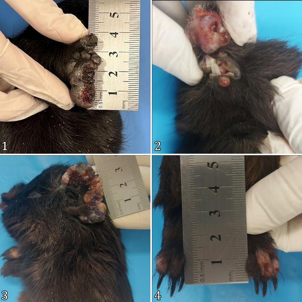
On clinical examination, all animals presented multiple crusted nodular lesions on the pinnae, snout, and digits of all limbs, as well as thickening of the pinnae and ulceration in affected areas (Fig. 1-4). Due to the severity of the lesions, the animals exhibited impaired locomotion, marked prostration, and signs of anorexia. Based on the chronicity and extent of the lesions, two guinea pigs were euthanized for further diagnostic investigation.
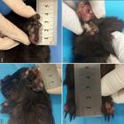
Macroscopic examination of cutaneous and mucosal lesions caused by Leishmania enriettii in guinea pigs (Cavia porcellus). (1) Right pinna showing irregular, nodular, and crusted lesions. (2) Nasal plane and lower lip with ulcerated nodular lesions. (3) Left pinna exhibiting irregular, nodular, and crusted lesions. (4) Digits with areas of alopecia and nodular swellings.
Cytological, histopathological, immunohistochemical and molecular analysis. Fine-needle aspiration was performed on the skin nodules, and the collected material was smeared onto glass slides and stained with rapid panoptic (LB, Laborclin, Brazil). The slides were examined under light microscopy for the presence of Leishmania amastigotes within macrophages.
Tissue samples from the skin, liver, spleen, lungs, and kidneys of the euthanized animals were fixed in 10% buffered formalin, routinely processed, and embedded in paraffin. Histological sections (4 μm thick) were stained with hematoxylin and eosin (HE) and analyzed by light microscopy. Immunohistochemical analysis was performed on skin sections using a monoclonal anti-mTXNPx antibody (Freire et al. 2022) to confirm the presence of Leishmania amastigotes.
Six tissue fragments, including two samples each from the liver, skin, and spleen of both euthanized animals, were inoculated into NNN/LIT biphasic medium and incubated at 25 °C. Cultures were monitored for promastigote growth for up to two months. These same samples were preserved in RNAlater and submitted to DNA extraction using the Gentra Puregene Cell and Tissue Kit (Qiagen, Valencia/CA, USA). Polymerase chain reaction (PCR) targeting the hsp70 gene (Garcia et al. 2004) was conducted to confirm the presence of Leishmania DNA and to determine the parasite species. Sanger sequencing was performed for molecular identification, and the resulting sequences were compared with reference strains available in GenBank.
Results
All three guinea pigs exhibited multiple nodular and crusted lesions on the pinnae, snout, and digits of all limbs. Thickening of the pinnae and ulceration in affected areas were also noted (Fig.1-4). The animals demonstrated marked locomotor impairment, increased prostration, and signs of anorexia. Due to the severity and chronicity of the lesions, two animals were euthanized for further diagnostic investigation.
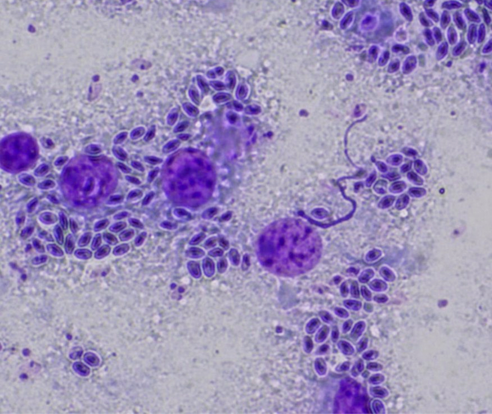
Cytological analysis of the skin nodules revealed numerous oval organisms with basophilic nuclei and adjacent kinetoplasts, consistent with Leishmania amastigotes, observed both free and within macrophages (Fig. 5). Multinucleated cells with cytoplasm densely packed with these forms were also present, along with abundant hypersegmented polymorphonuclear leukocytes.

Cytological analysis by needle aspiration of nodular lesions in guinea pigs. Macrophages infected with amastigote forms of Leishmania sp. Staining performed with rapid panoptic, magnification 1000x.
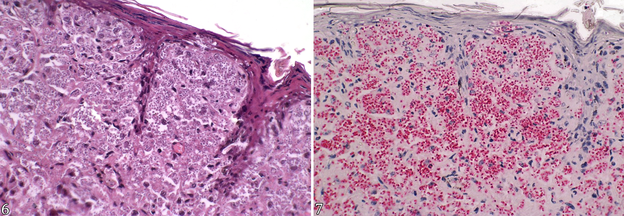
Histopathological analysis of the skin samples revealed irregular epidermal thickening, characterized by increased cellularity in the spinous layer (acanthosis), thickening of the granular layer, and both parakeratotic and orthokeratotic hyperkeratosis. Crust formation associated with extensive inflammatory infiltrates, composed of giant cells, histiocytes, plasma cells, scattered lymphocytes, and exudate, was also observed. In the dermis, a diffuse mixed inflammatory infiltrate predominated, consisting mainly of histiocytes, multinucleated giant cells, and sparse lymphocytes. Macrophages exhibited abundant cytoplasm densely filled with amastigotes. In some fields, amastigotes were also detected within the epidermis (Fig. 6). Deeper dermal layers contained collagen fibers and fibroblasts. Immunohistochemical analysis using the anti-mTXNPx monoclonal antibody confirmed the presence of Leishmania amastigotes in the skin. The staining was characterized by intense, granular, cytoplasmic labeling in parasitized macrophages within the dermis (Fig. 7). The immunoreactivity was focal and specific, with no background staining in non-parasitized tissues or epidermal layers.
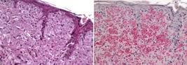
Histopathological and immunohistochemical analysis of skin samples from guinea pigs naturally infected with Leishmania enriettii. (6) Skin sample showing diffuse mixed inflammatory infiltrate, predominantly histiocytic, with few lymphocytes. (7) Immunohistochemistry using anti-mTXNPx monoclonal antibody (mAb), highlighting the labeling of amastigote forms in skin lesions. HE, obj. 40x.
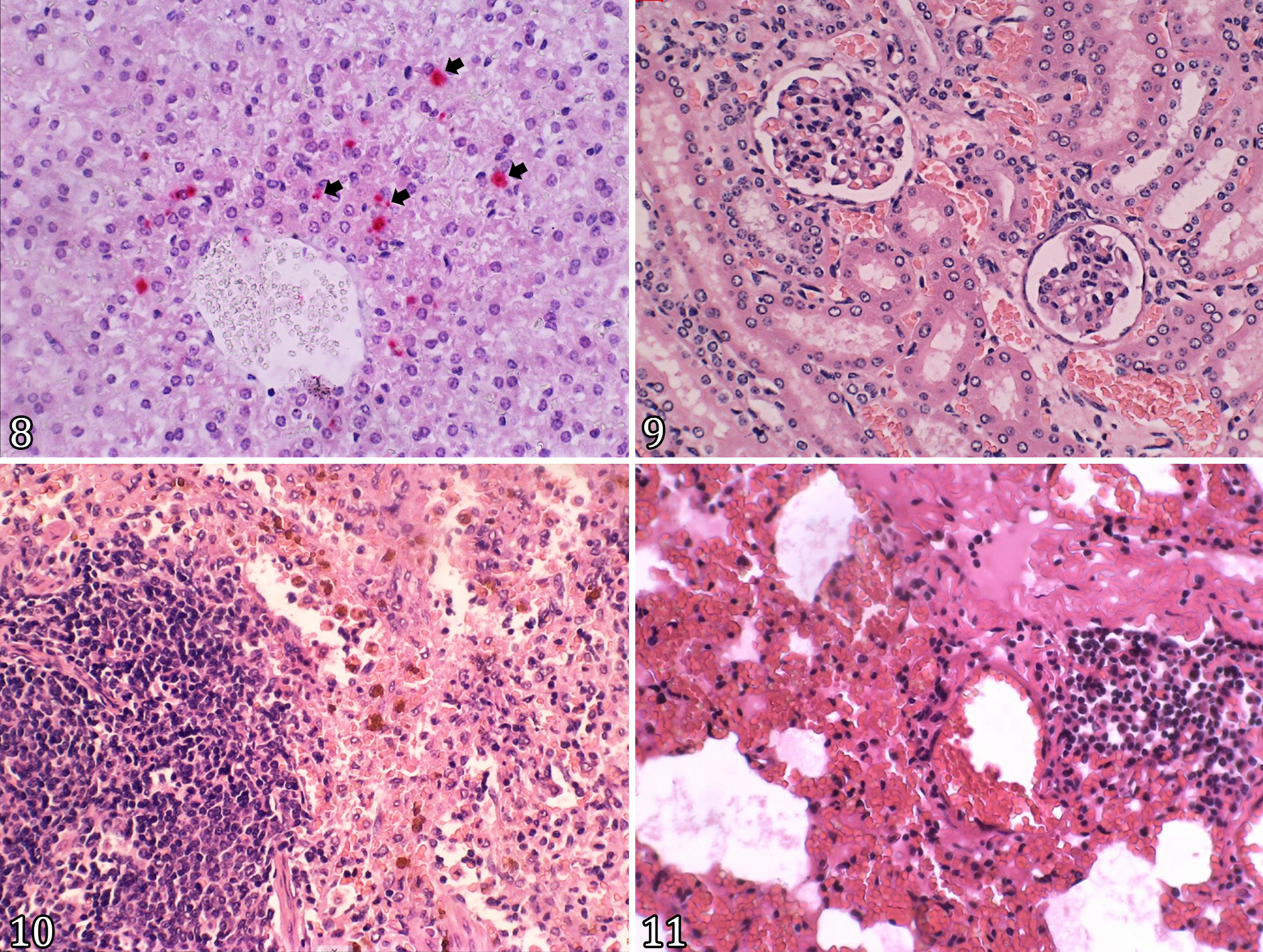
Liver sections exhibited preserved lobular architecture with mild lymphocytic inflammatory infiltrates confined to the portal tracts. Although typical amastigote forms were not observed, immunohistochemical labeling was detected in cells adjacent to centrilobular veins, suggesting antigenic presence in hepatic tissue (Fig. 8). The kidneys showed preserved cortical and medullary architecture, with discrete foci of congestion and interstitial hemorrhage (Fig. 9). The spleen exhibited moderate red pulp congestion, with numerous hemosiderin-laden macrophages (Fig. 10). In the lungs, alveolar hemorrhage and moderate capillary congestion were noted (Fig. 11). No amastigote forms were identified in these organs.
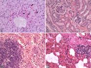
Histological sections of liver, kidney, spleen, and lungs from guinea pigs naturally infected with Leishmania enriettii. (8) Antigenic reactions (arrows) observed near the centrilobular vein in the liver through immunohistochemistry using the anti-mTXNPx monoclonal antibody (mAb). (9) Well-preserved cortical and medullary architecture of the kidney, with discrete foci of congestion and interstitial hemorrhage. (10) Moderate congestion of the red pulp in the spleen. (11) Alveolar hemorrhage and moderate capillary congestion in the lung. No amastigotes were detected in these tissues. HE, obj. 40x.
Leishmania promastigotes were successfully isolated from one liver sample. The remaining cultures remained negative after two months of incubation. Bacterial and fungal contamination was observed in three samples (one liver and two skin), preventing further culture.
PCR analysis targeting the hsp70 gene confirmed the presence of Leishmania DNA in skin, spleen, and liver samples. Sanger sequencing identified the parasite as Leishmania (Mundinia) enriettii (GenBank accession number: PV030921).
Discussion
This study provides a comprehensive assessment of the clinical, pathological, and molecular aspects of natural Leishmania enriettii infection in guinea pigs (Cavia porcellus), contributing significantly to the understanding of this parasite’s geographical distribution and clinical presentation. The detection of cases in Ampére, in the southwestern region of Paraná State, expands the known geographic range of L. enriettii in Brazil and brings it closer to the Argentine province of Misiones, suggesting potential transboundary circulation or regional underreporting. Until now, reports of L. enriettii in Brazil were limited to central Paraná, with isolated occurrences in the states of São Paulo and Rio Grande do Sul (Luz et al.1967, Machado et al. 1994, Thomaz-Soccol et al. 1996, Ecco et al. 2000, Rosa et al. 2020, Soccol et al. 2021).
Histopathological analysis revealed cutaneous inflammatory patterns consistent with previous descriptions of natural L. enriettii infections in guinea pigs, particularly the presence of a mixed inflammatory infiltrate with a predominance of histiocytic cells heavily parasitized by amastigotes (Thomaz-Soccol et al. 1996, Soccol et al. 2021). However, marked differences were observed when comparing systemic findings in this study to those reported in experimental infections using sand fly salivary gland extracts (SGE). For example, Alves-Sobrinho et al. (2022) described pronounced visceral lesions in the lungs and spleen, with parasitized macrophages and extensive granulomatous inflammation. In contrast, in the naturally infected animals examined here, although Leishmania DNA was detected in the liver and spleen by molecular methods, the parasitic burden appeared to be low, as no amastigotes were observed microscopically and the tissue architecture was preserved. These differences may reflect a lower parasite load acquired through natural infection or a more advanced stage of disease resolution at the time of necropsy. The absence of significant histopathological changes in the lungs and spleen may therefore indicate a subclinical or regressive infection. This finding underscores the complexity and variability of the host immune response in natural Leishmania infections.
Regarding the skin, histological findings showed a predominance of parasitized histiocytic cells but less tissue necrosis and fewer complex granulomas compared to experimental models involving high parasite inocula and/or SGE exposure (Paranaíba et al. 2015, Pinheiro et al. 2018). These observations reinforce the idea that the immunological context and route of infection play crucial roles in shaping the clinical presentation and parasite distribution within host tissues.
The identification of new infection foci in domestic guinea pigs in areas not previously considered endemic may indicate broader, silent circulation of L. enriettii, which likely goes undetected due to limited clinical suspicion and insufficient veterinary surveillance. Ampére is a transitional landscape between agricultural areas and remnants of native forest, located near the Argentine border, conditions that may facilitate the movement of vectors and wild hosts. However, the lack of systematic entomological studies in this region hinders the identification of potential vectors. Although Pintomyia monticola has been experimentally infected with L. enriettii (Luz et al. 1967), its vector competence and ecological relevance remain unknown (Akhoundi et al. 2016). This highlights the urgent need for targeted investigations of local sand fly fauna, particularly in peri-domestic and rural environments where guinea pigs are commonly raised. The absence of a clearly established entomological link continues to hamper a complete understanding of the L. enriettii transmission cycle.
The status of C. porcellus as the only confirmed reservoir of L. enriettii in Brazil should be interpreted with caution (Lainson 1997). As an exotic rodent species maintained as a companion animal in Brazil, its exclusive role in the natural transmission cycle raises the possibility that native wild rodents may also serve as natural hosts but remain undetected. The consistent identification of infections only in domestic guinea pigs (Machado et al. 1994, Thomaz-Soccol et al. 1996, Soccol et al. 2021, Ecco et al. 2000, Ribeiro et al. 2023) may reflect a sampling and surveillance bias favoring captive or pet animals. It is plausible that other native caviid rodents, such as Cavia aperea (Brazilian guinea pig) or Galea spixii (Spix’s yellow-toothed cavy), may also act as reservoirs but have not yet been investigated. Serological and molecular screening of wild rodents in areas with confirmed infections in domestic guinea pigs may provide critical insights into alternative reservoir hosts, with important implications for public health surveillance.
The findings of this study also contribute directly to expanding the differential diagnosis of dermatopathies in exotic small mammals such as guinea pigs. Nodular crusted lesions on the pinnae, snout, and limbs are frequently attributed to scabies, dermatophytosis, or cutaneous neoplasms (Soccol et al. 2021). However, in southern and southeastern Brazil, especially in areas where guinea pig breeding is common, cutaneous leishmaniasis caused by L. enriettii should be considered a relevant diagnostic hypothesis. The combined use of cytology, histopathology, immunohistochemistry, and PCR proved effective for diagnostic confirmation and can be integrated into the routine workflows of regional veterinary laboratories. In this context, the establishment of standardized diagnostic protocols for exotic pets such as C. porcellus is essential to reduce misdiagnoses and promote consistent clinical management. Moreover, implementing clinical guidelines that include leishmaniasis as a potential cause of refractory skin disease could improve case detection and lead to a more accurate understanding of the parasite’s true distribution.
Conclusions
This study provides a detailed characterization of Leishmania enriettii infection in naturally infected guinea pigs, highlighting key clinical, histopathological, and molecular features associated with the disease. The identification of cases in the southwestern region of Paraná State expands the known geographic distribution of this parasite and raises critical questions regarding its ecology, transmission dynamics, and environmental maintenance.
Although no human cases have been documented to date, the zoonotic potential of L. enriettii remains uncertain and should not be dismissed. The growing number of natural infection reports in domestic guinea pigs, coupled with the absence of a clearly identified vector or wild reservoir, underscores the need for ongoing surveillance and targeted research, particularly in transitional and peri-domestic areas where human-animal-vector interactions are likely.
The diagnostic approach employed in this study, combining cytological, histopathological, immunohistochemical, parasitological, and molecular methods, proved effective in confirming the infection and may serve as a reference for veterinary diagnostic protocols. The integration of complementary techniques is particularly valuable for distinguishing cutaneous leishmaniasis from other dermatopathies with overlapping clinical features in exotic pets. Furthermore, the findings support the need for active epidemiological surveillance in regions where exotic rodents such as Cavia porcellus are bred or kept, especially given their potential role as sentinel hosts or reservoirs.
Expanding investigations to include native rodent species and local sand fly fauna will be essential for clarifying the transmission pathways of L. enriettii and assessing its relevance to both animal and public health. Particular attention should be given to eco-epidemiological studies in border regions, where the movement of vectors and hosts may facilitate parasite dispersal and complicate control efforts.
Acknowledgments
To the Fiocruz Network of Technological Platforms at Instituto René Rachou - Fiocruz Minas for the DNA sequencing facility, and their assistance and DNA sequencing services.
References
-
Akhoundi M, Kuhls K, Cannet A, Votýpka J, Marty P, Delaunay P, Sereno D. A historical overview of the classification, evolution, and dispersion of Leishmania parasites and sandflies. PLoS Negl Trop Dis 2016; https://doi.org/10.1371/journal.pntd.0004349
» https://doi.org/10.1371/journal.pntd.0004349 -
Alves-Sobrinho EV, Pinheiro LJ, Paranaíba LF, Fontes IC, Parreiras PM, Gontijo NF, Tafuri WL, Laurenti MD, Soares RP. Leishmania enriettii visceralises in the trachea, lungs, and spleen of Cavia porcellus Mem Inst Oswaldo Cruz 2022; https://doi.org/10.1590/0074-02760220065
» https://doi.org/10.1590/0074-02760220065 -
Bruschi F, Gradoni L. The Leishmaniases: old neglected tropical diseases. 2018; https://doi.org/10.1007/978-3-319-72386-0
» https://doi.org/10.1007/978-3-319-72386-0 -
Ecco R, Langohr IM, Schossler JEW, Barros SS, Barros CSL. Leishmaniose cutânea em cobaias (Cavia porcellus). Ciência Rural 2000; https://doi.org/10.1590/S0103-84782000000300027
» https://doi.org/10.1590/S0103-84782000000300027 -
Freire ML, Rego FD, Lopes KF, Coutinho LA, Grenfell RFQ, Avelar DM, Cota G, Pascoal-Xavier MA, Oliveira E. Anti-mitochondrial tryparedoxin peroxidase monoclonal antibody-based immunohistochemistry for diagnosis of cutaneous leishmaniasis. Front Microbiol 2022; https://doi.org/10.3389/fmicb.2021.790906
» https://doi.org/10.3389/fmicb.2021.790906 -
Garcia L, Kindt A, Bermudez H, Llanos-Cuentas A, De Doncker S, Arevalo J, Quispe Tintaya KW, Dujardin J-C. Culture-independent species typing of neotropical Leishmania for clinical validation of a PCR-based assay targeting heat shock protein 70 genes. J Clin Microbiol 2004; https://doi.org/10.1128/jcm.42.5.2294-2297.2004
» https://doi.org/10.1128/jcm.42.5.2294-2297.2004 -
Lainson R. On Leishmania enriettii and other enigmatic Leishmania species of the Neotropics. Mem Inst Oswaldo Cruz 1997; https://doi.org/10.1590/S0074-02761997000300014
» https://doi.org/10.1590/S0074-02761997000300014 - Luz E, Giovannoni M, Borba AM. Infecção de Lutzomyia monticola por Leishmania enriettii Ann Fac Med Univ Fed Paraná 1967;9:121-128.
-
Machado MI, Milder RV, Pacheco RS, Silva M, Braga RR, Lainson R. Naturally acquired infections with Leishmania enriettii Muniz and Medina 1948 in guinea-pigs from São Paulo, Brazil. Parasitology 1994; https://doi.org/10.1017/S0031182000076241
» https://doi.org/10.1017/S0031182000076241 - Muniz J, Medina H. Cutaneous leishmaniasis of the guinea pig, Leishmania enriettii n. sp. Hospital (Rio J) 1948;33(1):7-25. PMid:18908199
-
Neal RA, Miles RA. Effect of sodium stibogluconate on infections of Leishmania enriettii, with observations on the interaction of drug and immune response. Ann Trop Med Parasitol 1977; https://doi.org/10.1080/00034983.1977.11687157
» https://doi.org/10.1080/00034983.1977.11687157 -
Paranaíba LF, Assis RR, Nogueira PM, Torrecilhas AC, Campos JH, Silveira ACO, Martins-Filho OA, Pessoa NL, Campos MA, Parreiras PM, Melo MN, Gontijo NF, Soares RPP. Leishmania enriettii: biochemical characterisation of lipophosphoglycans (LPGs) and glycoinositolphospholipids (GIPLs) and infectivity to Cavia porcellus Parasit & Vectors 2015; https://doi.org/10.1186/s13071-015-0633-8
» https://doi.org/10.1186/s13071-015-0633-8 -
Paranaíba LF, Pinheiro LJ, Torrecilhas AC, Macedo DH, Menezes-Neto A, Tafuri WL, Soares RP. Leishmania enriettii (Muniz & Medina, 1948): A highly diverse parasite is here to stay. PLoS Pathog 2017; https://doi.org/10.1371/journal.ppat.1006303
» https://doi.org/10.1371/journal.ppat.1006303 -
Pinheiro LJ, Paranaíba LF, Alves AF, Parreiras PM, Gontijo NF, Soares RP, Tafuri WL. Salivary gland extract modulates the infection of two Leishmania enriettii strains by interfering with macrophage differentiation in the model of Cavia porcellus Front Microbiol 2018; https://doi.org/10.3389/fmicb.2018.00969
» https://doi.org/10.3389/fmicb.2018.00969 -
Ribeiro C, Koubiach KN, Faccini LS, Teixeira MC, Schüür FA, Thomaz-Soccol V, Barros CSL, Coelho ACB. Outbreak of leishmaniasis caused by Leishmania enriettii in guinea pigs (Cavia porcellus). Pesq Vet Bras 2023; https://doi.org/10.1590/1678-5150-PVB-7241
» https://doi.org/10.1590/1678-5150-PVB-7241 -
Rosa LD, Soares AG, Marcili A, Diaz JDS, Wolkmer P, Bassuino DM, Schaefer PGJ, Henrich K, Rossato CK. Cutaneous leishmaniasis in Cavia porcellus (guinea pig): case report. Arq Bras Med Vet Zootec 2020; https://doi.org/10.1590/1678-4162-11459
» https://doi.org/10.1590/1678-4162-11459 -
Soccol VT, Castro EA, Reifur L, Teixeira VN, Lange RR, Luz E. Guinea pigs naturally infected by Leishmania enriettii: clinical analyses, parasite isolation and identification. Braz Arch Biol Technol 2021; https://doi.org/10.1590/1678-4324-75years-2021210095
» https://doi.org/10.1590/1678-4324-75years-2021210095 -
Thomaz-Soccol V, Pratlong F, Langue R, Castro E, Luz E, Dedet JP. New isolation of Leishmania enriettii Muniz and Medina, 1948 in Paraná State, Brazil, 50 years after the first description, and isoenzymatic polymorphism of the L. enriettii taxon. Ann Trop Med Parasitol 1996; https://doi.org/10.1080/00034983.1996.11813074
» https://doi.org/10.1080/00034983.1996.11813074
-
Data Availability Statement
All data supporting the findings of this study are included within the article. No additional datasets were generated or analyzed.
All data supporting the findings of this study are included within the article. No additional datasets were generated or analyzed.