 Granulomatous ventriculitis caused by Pythium insidiosum in a rosella (Platycercus eximius)
ABSTRACT:
Pythiosis is an infectious disease caused by the oomycete Pythium insidiosum and can affect various systems/tissues, including cutaneous, digestive and respiratory tracts. The disease has been reported in horses, cattle, sheep, goats, dogs, and cats; in birds, there are only two cases. This report describes the anatomopathological and immunohistochemical findings of pythiosis in a captive rosella (Platycercus eximius) that presented with weight loss and death. Macroscopically, multiple white nodules up to 0.5 cm in diameter were observed in the ventricular wall. Multifocal severe heterophilic and granulomatous ventriculitis was observed, associated with hyphae measuring 2 to 6 µm in diameter, with nearly parallel walls, occasionally septate and branched. The hyphae were strongly outlined in black in Grocott-Gomori silver methenamine (GMS), lightly stained by periodic acid-Schiff (PAS), and were positive by immunohistochemistry (IHC) using a polyclonal anti-Pythium insidiosum antibody. The diagnosis of ventriculitis caused by P. insidiosum in a rosella was established by anatomopathological findings and confirmed by IHC. This seems to be the first report of pythiosis in psittacines and the third in birds.
INDEX TERMS:
Digestive infection; oomycete; pathology; psittaciformes; pythiosis
RESUMO:
A pitiose é uma doença infecciosa causada pelo oomiceto Pythium insidiosum e pode afetar diversos sistemas/tecidos, incluindo os tratos cutâneos, digestivos e respiratórios. A enfermidade já foi relatada em cavalos, bovinos, ovinos, caprinos, cães, gatos e, em aves, existem apenas dois casos descritos. Este relato descreve os achados anatomopatológicos e imuno-histoquímicos de pitiose em uma rosela (Platycercus eximius) mantida em cativeiro, que apresentou emagrecimento e veio a óbito. Macroscopicamente, observaram-se múltiplos nódulos brancos de até 0,5 cm de diâmetro na parede ventricular. Histologicamente, foi observada ventriculite granulomatosa e heterofílica multifocal grave associada à presença de hifas medindo de 2 a 6 µm de diâmetro, com paredes quase paralelas, ocasionalmente septadas e ramificadas. As hifas foram fortemente delineadas em preto na coloração de prata metenamina de Grocott-Gomori (GMS), levemente coradas pelo ácido periódico de Schiff (PAS) e foram positivas na imuno-histoquímica (IHQ) utilizando anticorpo policlonal anti-Pythium insidiosum. O diagnóstico de ventriculite causada por P. insidiosum em rosela foi estabelecido com base nos achados anatomopatológicos e confirmado por IHQ. Este é o primeiro relato de pitiose em psitacídeos e o terceiro em aves.
TERMOS DE INDEXAÇÃO:
Infecção digestiva; oomiceto; patologia; psittaciformes; pitiose
Introduction
Pythiosis is an infectious disease caused by the oomycete Pythium insidiosum, a species known to infect humans and animals in tropical, subtropical, and temperate regions (Mendoza et al. 1996). The microorganism P. insidiosum is an aquatic oomycete found in swamps and ponds, where it exists as either mycelium or biflagellate zoospores. The zoospore is the infectious stage that moves, adheres to, and penetrates host tissue, subsequently progressing slowly into the bloodstream (Mendoza et al. 1993).
Animals with pythiosis may present clinical forms characterized by cutaneous/subcutaneous, gastrointestinal, respiratory, and disseminated involvement (Yolanda & Krajaejun 2022). Microscopically, pythiosis causes inflammatory lesions that range from eosinophilic granulomatous and pyogranulomatous (Martins et al. 2012).
The disease affects various species, including horses (Brown & Roberts 1988, Martins et al. 2012, Souto et al. 2022), dogs (Martins et al. 2012, Frade et al. 2017, Silva et al. 2024), cattle (Martins et al. 2012, Maia et al. 2020), sheep (Tabosa et al. 2004), cats (Rakich et al. 2005), goats (Carmo et al. 2015), humans (Krajaejun et al. 2006), and rarely birds (Pesavento et al. 2008, Souto et al. 2019). In wild animals, pythiosis has been reported in camels (Wellehan et al. 2004), Bengal tigers (Buergelt et al. 2006), and jaguars (Camus et al. 2004).
In birds, infection by P. insidiosum has been reported in a white-faced ibis (Plegadis chihi), presenting the cutaneous form (Pesavento et al. 2008), and in an ostrich (Struthio camelus) with gastrointestinal involvement affecting the esophagus (Souto et al. 2019).
The post mortem diagnosis of pythiosis is primarily obtained through histopathological examination (Pesavento et al. 2008, Souto et al. 2019), followed by additional techniques such as immunohistochemistry (IHC) (Pesavento et al. 2008, Souto et al. 2019), polymerase chain reaction (PCR) (Pesavento et al. 2008), and microbiological culture (Carrera et al. 2013).
The aim of the present study was to report the anatomopathological and immunohistochemical findings of ventriculitis caused by P. insidiosum in a rosella (Platycercus eximius).
Case Report
Ethical approval. The animal in this case report died of natural causes and was subsequently submitted for necropsy at the “Laboratório de Patologia Animal” (Laboratory of Animal Pathology - LAPA) of the “Universidade do Estado de Santa Catarina” (UDESC). Therefore, approval by the Animal Ethics Committee was not required.
A rosella (Platycercus eximius), kept in captivity at the Santa Catarina State Zoo, southern Brazil, presented with progressive weight loss and died approximately four days later. At necropsy, the bird was thin and multiple white nodules, up to 0.5 cm in diameter, were observed on the wall of the ventriculus. Samples from the proventriculus, ventriculus, lungs, liver, heart, kidneys, small and large intestines, and brain were fixed in 10% buffered formalin and sent to histopathological examination and stained with hematoxylin and eosin (HE), Grocott-Gomori methenamine silver (GMS), and periodic acid-Schiff (PAS).
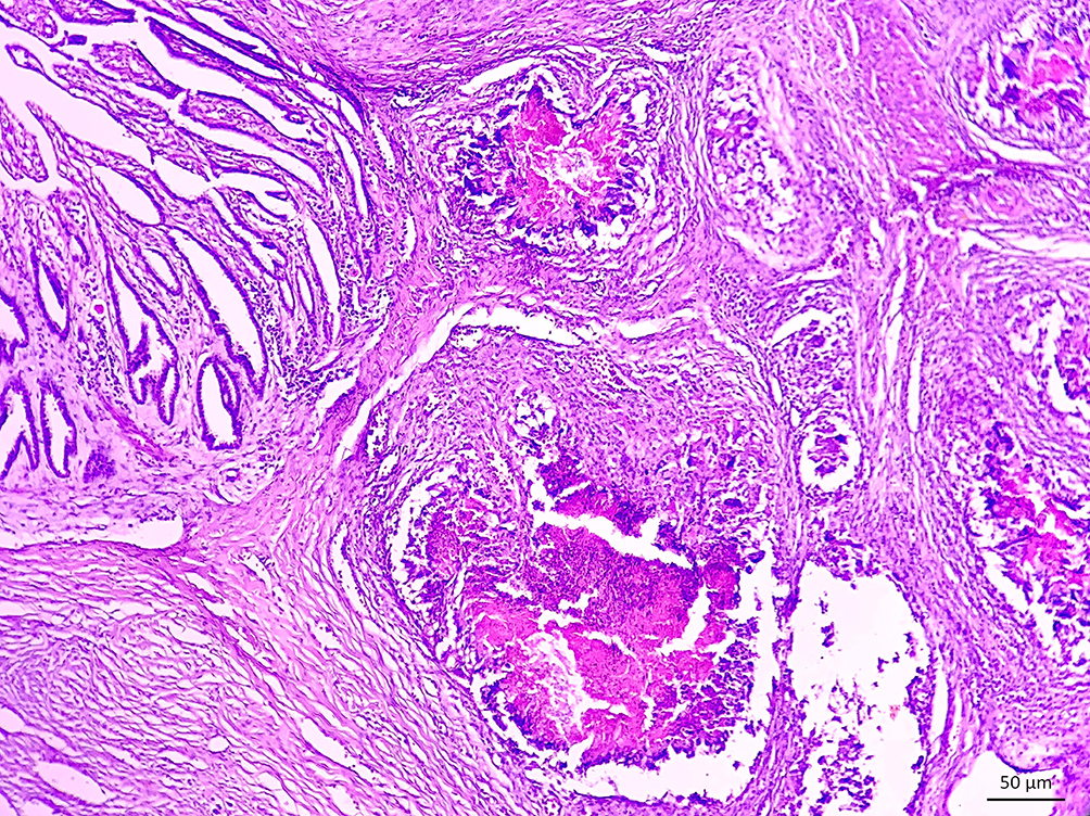
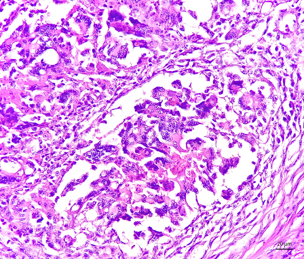
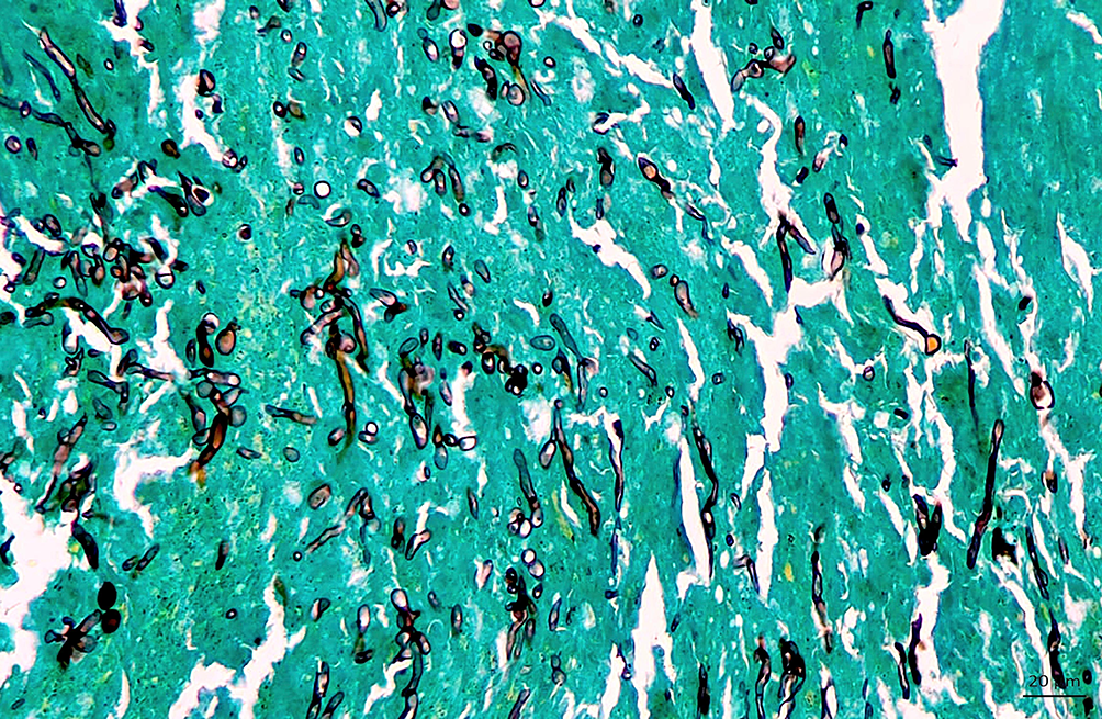
Histopathological evaluation revealed multifocal severe granulomatous and heterophilic ventriculitis, characterized by multifocal areas of heterophilic granulomas (Fig. 1), surrounded by a marked inflammatory infiltrate composed of macrophages and multinucleated giant cells, along with heterophils, lymphocytes, and plasma cells (Fig. 2). Hyphae measuring 2 to 6 µm in diameter with nearly parallel walls, occasionally septate and branched were observed, with better visualization in the GMS stain (Fig. 3) and weak staining in the PAS. No changes were observed in the other tissues examined.
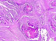
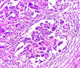
The heterophilic granuloma areas are surrounded by degenerated heterophils, macrophages, multinucleated giant cells, lymphocytes, and plasma cells. HE, obj. 20x.

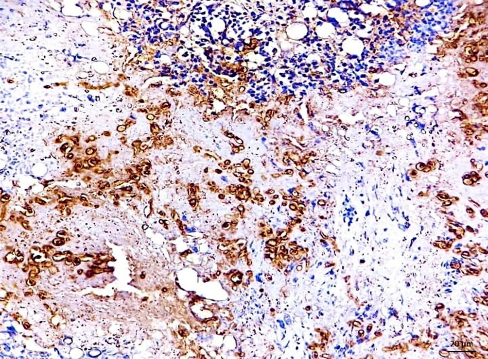
Histological sections of the ventriculus were subjected to immunohistochemical (IHC) analysis following the protocol described by Martins et al. (2012). Sections were deparaffinized, rehydrated, and endogenous peroxidase activity was blocked with 3% H₂O₂. Antigen retrieval was performed by microwave heating in TRIS-EDTA buffer (pH 9.0). The sections were then incubated with the primary antibody (1:1,000) for 60 min at 37 °C, followed by a biotinylated anti-rabbit secondary antibody. The reaction was visualized with 3,3′-diaminobenzidine (DAB; Sigma-Aldrich, St. Louis/MO, USA) and counterstained with hematoxylin. Fungal hyphae were strongly immunolabeled with the anti-Pythium insidiosum antibody (Fig. 4).
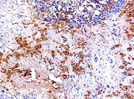
In IHC with polyclonal antibody against Pythium insidiosum, the hyphae show strong immunostaining (DAB; DakoCytomation). Obj. 40x.
Discussion
The diagnosis of pythiosis was based on anatomopathological and immunohistochemical findings. Pythiosis is a rare disease in birds (Pesavento et al. 2008, Souto et al. 2019), making this, to the authors’ knowledge, the third documented case and the first to describe granulomatous ventriculitis in psittacines.
Regarding the probable source of infection, it is believed to have occurred through the ingestion of water contaminated with zoospores of Pythium insidiosum. However, there is no information about the source of water consumed by the rosella. Pythiosis has been described in wild birds with access to agricultural fields and in animals kept in captivity with access to lakes (Pesavento et al. 2008, Souto et al. 2019).
In the gastrointestinal form of pythiosis, the main clinical signs described include vomiting, diarrhea with blood, and weight loss (Frade et al. 2017). In some cases, animals may exhibit subtle clinical signs, with loss of body condition being the only evident alteration (Rakich et al. 2005, Silva et al. 2024), similar to what was observed in this case. Changes in the rosella’s ventricular wall may have impaired digestive function, leading to the observed weight loss and death.
The microscopic characteristics of the oomycete and the granulomatous inflammation observed in this bird are similar to those described in mammals (Souto et al. 2022). In horses, pythiosis presents yellow-white masses known as “kunkers,” which are microscopically characterized by eosinophilic infiltration (Martins et al. 2012); however, these structures are not seen in ruminants and carnivores (Martins et al. 2012) or in birds (Pesavento et al. 2008, Souto et al. 2019).
The main fungal infections in birds associated with lesions in the gastrointestinal tract are caused by Candida spp., and Macrorhabdus ornithogaster (Kheirandish & Salehi 2011, Vasconcelos et al. 2011). In candidiasis, changes are primarily observed in the upper digestive tract, such as yellow and white necrotic plaques, elevated and dry lesions on the tongue, as well as thickening and wrinkling of the mucosa of the oral cavity, ingluvies, and esophagus (Asrani et al. 1993, Vasconcelos et al. 2011). In M. ornithogaster infection, the ventriculus is affected, associated with proventricular dilation and thickening of the wall, sometimes accompanied by ulcerations and hemorrhages in the proventricular mucosa (Marlier et al. 2006, Kheirandish & Salehi 2011). Both the macroscopic patterns described previously differ from the nodular pattern observed on the ventricular wall in this bird.
Histologically, candidiasis presents mucosal hyperplasia and hyperkeratinization, accompanied by yeasts and intralesional pseudohyphae (Vasconcelos et al. 2011). In cases of megabacteriosis (infection by M. ornithogaster), lymphoplasmacytic inflammation is particularly observed at the proventriculus-ventriculus junction, along with rupture of the koilin membrane (Kheirandish & Salehi 2011), associated with long, rod-shaped structures, arranged parallel, measuring 1.5-3 µm in diameter and 20-90 µm in length (Segabinazi et al. 2004).
Although aspergillosis primarily affects the respiratory system, forming white-yellow granulomas (Tokarzewski et al. 2007, Spanamberg et al. 2012, Melo et al. 2020), these may also be observed covering the serosa of organs within the coelomic cavity (Shukla et al. 2013). Microscopically, aspergillosis is characterized by granulomatous inflammatory infiltrates associated with hyphae that have basophilic, parallel, septate walls, with dichotomous branching (Spanamberg et al. 2012).
Morphologically, infections caused by Mucoromycota and the oomycete Lagenidium spp. are important differential diagnoses for pythiosis (Grooters et al. 2003). While there are no known reports of Lagenidium spp. infecting birds, its morphological features share similarities with those seen in pythiosis (Grooters et al. 2003).
Mucoromycota can cause lesions in the lungs, air sacs, myocardium, and kidneys. In all these areas, the presence of thrombosing vasculitis is a consistent observation. The fungal hyphae, besides being found inside the thrombi, frequently invade the surrounding tissues. Infected organs exhibit vascular and parenchymal lesions dominated by necrosis, hemorrhage, edema, and infiltration of heterophils (Carrasco et al. 1998). The fungal hyphae are broad and irregular (Severo et al. 2010).
The histochemical and immunohistochemical characteristics observed in this case were crucial for identifying the etiological agent. Given the weak staining of the hyphae with PAS, the black impregnation with GMS, and the positive immunolabeling with a polyclonal antibody for Pythium insidiosum. Immunohistochemistry is frequently used to diagnose pythiosis in animals (Brown & Roberts 1988, Martins et al. 2012). PCR is among the diagnostic methods applicable to cases of pythiosis. In our report, no frozen biological material was available; however, although PCR can be performed on formalin-fixed paraffin-embedded (FFPE) tissues, it was not applied, since immunohistochemistry had already proven sufficient for the identification of the agent. In a case involving a bird (Pesavento et al. 2008), PCR was used for diagnosis, as well as in studies with cattle and horses (Santos et al. 2011), sheep (Carrera et al. 2013), dogs (Silva et al. 2024), and cats (Soares et al. 2019).
Conclusion
Heterophilic granulomatous ventriculitis caused by Pythium insidiosum resulted in the death of a rosella, affecting the digestive tract with weight loss, and should be considered as a differential diagnosis for the main fungal infections affecting the digestive system of birds.
Acknowledgements
This study was financed by the “Coordenação de Aperfeiçoamento de Pessoal de Nível Superior” (CAPES) (Code 001).
References
-
Asrani RK, Paul Gupta RK, Sadana JR, Pandita A. Experimental candidiasis in Japanese quail: pathological changes. Mycopathologia 1993; https://doi.org/10.1007/BF01103575
» https://doi.org/10.1007/BF01103575 -
Brown CC, Roberts ED. Intestinal pythiosis in a horse. Aust Vet J 1988; https://doi.org/10.1111/j.1751-0813.1988.tb07369.x
» https://doi.org/10.1111/j.1751-0813.1988.tb07369.x -
Buergelt C, Powe J, White T. Abdominal pythiosis in a Bengal tiger (Panthera tigris tigris). J Zoo Wildl Med 2006; https://doi.org/10.1638/05-003.1
» https://doi.org/10.1638/05-003.1 -
Camus AC, Grooters AM, Aguilar RF. Granulomatous pneumonia caused by Pythium insidiosum in a Central American jaguar, Panthera onca J Vet Diagn Invest 2004; https://doi.org/10.1177/104063870401600612
» https://doi.org/10.1177/104063870401600612 -
Carmo PMS, Portela RA, Silva TR, Oliveira-Filho JC, Riet-Correa F. Cutaneous pythiosis in a goat. J Comp Pathol 2015; https://doi.org/10.1016/j.jcpa.2014.11.005
» https://doi.org/10.1016/j.jcpa.2014.11.005 -
Carrasco L, Gómez-Villamandos JC, Jensen HE. Systemic candidosis and concomitant aspergillosis and zygomycosis in two Amazon parakeets (Amazona aestiva). Mycoses 1998; https://doi.org/10.1111/j.1439-0507.1998.tb00342.x
» https://doi.org/10.1111/j.1439-0507.1998.tb00342.x -
Carrera MV, Peixoto RM, Gouveia GV, Pessoa CRM, Jesus FPK, Santurio JM, Botton SA, Costa MM. Pitiose em ovinos nos estados de Pernambuco e Bahia. Pesq Vet Bras 2013; https://doi.org/10.1590/S0100-736X2013000400011
» https://doi.org/10.1590/S0100-736X2013000400011 -
Frade MTS, Diniz PVN, Olinda RG, Maia LA, Galiza GJN, Souza AP, Nóbrega Neto PI, Dantas AFM. Pythiosis in dogs in the semiarid region of Northeast Brazil. Pesq Vet Bras 2017; https://doi.org/10.1590/S0100-736X2017000500010
» https://doi.org/10.1590/S0100-736X2017000500010 -
Grooters AM, Hodgin EC, Bauer RW, Detrisac CJ, Znajda NR, Thomas RC. Clinicopathologic findings associated with Lagenidium sp. infection in 6 dogs: initial description of an emerging oomycosis. J Vet Intern Med 2003; https://doi.org/10.1111/j.1939-1676.2003.tb02494.x
» https://doi.org/10.1111/j.1939-1676.2003.tb02494.x -
Kheirandish R, Salehi M. Megabacteriosis in budgerigars: diagnosis and treatment. Comp Clin Pathol 2011; https://doi.org/10.1007/s00580-010-1026-1
» https://doi.org/10.1007/s00580-010-1026-1 -
Krajaejun T, Sathapatayavongs B, Pracharktam R, Nitiyanant P, Leelachaikul P, Wanachiwanawin W, Chaiprasert A, Assanasen P, Saipetch M, Mootsikapun P, Chetchotisakd P, Lekhakula A, Mitarnun W, Kalnauwakul S, Supparatpinyo K, Chaiwarith R, Chiewchanvit S, Tananuvat N, Srisiri S, Suankratay C, Kulwichit W, Wongsaisuwan M, Somkaew S. Clinical and epidemiological analyses of human pythiosis in Thailand. Clin Infect Dis 2006; https://doi.org/10.1086/506353
» https://doi.org/10.1086/506353 -
Maia LA, Souto EPF, Frade MTS, Pimentel LA, Azevedo EO, Kommers GD, Riet-Correa F, Dantas AFM. Pythiosis in cattle in Northeastern Brazil. Pesq Vet Bras 2020; https://doi.org/10.1590/1678-5150-PVB-6532
» https://doi.org/10.1590/1678-5150-PVB-6532 -
Marlier D, Leroy C, Sturbois M, Delleur V, Poulipoulis A, VindeVogel H. Increasing incidence of megabacteriosis in canaries (Serinus canarius domesticus). Vet J 2006; https://doi.org/10.1016/j.tvjl.2005.07.006
» https://doi.org/10.1016/j.tvjl.2005.07.006 -
Martins TB, Kommers GD, Trost ME, Inkelmann MA, Fighera RA, Schild AL. A comparative study of the histopathology and immunohistochemistry of pythiosis in horses, dogs and cattle. J Comp Pathol 2012; https://doi.org/10.1016/j.jcpa.2011.06.006
» https://doi.org/10.1016/j.jcpa.2011.06.006 -
Melo AM, Silva-Filho RP, Poester VR, von Groll A, Fernandes CG, Stevens DA, Sabino R, Xavier MO. Aspergillosis in free-ranging aquatic birds. Med Mycol Case Rep 2020; https://doi.org/10.1016/j.mmcr.2020.04.005
» https://doi.org/10.1016/j.mmcr.2020.04.005 -
Mendoza L, Ajello L, McGinnis MR. Infections caused by the oomycetous pathogen Pythium insidiosum J Vet Intern Med 1996; https://www.researchgate.net/publication/282207830
» https://www.researchgate.net/publication/282207830 -
Mendoza L, Hernandez F, Ajello L. Life cycle of the human and animal oomycete pathogen Pythium insidiosum J Clin Microbiol 1993; https://doi.org/10.1128/jcm.31.11.2967-2973.1993
» https://doi.org/10.1128/jcm.31.11.2967-2973.1993 -
Pesavento PA, Barr B, Riggs SM, Eigenheer AL, Pamma R, Walker RL. Cutaneous pythiosis in a nestling white-faced ibis. Vet Pathol 2008; https://doi.org/10.1354/vp.45-4-538
» https://doi.org/10.1354/vp.45-4-538 -
Rakich PM, Grooters AM, Tang K-N. Gastrointestinal pythiosis in two cats. J Vet Diagn Invest 2005; https://doi.org/10.1177/104063870501700310
» https://doi.org/10.1177/104063870501700310 -
Santos CEP, Santurio JM, Marques LC. Pitiose em animais de produção no Pantanal Matogrossense. Pesq Vet Bras 2011; https://doi.org/10.1590/S0100-736X2011001200008
» https://doi.org/10.1590/S0100-736X2011001200008 -
Segabinazi SD, Flôres ML, Kommers GD, Barcelos AS, Veit DC, Eltz RD. Megabacteriose em emas (Rhea americana) no Estado do Rio Grande do Sul, Brasil. Ciência Rural 2004; https://doi.org/10.1590/S0103-84782004000300050
» https://doi.org/10.1590/S0103-84782004000300050 -
Severo CB, Guazzelli LS, Severo LC. Capítulo 7 - Zigomicose. J Bras Pneumol 2010; https://doi.org/10.1590/S1806-37132010000100018
» https://doi.org/10.1590/S1806-37132010000100018 -
Shukla S, Parihar S, Shrivastava N, Dar LM, Hussian SA. Outbreak of Aspergillus fumigatus in an organized Emu farm in Indore, India. J Adv Vet Res 2013; https://www.advetresearch.com/index.php/AVR/article/view/104/103
» https://www.advetresearch.com/index.php/AVR/article/view/104/103 -
Silva EMS, Martins KPF, Pereira AHB, Gris AH, Maruyama FH, Nakazato L, Colodel EM, Oliveira LGS, Boabaid FM. Esophageal and gastric pythiosis in a dog. Ciência Rural 2024; https://doi.org/10.1590/0103-8478cr20230272
» https://doi.org/10.1590/0103-8478cr20230272 -
Soares LMC, Schenkel DM, Rosa JMA, Azevedo LS, Tineli TR, Dutra V, Colodel EM. Feline subcutaneous pythiosis. Ciência Rural 2019; https://doi.org/10.1590/0103-8478cr20180448
» https://doi.org/10.1590/0103-8478cr20180448 -
Souto EPF, Kommers GD, Souza AP, Miranda Neto EG, Assis DM, Riet-Correa F, Galiza GJN, Dantas AFM. A retrospective study of pythiosis in domestic animals in northeastern Brazil. J Comp Pathol 2022; https://doi.org/10.1016/j.jcpa.2022.05.002
» https://doi.org/10.1016/j.jcpa.2022.05.002 -
Souto EPF, Pessoa CRM, Pessoa AFA, Trost ME, Kommers GD, Correa FR, Dantas AFM. Esophageal pythiosis in an ostrich (Struthio camelus). Arq Bras Med Vet Zootec 2019; https://doi.org/10.1590/1678-4162-10666
» https://doi.org/10.1590/1678-4162-10666 -
Spanamberg A, Casagrande RA, Ferreiro L, Rolim VM, Souza SO, Gonçalves ICM, Oliveira LGS, Wouters F, Wouters ATB, Fontana CS, Driemeier D. Aspergilose em trinca-ferros (Saltator similis) competidores de canto. Acta Scient Vet 2012; https://www.redalyc.org/pdf/2890/289023924024.pdf
» https://www.redalyc.org/pdf/2890/289023924024.pdf -
Tabosa IM, Riet-Correa F, Nobre VMT, Azevedo EO, Reis-Júnior JL, Medeiros RMT. Outbreaks of pythiosis in two flocks of sheep in northeastern Brazil. Vet Pathol 2004; https://doi.org/10.1354/vp.41-4-412
» https://doi.org/10.1354/vp.41-4-412 -
Tokarzewski S, Ziółkowska G, Łopuszyński W, Nozdryn-Płotnicki Z. Aspergillus fumigatus infection in a pigeon flock. Bull Vet Inst Pulawy 2007; https://aviexpert.pl/images/2007-Aspergillus-fumigatus-infection-in-a-pigeon-flock.pdf
» https://aviexpert.pl/images/2007-Aspergillus-fumigatus-infection-in-a-pigeon-flock.pdf -
Vasconcelos TCB, Longa CS, Albuquerque DDA, Costa CHC, Bruno SF. Aspectos clínicos e anatomopatológicos de aspergilose e candidíase em calopsita (Nymphicus hollandicus): relato de caso. Rev Port Ciênc Vet 2011; https://www.researchgate.net/publication/311768329
» https://www.researchgate.net/publication/311768329 -
Wellehan JFX, Farina LL, Keoughan CG, Lafortune M, Grooters AM, Mendoza L, Brown M, Terrell SP, Jacobson ER, Heard DJ. Pythiosis in a dromedary camel (Camelus dromedarius). J Zoo Wildl Med 2004; http://doi.org/10.1638/03-098
» https://doi.org/10.1638/03-098 -
Yolanda H, Krajaejun T. Global distribution and clinical features of pythiosis in humans and animals. J Fungi 2022; http://doi.org/10.3390/jof8020182
» https://doi.org/10.3390/jof8020182
The authors declare that all data used are available in this article.