 CHEMICAL PROFILE, ANTIBACTERIAL, ANTIFUNGAL AND INSECTICIDE POTENTIAL OF ESSENTIAL OIL FROM THE LEAVES OF Varronia dardani (Taroda) J. S. Mill
Abstract
The essential oil (EO) of Varronia dardani (Taroda) J.S. Mill (Cordiaceae), a plant widely distributed in northeastern Brazil, was analyzed for its chemical profile and evaluated for its antifungal, antibacterial, and insecticidal activities. The EO was obtained by hydrodistillation and analyzed by gas chromatography coupled to a mass spectrometer (GC-MS) and gas chromatography with a flame ionization detector (GC-FID). Limonene (27.49%) and dehydro-1,8-cineole (13.23%) were identified as the major constituents. The EO exhibited strong bactericidal activity, with minimum inhibitory concentration (MIC) and minimum bactericidal concentration (MBC) values of 1.5 mg mL−1 against Staphylococcus aureus and 200 mg mL−1 against Escherichia coli, and Pseudomonas aeruginosa. For Candida albicans and Candida parapsilosis, the MIC was 48 mg mL−1, indicating a fungistatic effect. In larvicidal assays, the EO showed LC50 and LC90 (lethal concentrations required to kill 50 and 90% of the larvae) values of 1.20 and 2.57 mg mL−1 after 1 h, which decreased to 0.92 and 1.92 mg mL−1 after 2 h. These findings demonstrate that EO is a promising natural antimicrobial agent and an effective larvicide against A. aegypti larvae.
Keywords:
Varronia dardani
; hydrodistillation; terpenoids; antimicrobial activity;
Aedes aegypti
INTRODUCTION
The genus Varronia, one of the largest within the family Cordiaceae, includes species that are widely used in folk medicine to treat respiratory, digestive, inflammatory, and infectious conditions.1 In addition to the traditional uses, several pharmacological studies2 have demonstrated anti-inflammatory, antioxidant, antimicrobial, antinociceptive and cytotoxic properties in species of the genus. Moreover, investigations involving Varronia species have revealed promising potential for the development of novel therapeutic agents and for the control of different pathogenic microorganisms.1 Insecticidal properties of essential oils (EOs) from Varronia spp. have also been reported3, 4 against pests of the orders Coleoptera and Hymenoptera.
Varronia dardani (Taroda) J. S. Mill, popularly known as “moleque duro”, is a shrubby plant endemic to the Caatinga biome and distributed across northeastern Brazil, where the climate is predominantly semiarid.2, 5 Although only a few studies have addressed its pharmacological potential, previous investigations6 have reported that the EO from V. dardani leaves exhibits antibiofilm activity against microorganisms of dental relevance, including Enterococcus faecalis and Streptococcus salivarius. In addition, high- and medium-performance liquid chromatography analyses of the ethanolic extract of V. dardani leaves identified chalcones, flavanones and flavonoids, with the extract displaying spasmolytic activity on rat aorta and trachea, guinea-pig ileum and rat uterus.7
Despite these findings, the pharmacological and insecticidal potential of the EO of V. dardani leaves remains poorly explored, and studies focusing on its antimicrobial and larvicidal properties are still scarce. Therefore, this study aims to characterize the volatile chemical constituents of the EO from Varronia dardani leaves and to evaluate its antibacterial, antifungal and insecticidal activities.
EXPERIMENTAL
Plant material
The leaves of Varronia dardani were collected during its flowering stage in April 2022 in the city of Patu, Rio Grande do Norte State, Brazil (6°08’12” S, 37°38’ W). A voucher specimen is deposited in the Dárdano de Andrade Lima Herbarium of the Federal University of Rural Semi-Arido (UFERSA), Rio Grande do Norte, under number MOSS 15627. The accepted plant name was confirmed through the World Flora Online (WFO),8 with Cordia dardanoi Taroda listed as a synonym.
Essential oil of leaves from Varronia dardani obtention
Fresh leaves of V. dardani (2.5 kg) were added to a 5.0 L flask, along with 2.5 L of distilled water. The flask was then connected to a Clevenger-type apparatus and subjected to a condensate then subjected to a hydrodistillation process for approximately 2 h. The EO was then separated from the hydrolat and dried with anhydrous sodium sulfate (Na2SO4). The resulting yellow fluid EO with a characteristic odor, was stored in a clean, dry bottle and kept under refrigeration at 3 °C until analysis and testing. The EO yield was 0.35% (w/w), calculated from the weight of fresh leaves.
Analysis of the chemical constituents of essential oil of leaves from Varronia dardani
The EO was analyzed using gas chromatography coupled to a mass spectrometer (GC-MS) and gas chromatography with a flame ionization detector (GC-FID). Both analyses were performed using a Shimadzu QP2010 Ultra (GC-MS) and a GC-2010 Plus (GC-FID) system under the following conditions: an Rtx-5 column (5% phenyl-95% dimethylpolysiloxane, 30 m × 0.25 mm × 0.25 μm) was used for both. Splitless injection was performed at 250 °C, and the oven temperature was programmed from 60 to 240 °C at a rate of 3 °C min−1, holding at 240 °C for 7 min. The carrier gas flow rate was 1.2 mL min−1, using helium (He) for GC-MS and hydrogen (H2) for GC-FID. The interface temperature was 240 °C; mass spectra were recorded from 40 to 450 m/z at a scan interval of 0.5 s and electron impact ionization at 70 eV. Individual content was determined based on the peak area of each compound relative to the total peak area in the chromatogram. Each component was identified based on the retention index (RI) considering a homologous series of n-alkanes (C7-C30), corrected by linear regression, and by comparison of the specific fragmentation pattern of each component with mass spectra in the virtual database library (Nist/Epa/Nih Library, 2011) as well as by visual comparison with mass spectra reported in the literature.9
Antimicrobial assay
In the experiments, standard strains of Escherichia coli (ATCC25922) (American Type Culture Collection), Pseudomonas aeruginosa (ATCC27853), Staphylococcus aureus (ATCC25923), Candida albicans (ATCC90028) and Candida parapsilosis (ATCC22019) were provided by the Laboratory of Antimicrobial Assays (LEAN) of the Federal University of Rio Grande do Norte (UFRN), Natal, Brazil. The bacterial and fungal microorganisms were maintained in Mueller-Hinton agar (Difco Laboratories, Detroit, Michigan, USA) and Sabouraud dextrose agar (Difco Laboratories, Detroit, Michigan, USA) at 4 °C, respectively. Prior to each experiment, the strains were seeded on agar plates. For the preparation of the inoculum, adjusted to the 0.5 scale of the McFarland standard, colonies were resuspended in sterile 0.85% saline solution for bacterial strains and 0.9% saline solution for fungal strains. Sensitivity testing was performed by broth microdilution according to the Clinical and Laboratory Standards Institute (CLSI).10
Minimum inhibitory concentration assay
Minimum inhibitory concentration (MIC) was determined through the broth microdilution technique in a 96-well flat-bottom microplate for cellular culture.11 The OE samples were solubilized in 5% DMSO (dimethyl sulfoxide) in distilled water to obtain emulsions at the necessary concentrations for the tests, starting at a maximum concentration of 200 mg mL−1 for antibacterial tests in Mueller-Hinton medium (INLAB®, São Paulo, Brazil) and 48 mg mL−1 in Roswell Park Memorial Institute (RPMI) 1640 medium with L-glutamine and without sodium bicarbonate (INLAB®, São Paulo, Brazil) for antifungal tests. Concurrently, microbial viability control (wells containing the culture medium and microbial inoculum), medium sterility control (wells containing only the culture medium), interference of the solubilizing agents in negative control (wells containing 5% DMSO, medium, and inoculum) and controls with gentamicin 10 μg mL−1 and amphotericin B 16 μg mL−1 (for bacterial and fungal strains, respectively) were performed. Except for the medium sterility control, all wells received 100 μL of the microbial inoculum adjusted to the 0.5 scale of the McFarland standard. The microplates were incubated at 37 °C for 24 and 48 h for bacterial and fungal assays, respectively. MIC was defined as the lowest concentration of the product in which there was visible inhibition of bacterial and fungal growth in the wells compared to the controls. The reading occurred 2 h after the detection of staining in the wells following the addition of 0.5% of 2,3,5-triphenyl tetrazolium chloride for the plates inoculated with bacteria and 0.01% of resazurin for the yeast assays. The experiment was conducted in triplicate.
Minimum bactericidal concentration and minimum fungicide concentration assays
After determining the MIC, to perform the minimum bactericidal concentration (MBC) and minimum fungicidal concentration (MFC) assays, 10 μL aliquots of the supernatant from wells showing complete inhibition of bacterial and fungal growth were spot-seeded on agar plates containing Mueller-Hinton agar and Sabouraud dextrose culture medium for bacterial and fungal strains, respectively.12 The agar plates were incubated for 24 and 48 h at 35 ± 2 °C, for bacterial and fungal strains, respectively. The MBC and MFC were defined as the lowest concentration of the product that showed no bacterial and fungal growth in the culture medium.13 The experiment was conducted in triplicate.
Aedes aegypti colony
Fourth instar (Aedes aegypti) larvae were used in all larvicidal assays. The larvae of A. aegypti (Rockefeller strain) were obtained from the Toxicology Laboratory of the Department of Antibiotics at the Federal University of Pernambuco (UFPE) and were kept at a temperature of 27 ± 1 °C, relative humidity (RH) of 70 ± 5% and 12-h photoperiod (light/dark). The A. aegypti eggs were placed in containers with mineral water (pH 7.0) and cat food (Whiskas®) to allow the larvae to hatch.
Larvicidal assay
The evaluation of the larvicidal activity of the essential oil was carried out according to Carvalho et al.14 and Dantas et al.15 with modifications in 24-well cell culture plates. Here, the essential oil was solubilized in Tween 80 (1%, v/v) to prepare test solutions at concentrations 0.25, 0.5, 1, 2, 3, 4, and 5 mg mL−1. Following, the larvicidal assay was performed with six replicates using 2 mL of the oil-water emulsion and ten A. aegypti larvae in four instars totaling 60 larvae per concentration. Control solution was prepared with 1% Tween 80. Three independent assays were performed at different dates. Larvae mortality was observed after periods of 0.5, 1, 2, 3, 4, and 24 h, which was conducted at a temperature of 27 ± 1 °C, RH of 70 ± 5% and a 12-h photoperiod. The results were subjected to probit analysis using BioStat 7 (registration No. 30915489, AnalystSoft Inc., Walnut, CA, USA, 2020) to obtain the lethal concentrations required to kill 50 and 90% of the larvae (LC50 and LC90, respectively).
RESULTS AND DISCUSSION
Chromatographic analysis of essential oil of leaves from Varronia dardani
About 25 compounds were identified, representing 99.9% of the chemical composition of the EO. As can be observed, the oil consists mostly of monoterpenes (78.49%). In the quantitative analysis, the main chemical constituents present in the EO, in order of abundance, were limonene (27.49%), dehydro-1,8-cineole (13.23%), linalool (10.24%), (2Z,6E)-farnesyl acetate (6.67%), (2Z,6Z)-farnesol (6.34%), α-terpineol (6.27%) and terpinen-4-ol (5.77%). The chemical constituents were identified and quantified using GC-MS and GC-FID, as presented in Table 1 and the Supplementary Material.
Although EO of leaves from V. dardani has a known chemical composition, the EO from the region of Patu (Rio Grande do Norte, Brazil) showed a different chemical composition to that studied by Veloso et al.6 in Serra Branca (Paraíba, Brazil), which showed as major compounds δ-cadinene (8.71%), thymol (7.43%), carvacrol (5.92%), γ-cadinene (5.66%), p-cymene (5.25%) and δ-cadinol (5.20%). The survey conducted by Ramos et al.16 indicates that the production and chemical composition of volatiles in plants are intrinsically associated with the environment in which the species is located, and factors such as biotic, abiotic, and biochemical/genetic influence the quality and quantity of the chemical components present in the EO.
Effect of essential oil of leaves from Varronia dardani on antimicrobial activities
The antimicrobial potential of the EO was evaluated by determining MIC, MBC and MFC. Among the tested concentrations, the EO presented inhibitory activity against 100% of the bacterial isolates at concentrations of 200 and 1.5 mg mL−1 (Table 2). In the evaluation of the bactericidal action, the results show that 100% exhibited no growth at the concentrations of 200 and 1.5 mg mL−1, demonstrating that the EO was effective in causing cell death of the etiological agents. The strain of S. aureus (ATCC25923) showed the best sensitivity to the oil sample, with the same inhibitory and bactericidal concentration (1.5 mg mL−1).
Evaluation of minimum inhibitory concentration (MIC), minimum bactericidal concentration (MBC) and minimum fungicide concentration (MFC) of essential oil (EO) of leaves from Varronia dardani on bacterial species and yeasts
Research on essential oils (EOs) has demonstrated their broad antimicrobial potential. Researchers such as Bona et al.17 report that most EOs probably have antimicrobial potential, denaturing and causing coagulation of proteins by affecting the cell wall structure of bacterial and fungal agents. Furthermore, they are responsible for altering the permeability of the plasma membrane, which leads to the disruption of vital cell processes such as electron transport, protein translocation, phosphorylation, and other reactions, resulting in the loss of chemiosmotic control and consequent cell death. However, given the amount and variety of phytoconstituents present in these plant products, it becomes difficult to attribute a single specific mechanism of action, although it is a widely discussed subject among the scientific community.18, 19
Although limonene is the major constituent of the essential oil, it was not tested individually. This study aimed to evaluate the biological activity of the whole essential oil, maintaining its natural synergistic composition, since interactions among monoterpenes may enhance or modify antimicrobial effectiveness.20 Additionally, the amount of essential oil obtained was insufficient to allow the isolation and individual testing of its major components. For these reasons, only the complete essential oil was evaluated in the antimicrobial assays.
Currently, Tian et al.,21 using EO of Zingiber striolatum against bacteria and yeasts, found MIC values ranging from 3.12 to 0.78 mg mL−1, which the S. aureus strain presented the lowest MIC value (0.78 mg mL−1), similar to the result of the present study in which S. aureus also presented the lowest MIC value (1.5 mg mL−1). In another study published by Aelenei et al.,22 the researchers evaluated samples of E. coli (ATCC25922), S. aureus (ATCC 33591, ATCC43300 and ATCC6538), S. epidermidis (ATCC12228) and P. aeruginosa (ATCC9027 and ATCC27853), noting that the MIC value (5.44 mg mL−1) was the same for all bacteria tested against the EO of coriander (Coriandrum sativum), thus proving the antimicrobial potential of the plant sample, similar to the finding in this study, although the plant products are from different sources.
Moreover, Owen et al.,23 evaluating the antibacterial activity of the EO of cumin (Cuminum cyminum), oregano (Origanum compactum) and rosewood (Aniba rosaeodora) against clinical isolates (methicillin-resistant S. aureus, MRSA; methicillin-sensitive S. aureus, MSSA; E. coli and P. aeruginosa), found that the different oils showed antimicrobial activity against all isolates tested. It is noteworthy that among the findings of Owen et al.,23 P. aeruginosa was the species that showed the highest MIC for cumin (18.6 mg mL−1), oregano (9.20 mg mL−1), and rosewood (35. 20 mg mL−1), corroborating with the result found in the present study, when the EO was tested for P. aeruginosa (ATCC27853), showing higher MIC when compared to the other microorganisms except from E. coli (ATCC25922). This finding may suggest that the EO was probably less efficient against these Gram-negative bacteria, considering that these microorganisms have complex cell wall compared to Gram-positive bacteria,24 in addition to P. aeruginosa presenting an extracellular envelope consisting of alginate, enabling biofilm formation and, consequently, possible development of resistance to antimicrobials.25
Furthermore, Macedo26 analyzed the antibacterial activity of Lippia grata Schauer EO against clinical isolates of P. aeruginosa, which resulted in total growth inhibition ranges in MIC from 50 to 6.25 mg mL−1. In the same study, the bacteriostatic character of the oil was observed in all strains tested, different from the present study, which, although presenting the maximum MIC value (200 mg mL−1) for P. aeruginosa, the EO was able to totally kill the bacteria, presenting a bactericidal character.
The antifungal activity of the EO sample against Candida spp. strains showed no variation, with the maximum concentration (48 m mL−1) being the only one able to totally inhibit the growth of fungal agents (Table 2). Regarding the MFC of the EO sample on Candida spp. strains, visible growth was observed in both strains studied, confirming that the substance acted only as fungistatic. Altogether, even the high use of fungistatic agents promotes the development of resistant strains, they are as efficient as fungicides from a clinical point of view and are widely used, preventing significant increases in life-threatening infections.27
Differently, Vieira et al.28 reported high sensitivity of several species of the Candida species against the EO of cumin (Cuminum cyminum) with the MIC values ranging from 2.188 to 4.375 mg mL−1 and fennel (Foeniculum vulgare) ranging from 8.75 to 4.375 mg mL−1. In contrast, Saraiva Filho29 did not observe antifungal activity when analyzing the EO of Zanthoxylum petiolare against C. tropicalis and C. albicans.
Altogether, different results of EO assays against microbial agents can be associated with several factors, such as the microbial species evaluated,30 part of the plant from which the plant product was extracted31 and the content and chemical composition of the plant product.32 Moreover, other factors such as plant age, growing conditions, storage, climatic, ecological, physiological and genetic factors influence changes in the production of secondary metabolites.33
Larvicidal activity of the essential oil of leaves from Varronia dardani on A. aegypti
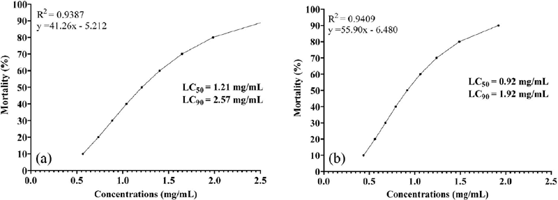
The essential oil from V. dardani promoted the mortality of A. aegypti larvae (Table 3). The lethal concentrations (mg mL−1) required to kill 50% (LC50) and 90% (LC90) of larvae after 1 h were 1.21 [1.04-1.38] and 2.57 [2.19-3.15] mg mL−1, respectively (Figure 1a) and after 2 h were 0.92 [0.62-1.27] and 1.92 [1.37-3.72] mg mL−1, respectively (Figure 1b).
Mortality of A. aegypti larvae exposed to V. dardani essential oil after 0.5, 1, 2, 3, 4 and 24 h of exposure

Larvicidal activity of V. dardani essential oil on A. aegypti L4 larvae. Lethal concentrations (LC50) and LC90) at (a) 1 h and (b) 2 h
The larvicidal activity of the essential oil could be attributed to major components such as limonene, linalool and 1,8-cineole. These secondary metabolites present in plant essential oils are cited in the literature34, 35, 36 as the active principles of larvicidal activity against A. aegypti.
The larvicidal efficacy of V. dardani essential oil on A. aegypti is higher than that of essential oils from other plant species since the LC50 and LC90 values for 1 and 2 h are lower. Costa et al.37 studied the larvicidal effect of Eugenia stipitata essential oil and determined the LC50 and LC90 values of 4.93 and 17.41 mg mL-1 after 1 h, respectively. Silva et al.38 determined that the essential oil of Croton tetradenius is a larvicidal agent with LC50 of 5.25 mg mL−1 and LC90 of 6.72 mg mL−1 after 2 h of exposure.
CONCLUSIONS
The EO of leaves from Varronia dardani demonstrated significant antibacterial potential activity against Gram-positive and Gram-negative bacteria and showed fungistatic effects against Candida species. The EO also exhibited remarkable larvicidal potential against Aedes aegypti fourth-instar larvae, with low LC50 and LC90 values after short exposure periods. Monoterpenes were the predominant constituents of the oil, supporting the hypothesis that these compounds play an essential role in the biological properties observed.
Overall, the results indicate that V. dardani EO is a promising natural source of antimicrobial agents and a potential larvicidal alternative for vector control programs. Future studies should focus on isolating the major and minor constituents of the EO, evaluating their individual and synergistic biological effects, and exploring the molecular mechanisms involved in antimicrobial and larvicidal activity. Additionally, in vivo assays and toxicity studies are essential to support the safe and effective application of V. dardani EO in therapeutic and environmental contexts.
SUPPLEMENTARY MATERIAL
Complementary material for this study is available at http://quimicanova.sbq.org.br/, as a PDF file, with free access.
DATA AVAILABILITY STATEMENT
All data generated or analyzed during this study are included in this published article.
ACKNOWLEDGMENTS
We thank the CNPq, CAPES, and INCT BioNat.
REFERENCES
-
1 Oza, M. J.; Kulkarni, Y. A.; J. Pharm. Pharmacol. 2017, 69, 755. [Crossref]
» Crossref -
2 de Melo, J. I. M.; Biotemas 2012, 25, 109. [Crossref]
» Crossref -
3 de Oliveira, B. M. S.; Melo, C. R.; Santos, A. C. C.; Nascimento, L. F. A.; Nízio, D. A. C.; Cristaldo, P. F.; Blank, A. F.; Bacci, L.; Environ. Sci. Pollut. Res. 2019, 26, 6602. [Crossref]
» Crossref -
4 Fonseca, M.; Andrade, F.; Sena, N.; Martins, E.; Venzon, M.; Planta Med. 2023, 89, 1357. [Crossref]
» Crossref -
5 Varronia dardani (Taroda) J.S.Mill.; Flora e Funga do Brasil. [Link] accessed in January 2026
» Link -
6 Veloso, C. A. G.; de Souza, P. H. S.; Nóbrega, F. P.; de Medeiros, A. C. D.; Fechine, I. M.; de Melo, J. I. M.; Tavares, J. F.; da Silva, M. S.; Costa, V. C. O.; Brazilian Journal of Development 2020, 6, 12887. [Crossref]
» Crossref -
7 Veloso, C. A. G.; Figueiredo, I. A. D.; da Silva, G. R.; de Melo, J. I. M.; da Silva, M. S.; Tavares, J. F.; Cavalcante, F. A.; Costa, V. C. O.; Nat. Prod. Res. 2021, 35, 4197. [Crossref]
» Crossref -
8 Varronia dardani (Taroda) J.S.Mill.; WFO, The World Flora Online. [Link] accessed in January 2026
» Link - 9 Adams, R. P.; Identification of Essential Oil Components by Gas Chromatography/Quadrupole Mass Spectrometry, 4.1 ed.; Allured Publ. Corp.: Carol Stream, IL, USA, 2017.
- 10 Clinical and Laboratory Standards Institute (CLSI); CLSI Supplement M100: Performance Standards for Antimicrobial Susceptibility Testing, 30th ed.; CLSI: Wayne, PA, 2020.
- 11 Clinical and Laboratory Standards Institute (CLSI); CLSI Supplement M60: Performance Standards for Antifungal Susceptibility Testing of Yeasts, 2nd ed.; CLSI: Wayne, PA, 2020.
-
12 Sakita, K. M.; Conrado, P. C. V.; Faria, D. R.; Arita, G. S.; Capoci, I. R. G.; Rodrigues-Vendramini, F. A. V.; Pieralisi, N.; Cesar, G. B.; Gonçalves, R. S.; Caetano, W.; Hioka, N.; Kioshima, E. S.; Svidzinski, T. I. E.; Future Microbiol. 2019, 14, 519. [Crossref]
» Crossref -
13 Silva, D.; Diniz-Neto, H.; Cordeiro, L.; Silva-Neta, M.; Silva, S.; Andrade-Júnior, F.; Leite, M.; Nóbrega, J.; Morais, M.; Souza, J.; Rosa, L.; Melo, T.; Souza, H.; Sousa, A.; Rodrigues, G.; Oliveira-Filho, A.; Lima, E.; Int. J. Mol. Sci. 2020, 21, 1785. [Crossref]
» Crossref -
14 Carvalho, K. S.; e Silva, S. L. C.; de Souza, I. A.; Gualberto, S. A.; da Cruz, R. C. D.; dos Santos, F. R.; de Carvalho, M. G.; Parasitol. Res. 2016, 115, 3441. [Crossref]
» Crossref -
15 Dantas, J. O.; Araújo-Piovezan, T. G.; Santos, D. P.; Alves, A. E. O.; Pinheiro, S. S. C.; Ribeiro, G. T.; Ensaios e Ciência: Ciências Biológicas, Agrárias e da Saúde 2019, 23, 104. [Link] accessed in January 2026
» Link - 16 Ramos, Y. J.; Felisberto, J. R. S.; Oliveira, C. C.; de Pontes, E. D.; Machado, D. B.; Fonseca, I. C.; Moreira, D. L. In Avanços Científicos, Tecnológicos e de Inovação na Botânica, 1st ed.; de Francisco, A. L. O., org.; Atena Editora: Belo Horizonte, 2020, ch. 7.
-
17 Bona, T. D. M. M.; Pickler, L.; Miglino, L. B.; Kuritza, L. N.; Vasconcelos, S. P.; Santin, E.; Pesquisa Veterinária Brasileira 2012, 32, 411. [Crossref]
» Crossref -
18 Dorman, H. J. D.; Deans, S. G.; J. Appl. Microbiol. 2000, 88, 308. [Crossref]
» Crossref -
19 Carson, C. F.; Mee, B. J.; Riley, T. V.; Antimicrob. Agents Chemother. 2002, 46, 1914. [Crossref]
» Crossref -
20 Bassolé, I. H. N.; Juliani, H. R.; Molecules 2012, 17, 3989. [Crossref]
» Crossref -
21 Tian, M.; Liu, T.; Wu, X.; Hong, Y.; Liu, X.; Lin, B.; Zhou, Y.; Nat. Prod. Res. 2020, 34, 2621. [Crossref]
» Crossref -
22 Aelenei, P.; Rimbu, C. M.; Guguianu, E.; Dimitriu, G.; Aprotosoaie, A. C.; Brebu, M.; Horhogea, C. E.; Miron, A.; Lett. Appl. Microbiol. 2019, 68, 156. [Crossref]
» Crossref -
23 Owen, L.; White, A. W.; Laird, K.; Phytochem. Anal. 2018, 30, 121. [Crossref]
» Crossref -
24 Seibert, J. B.; Bautista-Silva, J. P.; Amparo, T. R.; Petit, A.; Pervier, P.; Almeida, J. C. S.;Azevedo, M. C.; Silveira, B. M.; Brandão, G. C.; de Souza, G. H. B.; Teixeira, L. F. M.; dos Santos, O. D. H.; Food Chem. 2019, 287, 61. [Crossref]
» Crossref -
25 Zamora, C. M. P.; Torres, C. A.; Nunez, M. B.; Molecules 2018, 23, 544. [Crossref]
» Crossref -
26 Macedo, C. A.: Atividade Antibacteriana e Antibiofilme da Lippia grata Frente a Isolados Clínicos de Pseudomonas aeruginosa; Dissertação de Mestrado, Universidade Federal do Rio Grande do Norte, Natal, Brasil, 2019. [Link] accessed in January 2026
» Link -
27 Monk, B. C.; Goffeau, A.; Science 2008, 321, 367. [Crossref]
» Crossref -
28 Vieira, J. N.; Gonçalves, C. L.; Villarreal, J. P. V.; Gonçalves, V. M.; Lund, R. G.; Freitag, R. A.; Silva, A. F.; Nascente, P. S.; Brazilian Journal of Biology 2019, 79, 432. [Crossref]
» Crossref -
29 Saraiva Filho, D. E.; Neves, A. M.; de Moraes, S. M.; de Souza, E. B.; Rodrigues, T. H. S.; dos Santos, H. S.; Mesquita, M. C. A.; Fontenelle, R. O. S.; Brazilian Journal of Development 2021, 7, 38904. [Crossref]
» Crossref -
30 Giordani, C.; Santin, R.; Cleff, M. B.; Rev. Bras. Plant. Med. 2015, 17, 175. [Crossref]
» Crossref -
31 Geetha, V.; Chakravarthula, S. N.; J. For. Res. 2018, 29, 373. [Crossref]
» Crossref -
32 Miranda, C. A. S. F.; Cardoso, M. G.; Batista, L. R.; Rodrigues, L. M. A.; Figueiredo, A. C. S.; Revista Ciência Agronômica 2016, 47, 213. [Link] accessed in January 2026
» Link -
33 de Oliveira, A. R. M. F.; Jezler, C. N.; Oliveira, R. A.; Costa, L. C. B.; Rev. Ceres 2012, 59, 241. [Crossref]
» Crossref -
34 Marques, D. M.; Rocha, J. F.; de Almeida, T. S.; Mota, E. F.; S. Afr. J. Bot. 2021, 143, 69. [Crossref]
» Crossref -
35 Bailão, E. F. L. C.; Pereira, D. G.; Romano, C. A.; Paz, A. T. S.; e Silva, T. M.; de Paula, J. R.; Gomes, C. M.; Borges, L. L.; S. Afr. J. Bot. 2022, 144, 257. [Crossref]
» Crossref -
36 Santos, E. F.; Anselmo, W. M.; de Lemos, E. E. P.; de Aguiar, J. C. R. O. F.; da Silva, A. C.; dos Santos, F. H. G.; Arruda, C. C. L.; Aguiar, J. V. C.; de Andrade, J. J. A.; da Rocha, S. K. L.; Araújo, L. A.; Pereira Júnior, P. G.; Albuquerque, C. F. O.; Sousa, E. S.; dos Santos, G. L.; da Conceição, T. Z.; de Andrade, L. A.; Soares, L. A. L.; Ferreira, M. R. A.; Navarro, D. M. A. F.; Molecules 2025, 30, 3116. [Crossref]
» Crossref -
37 Costa, W. K.; de Oliveira, J. R. S.; de Oliveira, A. M.; Santos, I. B. S.; da Cunha, R. X.; de Freitas, A. F. S.; da Silva, J. W. L. M.; Silva, V. B. G.; de Aguiar, J. C. R. O. F.; da Silva, A. G.; Navarro, D. M. A. F.; Lima, V. L. M.; da Silva, M. V.; Ind. Crops Prod. 2020, 144, 112059. [Crossref]
» Crossref -
38 Silva, P. B.; Santos, R. B. R.; da Cruz, R. C. D.; da Silva, D. C.; da Silva, P. S. D.; Biocatal. Agric. Biotechnol. 2023, 51, 102743. [Crossref]
» Crossref
Edited by
-
Executive Editor handled this article:
Rodrigo O. M. A. de Souza