Molecular methods applied to gynecological diseases: a laboratory review
INTRODUCTION
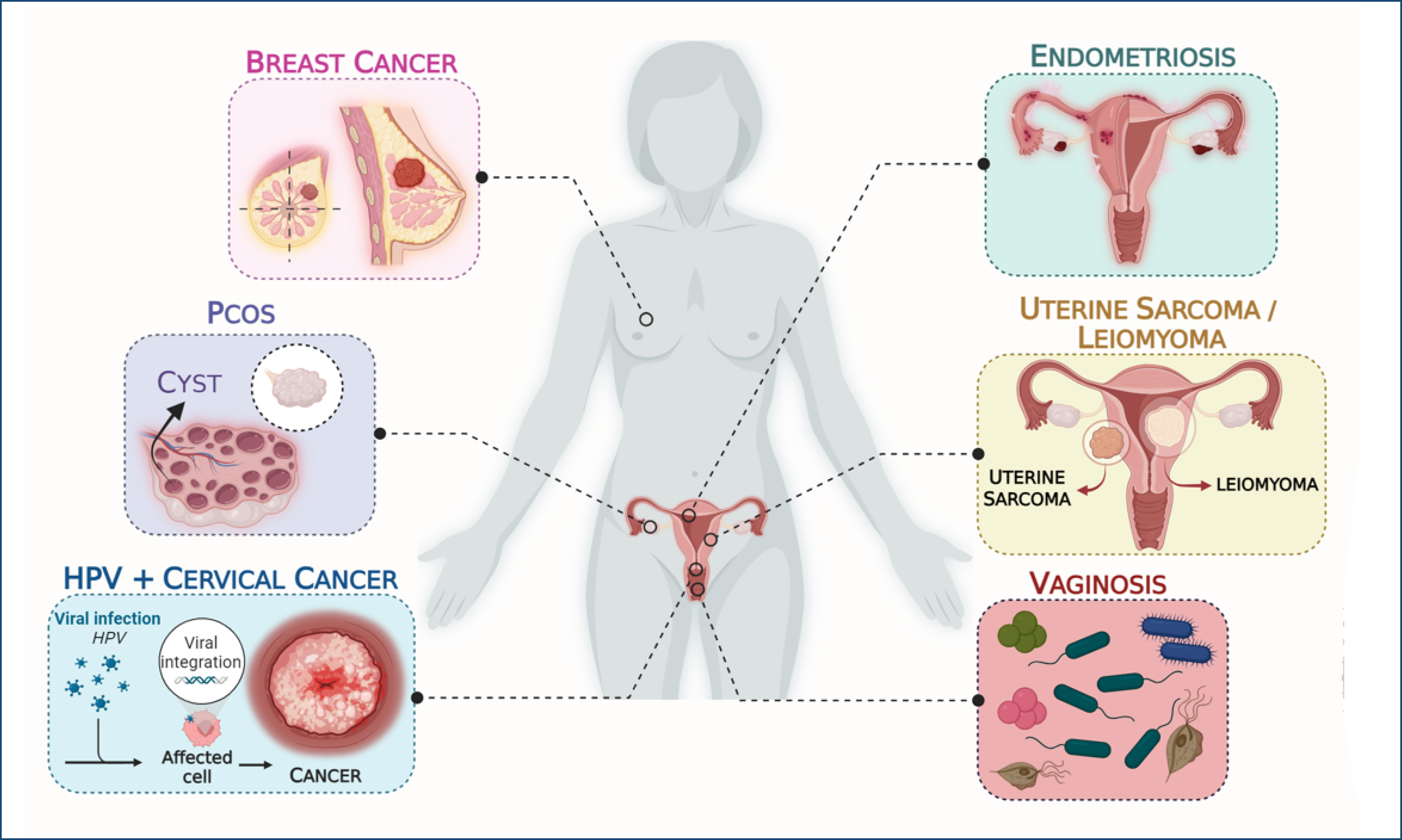
Gynecology is a medical specialty focused on the health of the female reproductive system1. Gynecological diseases can affect both the internal and external parts of the genital organs, with causes that include inflammatory conditions (such as endometriosis and pelvic inflammatory disease), infectious diseases like bacterial vaginosis (BV) and chlamydia (caused by bacteria, fungi, and viruses), hormonal issues (such as polycystic ovary syndrome [PCOS]), and genetic factors (such as gynecological cancers, which are multifactorial but have a genetic origin)1,2. Figure 1 presents the most common gynecological conditions that were covered in the present review.

Graphical representation of the most common gynecological diseases covered in the present study. The figure was developed by the authors using Biorender software.
Research in molecular biology has highlighted the significance of molecular tests in gynecology and obstetrics, yielding promising results2. Techniques such as polymerase chain reaction (PCR) are essential for detecting specific DNA or RNA sequences1,2. Next-generation sequencing (NGS) provides comprehensive genomic profiles, revealing genetic variations linked to diseases like gynecological cancer. Immunohistochemistry (IHC) has emerged as a valuable tool for visualizing specific proteins in tissue samples and standardizing molecular markers in cervical cancer2.
MOST COMMON GYNECOLOGICAL DISEASES
Vaginosis
BV is characterized by an imbalance in vaginal flora, specifically a decrease in beneficial lactobacilli and an increase in anaerobic bacteria. It is the most common cause of discharge, itching, and elevated vaginal pH in women of childbearing age. Gardnerella vaginalis, the primary bacterium linked to BV, contributes to this imbalance by producing substances that promote the growth of other anaerobic bacteria, leading to symptoms such as gray discharge with a "fishy" odor3.
For the molecular diagnosis of BV, several tests are commonly used. PCR detects the DNA of bacteria associated with BV, while DNA sequencing analyzes the complete genetic material of vaginal bacteria. Nucleic acid hybridization employs labeled DNA/RNA probes to identify specific target bacteria. Additionally, nucleic acid amplification tests (NAATs) can amplify and detect specific nucleic acids, helping differentiate BV from other vaginal infections3.
Polycystic ovary syndrome
PCOS is a common endocrine disorder affecting 5–18% of women of reproductive age. It is characterized by endocrine dysfunction, reproductive issues, hyperandrogenism, oligo- or anovulation, and the presence of polycystic ovaries on ultrasound, following the exclusion of other endocrine disorders1,4.
The exact cause of PCOS remains unclear, but some genetic factors may play a role. In addition, microRNAs (miRNAs) have emerged as potential genetic biomarkers due to their regulatory functions and ease of detection5.
In molecular biology, the analysis of gene expression through quantitative real-time PCR (qRT-PCR) and IHC has shed light on how certain genes behave in relation to PCOS and its development. Inhibiting the Notch gene signaling pathway by knocking down NOTCH1 and RBPJ genes resulted in a darkening of white adipose tissue (WAT), which improved insulin sensitivity and glucose tolerance. The Notch pathway inhibition using the drug dibenzazepine enhanced Ucp1 expression, suggesting that this signaling pathway may serve as a potential therapeutic target for addressing some consequences of PCOS4.
The genes ACACA, SREBP1, CPT1, and CD36 were also analyzed using qRT-PCR6.
Endometriosis
Endometriosis is a benign gynecological condition affecting 10–15% of women of childbearing age and 3–5% of postmenopausal women1. It is characterized by the presence of endometrial-like tissue outside the uterine cavity, leading to chronic inflammation that can impact pelvic tissues and organs1,7. This complex condition involves genetic and hormonal factors7.
There are three morphological classifications of endometriosis:
-
Peritoneal/superficial: This is the mildest form, affecting the abdominal walls and pelvic organs, with limited tissue penetration.
-
Deep infiltrative: This involves deeper tissue invasion (greater than 5 mm), often affecting the bladder and rectum, causing significant pain and potential organ damage.
-
Ovarian endometriomas: These are cysts formed by endometrial tissue that develop outside the uterus, usually in the ovaries.
The symptoms are multifactorial and may include painful periods, painful intercourse, chronic pelvic pain, and infertility due to the backflow of endometrial tissue during menstruation.
Recent studies using genetic analysis by PCR can help identify changes in some genes related to growth and immunological responses such as the PI3K, AKT, NLRP3, Caspase-1, GSDMD, and GSDMD-N genes, which demonstrate that they may be associated with inflammation and endometriosis while undergoing changes in their signaling pathway1,7.
Breast cancer
Breast cancer is frequently diagnosed in women and continues to see a rise in global incidence8. A combination of genetic and non-genetic factors contributes to its onset, including age, reproductive risk factors, exogenous female hormones, lifestyle elements, radiation exposure, mammographic density, and histologic lesions8,9. Genetic mutations play a crucial role in the development and progression of breast cancer. Mutations in specific genes, such as BRCA1 and BRCA2, are strongly linked to an increased risk of developing breast cancer, as these genes are involved in DNA repair and maintaining genomic stability9. When these genes are mutated, the ability of cells to repair damaged DNA is compromised, leading to the accumulation of genetic alterations that drive cancer development. Additionally, mutations in other genes, such as TP53, PIK3CA, and HER2, can also contribute to breast cancer by affecting key pathways involved in cell growth, division, and survival. Understanding these genetic mutations is essential for identifying individuals at high risk, as well as for the development of targeted therapies that can specifically address the underlying genetic causes of breast cancer. Approximately 8–10% of breast cancer cases are attributed to hereditary mutations, half of which are related to BRCA1 and BRCA28,9.
Human papillomavirus+cervical cancer
Human papillomavirus (HPV) causes a widespread sexually transmitted infection that can be contracted through both penetrative and non-penetrative genital skin-to-skin contact. The virus invades basal epithelial cells of mucocutaneous membranes, leading to a range of outcomes, from benign lesions to cancers, including oropharyngeal, cervical, vulvar, vaginal, and penile cancers. Cervical cancer, the fourth most common cancer among women globally, is strongly linked to HPV10.
The increased microbial diversity is correlated with HPV expression, as it contributes to increased cervical inflammation and vaginal bacterial infections, thereby favoring the development of precancerous lesions9. Additionally, the reduction of lactobacilli is associated with a higher prevalence of the oncogenic proteins E6 and E7 of HPV, which inhibit tumor suppressor genes. Of the more than 20 HPV types affecting the genital epithelium, types 16 and 18 are most commonly associated with cervical cancer10.
Various screening methods, including HPV DNA testing, precision tests, viral load quantification, and protein detection via IHC and Western blot, offer specificity and predictive advantages11.
Uterine sarcomas
Uterine sarcomas are a rare form of malignant neoplasm, comprising 3–7% of all uterine tumors and less than 1% of all gynecological cancers, with an annual incidence of approximately 0.30/100,000 women12. These tumors often originate in soft tissues, particularly the smooth muscle of the myometrium, with leiomyosarcoma (LMS) being the most common type, typically affecting women over 45 years old12. Due to their aggressive nature, the survival rate remains low, even with early diagnosis12.
Adenosarcoma, comprising a benign epithelial component and a malignant stromal component, accounts for 5% of uterine sarcomas. It has a lower malignant potential but can still present challenges, including high rates of local recurrence. Uterine sarcomas are histologically classified into carcinosarcomas (50%), endometrial stromal sarcomas (15%), and mixed epithelial/mesenchymal tumors due to their high malignancy12,13.
Leiomyoma
Leiomyomas (uLMs) are benign tumors that originate in the myometrium from smooth muscle cells of the uterus14. The causes of their development are not fully understood, but estrogen and hormonal contraceptives may promote their growth15. Affecting approximately 70% of women of reproductive age, uLMs are the most common benign genital tumors and typically regress after menopause14,15.
The classification of uLMs is based on their location:
-
Subserosal: Outer surface of the uterus; may be pedunculated or intraligamentary;
-
Submucosal: Located beneath the endometrium, potentially causing complications during pregnancy;
-
Cervical: Uterine cervix;
-
Intramural: Within the uterine wall, they can grow large enough to distort the uterine cavity.
The uLMs can develop as solitary tumors or in multiples, varying in size throughout the uterus1,15,16. Approximately 30% of women are asymptomatic, while the remaining 70% experience symptoms16. Common issues include excessive menstrual bleeding, abdominal pain, chronic constipation, and urinary incontinence14,16. These tumors may also affect fertility and increase the risk of complications during pregnancy15,16.
The diagnosis of uLMs involves a physical examination and symptom analysis, typically confirmed by ultrasound and imaging tests1,14,15. Treatment options mainly include hormone-based medications and hysterectomy. Recent molecular biology studies are exploring methods, such as qPCR. A relevant study, using qPCR, showed that the inactivation of the nuclear factor (NF)-κB signaling pathway, through the TRIM9 gene, was correlated with uLM cell growth and changes in apoptosis. The authors observed that changes in the cell behavior occur depending on the pathway being inactivated, highlighting its importance in understanding the etiology and offering potential for less invasive diagnostic and treatment approaches, with a focus on genetic factors14–16.
MOLECULAR METHODS APPLIED IN THE DIAGNOSIS AND FOLLOW-UP OF THE GYNECOLOGICAL DISEASES
Methods such as PCR enable the detection of specific pathogens like HPV and Chlamydia, aiding in the diagnosis of infections and identifying high-risk HPV strains linked to cervical cancer. DNA sequencing plays a crucial role in identifying genetic mutations, such as those in BRCA1 and BRCA2, which predispose individuals to ovarian and endometrial cancers.
Fluorescence in situ hybridization (FISH) detects chromosomal abnormalities in cancer cells, especially for cervical and endometrial cancers, while DNA microarrays allow large-scale gene expression profiling, which helps classify gynecological cancers and personalize treatment. Additionally, molecular biology tests provide rapid identification of infections like gonorrhea and Chlamydia. Finally, IHC and miRNA expression profiling aid in identifying biomarkers that inform cancer prognosis and treatment response.
All these molecular techniques have revolutionized gynecological care, improving diagnostic accuracy and treatment efficacy. However, this discussion will focus on more common, affordable, and accessible methods currently available.
Immunohistochemistry
The IHC technique is widely used in molecular diagnosis because it allows for the detection of specific antigens in tissues or the identification of infectious agents. This is achieved through the binding of these antigens to monoclonal antibodies, which facilitates the visualization and analysis of cellular structures. In certain cases, IHC is crucial for diagnosing oncological diseases by identifying target molecules17.
Genetic sequencing
Sequencing is a molecular biology technique used to determine the exact order of nucleotides in a given DNA or RNA molecule1. It is employed to identify and diagnose genetic diseases, perform gene cloning, conduct phylogenetic studies, and identify microorganisms. Recently, an NGS method has been developed. NGS allows for a higher volume of data analysis in a shorter amount of time. This technique is particularly useful for diagnosing uterine sarcomas, as these tumors are heterogeneous and involve specific genes—often gene fusions—that are linked to tumor phenotypes1.
Both methods share similar enzymatic principles, but NGS bypasses the need for separating enzymatic nucleotide incorporation from the sequence ladder and data acquisition. This innovation allows NGS to generate sequence data from tens of thousands to billions of templates simultaneously1.
Polymerase chain reaction methods and gene expression analysis
The PCR has progressed through three generations: conventional PCR, qRT-qPCR, and digital PCR (dPCR)18,19. These techniques serve to amplify and analyze DNA, presenting a swift and cost-effective approach recognized for its high sensitivity and accuracy in molecular detection16–19. Applications encompass mRNA detection, rare mutation identification, and the quantification of copy number variation19,20.
While evaluating HPV, qRT-PCR measures the viral load. Quantifying HPV viral load and detecting integration are proposed as predictive indicators for disease progression and severity11,19.
Gene expression analysis is a technique that consists of the quantification of messenger RNA (mRNA) levels and the detection of specific genes21. It has a great advantage in discovering the genetic origin of diseases aiming at their development, from the activation of the gene to its phenotype18,21.
Table 1 presents a summary of the gynecological diseases discussed, along with the techniques and molecular targets identified to date for diagnosing each condition and their potential therapies.
REFERENCES
- 1 Fernandes CE, Silva MF, Silva FAL, Pompei LM, Machado RB, Podgaec S. Tratado de Ginecologia: Febrasgo. 1st ed. Rio de Janeiro (RJ): Elsevier; 2019. p. 998. ISBN 978-85-352-3302-5.
- 2 Duarte AJS, Maciel GAR, Carvalho KC, Baracat EC, Júnior JMS. Investigação clínica e molecular em ginecologia. São Paulo (SP): Atheneu; 2014. p. 363. ISBN 978-85-388-0512-0.
-
3 Coleman JS, Gaydos CA. Molecular diagnosis of bacterial vaginosis: an update. J Clin Microbiol. 2018;56(9):e00342-18. https://doi.org/10.1128/JCM.00342-18
» https://doi.org/10.1128/JCM.00342-18 -
4 Marcondes RR, Maliqueo M, Fornes R, Benrick A, Hu M, Ivarsson N, et al. Exercise differentially affects metabolic functions and white adipose tissue in female letrozole- and dihydrotestosterone-induced mouse models of polycystic ovary syndrome. Mol Cell Endocrinol. 2017;448:66-76. https://doi.org/10.1016/j.mce.2017.03.025
» https://doi.org/10.1016/j.mce.2017.03.025 -
5 Maffazioli GDN. Avaliação do perfil de miRNAs circulantes em pacientes com síndrome dos ovários policísticos. 2021. Tese (Doutorado em Obstetrícia e Ginecologia) - Faculdade de Medicina, Universidade de São Paulo. São Paulo; 2021. https://doi.org/10.11606/T.5.2021.tde-16082021-130548
» https://doi.org/10.11606/T.5.2021.tde-16082021-130548 - 6 Giulia SA. Identificação e localização das procineticinas e seus receptores no ovário de ratas com síndrome dos ovários policísticos induzida por esteroides sexuais. 2018.
-
7 An M, Fu X, Meng X, Liu H, Ma Y, Li Y, et al. PI3K/AKT signaling pathway associates with pyroptosis and inflammation in patients with endometriosis. J Reprod Immunol. 2024;162:104213. https://doi.org/10.1016/j.jri.2024.104213
» https://doi.org/10.1016/j.jri.2024.104213 -
8 Nolan E, Lindeman GJ, Visvader JE. Deciphering breast cancer: from biology to the clinic. Cell. 2023;186(8):1708-28. https://doi.org/10.1016/j.cell.2023.01.040
» https://doi.org/10.1016/j.cell.2023.01.040 - 9 Petrucelli N, Daly MB, Pal T. BRCA1- and BRCA2-associated hereditary breast and ovarian cancer. 1998 Sep 4 [updated 2023 Sep 21]. In: Adam MP, Feldman J, Mirzaa GM, Pagon RA, Wallace SE, Amemiya A, editors. GeneReviews® [Internet]. Seattle (WA): University of Washington; 1993–2025. PMID: 20301425.
-
10 Waghe T, Acharya N. Advancements in the management of cervical intraepithelial neoplasia: a comprehensive review. Cureus. 2024;16(4):e58645. https://doi.org/10.7759/cureus.58645
» https://doi.org/10.7759/cureus.58645 -
11 Dreyer G, Visser C, Dreyer GJ, Botha MH, Merwe FH, Richter KL, et al. The performance of single and combination test strategies using visual inspection, cytology, high-risk HPV DNA and HPV16/18 to screen South African women with and without HIV-infection. Infect Agent Cancer. 2024;19(1):22. https://doi.org/10.1186/s13027-024-00586-3
» https://doi.org/10.1186/s13027-024-00586-3 -
12 Lewis D, Liang A, Mason T, Ferriss JS. Current treatment options: uterine sarcoma. Curr Treat Options Oncol. 2024;25(7):829-53. https://doi.org/10.1007/s11864-024-01214-3
» https://doi.org/10.1007/s11864-024-01214-3 -
13 Mbatani N, Olawaiye AB, Prat J. Uterine sarcomas. Int J Gynaecol Obstet. 2018;143(Suppl 2):51-8. https://doi.org/10.1002/ijgo.12613
» https://doi.org/10.1002/ijgo.12613 -
14 Keizer AL, Semmler A, Kok HS, Kesteren PJM, Huirne JAF, Hehenkamp WJK. Modifiable prognostic factors in uterine fibroid development: a systematic review of literature. J Obstet Gynaecol. 2024;44(1):2288225. https://doi.org/10.1080/01443615.2023.2288225
» https://doi.org/10.1080/01443615.2023.2288225 - 15 Souza RB, Lages L, Puppim PHB, Testoni MR, Dias LSA, Nascimento FH, et al. Leiomioma uterino – aspectos epidemiológicos, fisiopatológicos e manejo terapêutico / Uterine Leiomyoma - epidemiological and pathophysiological aspects and therapeutic management. Braz J Desenvolver. 2022.
-
16 Yang F, Liu H, Yu Y, Xu L. TRIM9 overexpression promotes uterine leiomyoma cell proliferation and inhibits cell apoptosis via NF-κB signaling pathway. Life Sci. 2020;257:118101. https://doi.org/10.1016/j.lfs.2020.118101
» https://doi.org/10.1016/j.lfs.2020.118101 -
17 McCombie WR, McPherson JD, Mardis ER. Next-generation sequencing technologies. Cold Spring Harb Perspect Med. 2019;9(11):a036798. https://doi.org/10.1101/cshperspect.a036798
» https://doi.org/10.1101/cshperspect.a036798 -
18 Ma W, Tang W, Kwok JSL, Tong AHY, Lo CWS, Chu ATW, et al. A review on trends in development and translation of omics signatures in cancer. Comput Struct Biotechnol J. 2024;23:954-71. https://doi.org/10.1016/j.csbj.2024.01.024
» https://doi.org/10.1016/j.csbj.2024.01.024 -
19 Jiang H. Latest research progress of liquid biopsy in tumor-a narrative review. Cancer Manag Res. 2024;16:1031-42. https://doi.org/10.2147/CMAR.S479338
» https://doi.org/10.2147/CMAR.S479338 -
20 Dickinson K, Sharma A, Agnihotram RV, Altuntur S, Park M, Meterissian S, et al. Circulating tumor DNA and survival in metastatic breast cancer: a systematic review and meta-analysis. JAMA Netw Open. 2024;7(9):e2431722. https://doi.org/10.1001/jamanetworkopen.2024.31722
» https://doi.org/10.1001/jamanetworkopen.2024.31722 -
21 Segundo-Val IS, Sanz-Lozano CS. Introdução à análise da expressão gênica. Métodos Mol Biol. 2016;1434:29-43. https://doi.org/10.1007/978-1-4939-3652-6_3
» https://doi.org/10.1007/978-1-4939-3652-6_3