 Description of the first instar larvae of four species of Cetoniinae (Coleoptera: Scarabaeidae) in northeastern China
ABSTRACT
Larval identification is crucial for the utilization and management of insect resources, yet morphological studies remain too limited to fulfill this requirement. Flower chafers represent one of the best-studied groups of holometabolous insects in terms of larval morphology, while their adults exhibit highly diverse habits. However, previous research has focused mainly on the larger third instar larvae, offering limited utility for early identification. In this study, we describe and compare the first instar larvae of Cetonia magnifica Ballion, 1871, Gametis jucunda (Faldermann, 1835), Protaetia brevitarsis (Lewis, 1879) and Lasiotrichius succinctus (Pallas, 1781) using scanning electron microscopy (SEM). Distinct morphological characters, including eyespots, nesium, process of haptomerum, scissorial teeth, and palidia on the raster, were highlighted, providing new data to improve early identification within Cetoniinae.
Keywords:
White grubs; Pollinators; Early-stage identification; SEM
Introduction
The flower chafers (Coleoptera: Scarabaeidae: Cetoniinae) represent one of the best-studied groups of holometabolous insects in terms of larval morphology (Šípek, 2010), while at the same time displaying some of the most diverse and complex adult ecologies among scarabs (Puker et al., 2014; Perissinotto and Clennell, 2021). They are known to function as flower visitors and potential pollinators (Vanin and Costa, 1984; Silva et al., 2023), to cause economic damage by feeding on ripe fruits (Liu et al., 2025), to prey opportunistically on hymenopteran larvae or soft-bodied nymphs of Auchenorrhyncha (Alpert and Ritcher, 1975; Micó et al., 2008; Perissinotto et al., 2014; Puker et al., 2015), and even to engage in facultative associations with ants (Cazier and Statham, 1962). In contrast to this adult ecological diversity, their larvae are morphologically conservative: all are typical “white grubs” that adopt a C-shaped posture in soil or decaying organic matter (Ritcher, 1966; Šípek and Král, 2012; Chen et al., 2025).
Larvae of Cetoniinae exhibit considerable morphological diversity (Zhang, 1984; Šípek and Král, 2012), yet descriptions exist for only about 5% of species, spanning tribes such as Cetoniini (Micó et al., 2000; Micó and Galante, 2003; Uliana et al., 2019), Gymnetini (Rodrigues et al., 2016; Ibarra-Polesel et al., 2017; Gasca-Álvarez et al., 2019; Ibarra-Polesel et al., 2022; Mendoza et al., 2024), Goliathini (Nogueira et al., 2009; Perissinotto and Orozco, 2013; Kouklík, 2017), Trichiini (Shabalin and Bezborodov, 2009; Šípek et al., 2012; Dong et al., 2021) and several others. As in other scarabaeoids, research has focused primarily on the larger third instar (Böving, 1936; Ritcher, 1966), likely due to limitations of earlier imaging techniques. In contrast, the earlier larval stages remain poorly studied, despite their recognized biological and taxonomic significance (Zalucki et al., 2002; Sims and Shapiro, 2014; Truman, 2019; Dent and Binks, 2020).
Cetonia magnificaBallion, 1871, Gametis jucunda (Faldermann, 1835), Protaetia brevitarsis (Lewis, 1879) and Lasiotrichius succinctus (Pallas, 1781) are co-distributed in northeastern China. Previous studies on the immature stages of these four sympatric species have primarily focused on the final instar larvae and they were described based on line drawings, with key structures: head, mouthparts, and raster, some lack mandibles and maxillae descriptions (Medvedev, 1952; Zhang, 1984; Sawada, 1991; Fang et al., 2000; Shabalin and Kalinina, 2006; Shabalin and Bezborodov, 2009). The third-instar larvae of L. succinctus were most comprehensively described using scanning electron microscopy (Dong et al., 2021). However, the first-instar larvae are entirely lacking which is crucial for early-stage identification within Cetoniinae.
In this study, we described and compared the first instar larvae of four sympatric Cetoniinae species: C. magnifica, G. jucunda, P. brevitarsis and L. succinctus using scanning electron microscopy (SEM) for the first time. Given their differing adult habits, the morphological distinctions identified may facilitate earlier management or utilization of these sympatric species.
Materials and methods
Insect collection, rearing and identification
The adults were collected under light traps, with the detailed collection information listed in Table 1. Adults were specifically maintained in plastic boxes containing moist fermented sawdust (Beetle-Password, Shenyang, China) under laboratory conditions ([21 ± 1]°C, 60 ± 5% relative humidity, 14 L:10 D photoperiod). An off-cut of a decayed wood log was provided for the potential boring and deposition. The first instar larvae were collected from the sawdust. The specimens collected were deposited at the Entomological Museum of Shenyang Agricultural University (SYAU).
Sample information and references describing the third-instar larvae of the four flower chafers.
Light and scanning electron microscopy
For morphological comparisons, at least ten larvae of each species were fixed in Dietrich’s solution (formalin: 95% ethanol: glacial acetic acid: distilled water=6: 15: 1: 80, v/v), which was heated up to 70°C and then left to stand for 12h under hood before being preserved in 75% ethanol (Jiang and Hua 2015). Photographs were taken with a Nikon D810 digital camera (Nikon Corporation, Tokyo, Japan). The draft drawing was improved with Adobe Photoshop CS4.
For scanning electron microscopy (SEM), more than ten specimens of each species were prepared and examined to exclude individual variations. These larvae were dissected and examined in 70% ethanol under a Leica EZ4HD stereoscopic zoom microscope. They were ultrasonically cleaned for two minutes and rinsed twice in 70% ethanol. The dissected organs were serially dehydrated in a graded ethanol, replaced by tertiary butanol, freeze-dried for 3 hours, sputter-coated with gold, and examined under a Hitachi S-3400N scanning electron microscope (Hitachi, Tokyo, Japan) at 5 kV. The nomenclature of larval morphology follows Ritcher (1966) and Lawrence et al. (2011).
Result
General larval morphology
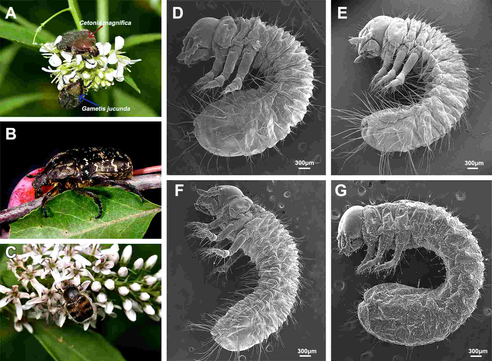
Larvae of the four species are typically scarabaeiform, bearing three pairs of thoracic legs on the C-shaped body (Fig. 1DG). The larval heads are heavily sclerotized, bearing a pair of antennae and a set of chewing mouthparts directed ventrally. The antennae are cylindrical and dumpy, consisting of four antennomeres.
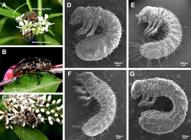
Adult and first instar larvae in habitus. (A) Adults of C. magnifica and G. jucunda in habitus; (B) Adult of P. brevitarsis in habitus; (C) Adult of L. succinctus in habitus; (D) Larva of C. magnifica; (E) Larva of G. jucunda; (F) Larva of P. brevitarsis; (G) Larva of L. succinctus.
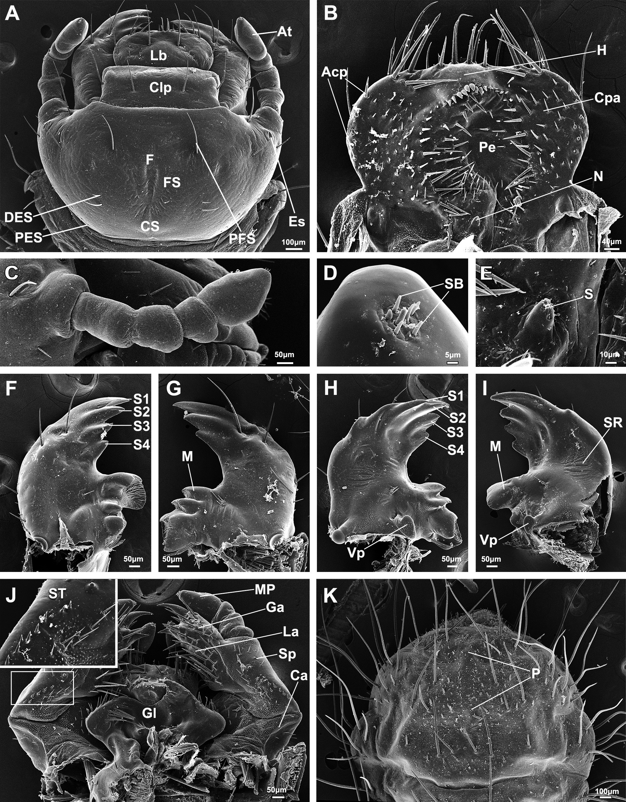
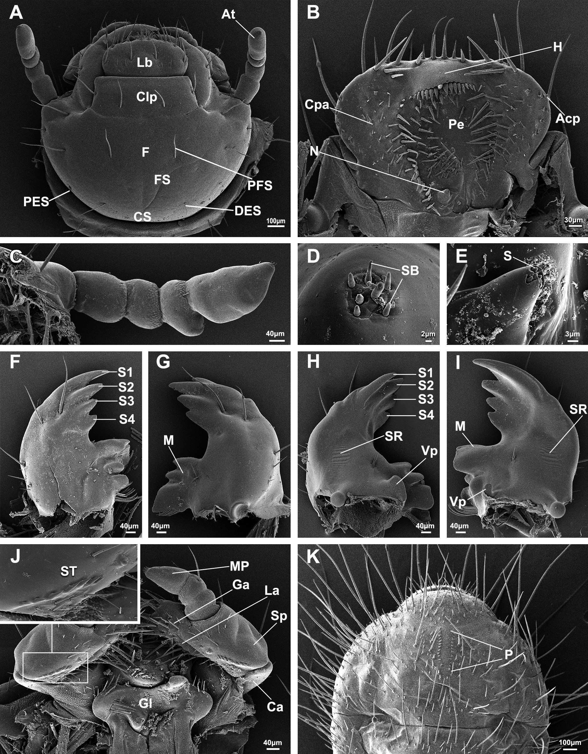
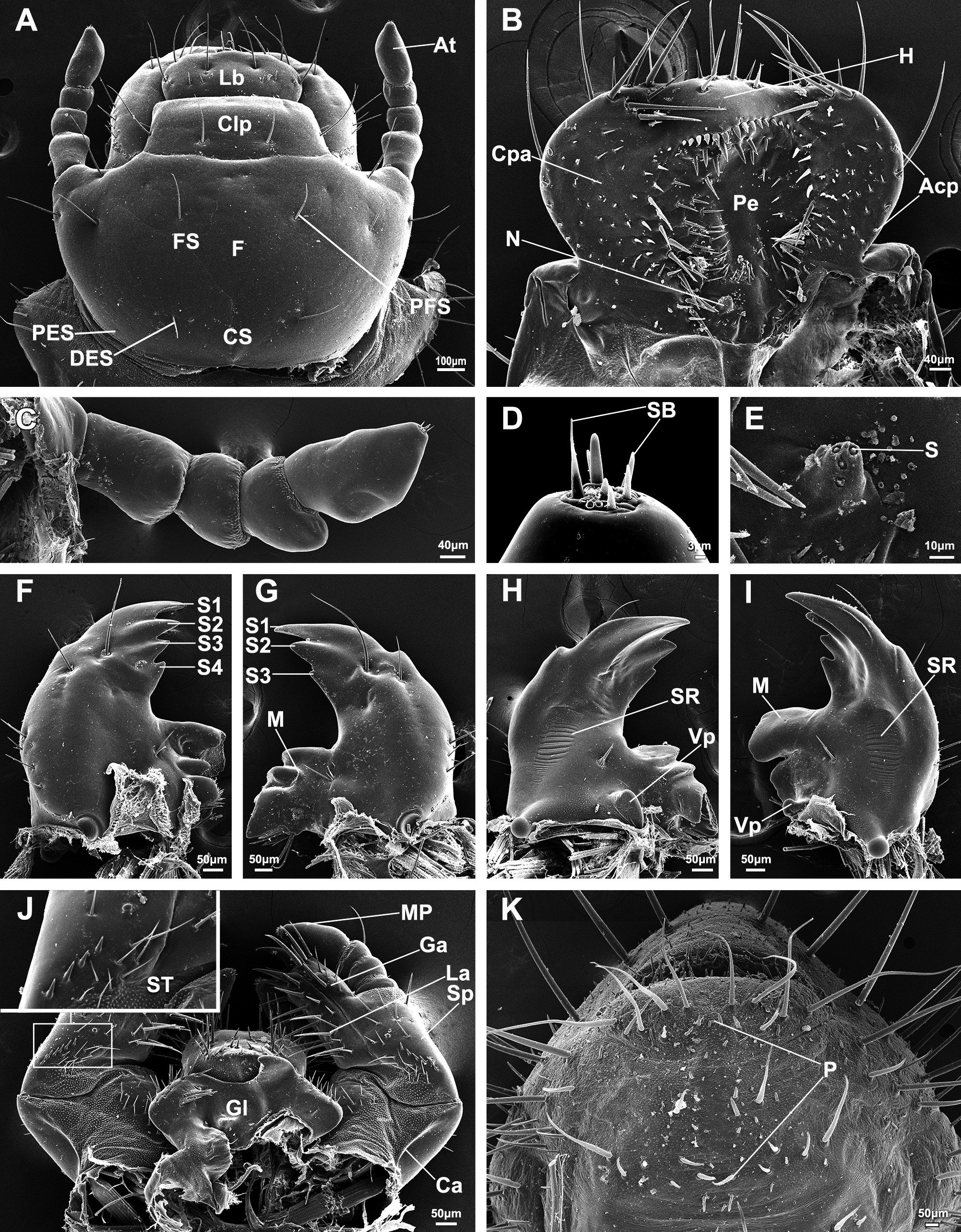
Mouthparts are of the biting-chewing type, composed of a labrum, a pair of mandibles, and a maxilla-labia-hypopharynx complex. The labrum is usually equipped symmetrically with two pairs of longer setae, articulated distally to the clypeus with two or three paired setae (Figs. 2A, 3A, 4 A and 5A). Epipharynx is generally fan-shaped or tri-lobed, bearing a haptomerum apically, paired chaetopariae and acanthopariae laterally and a haptolachus with a nesium proximally. Dexiotorma and laeotorma are usually transverse, sclerotized.
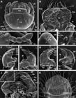
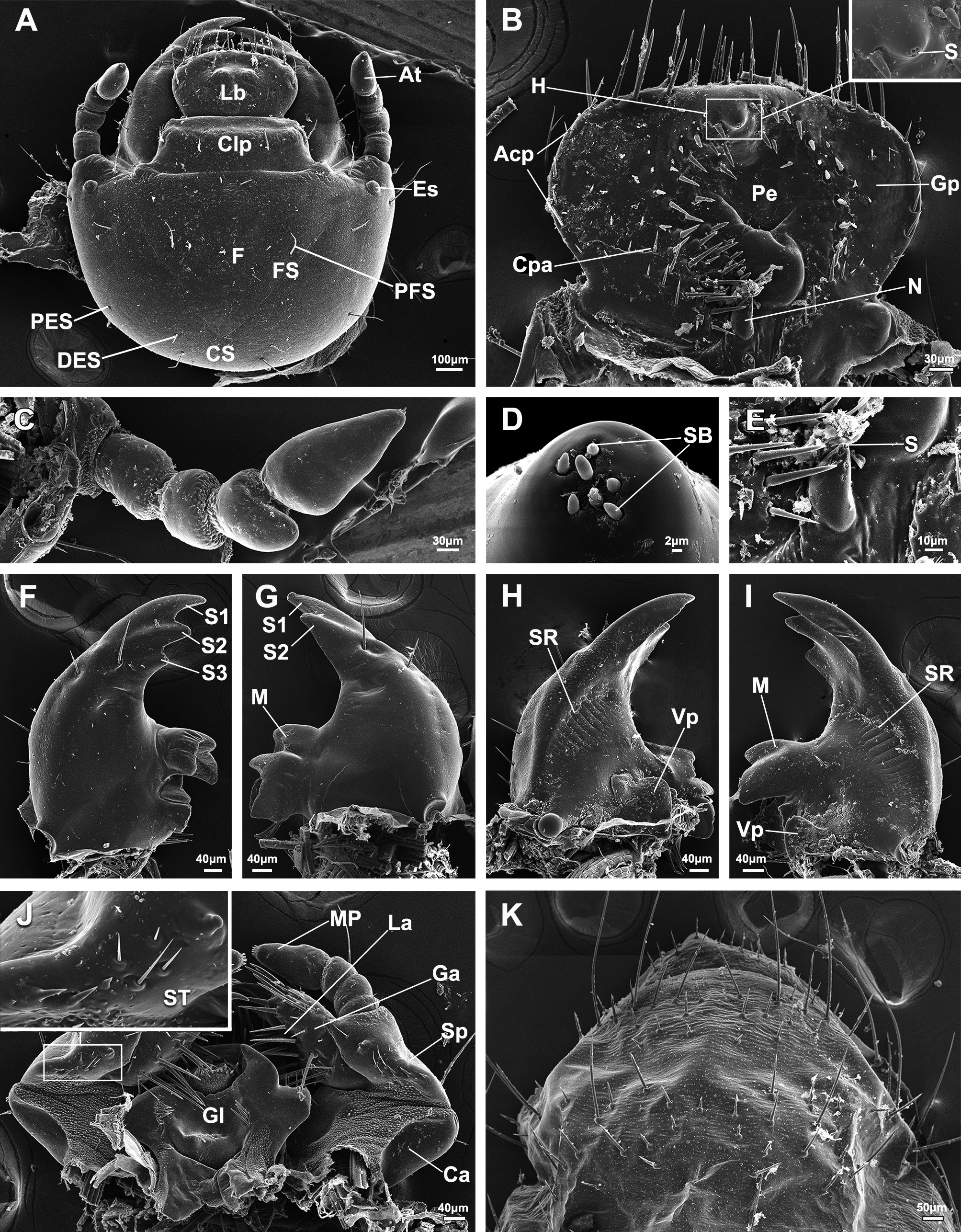
Detailed larval characters of C. magnifica. (A) Head; (B) Epipharynx; (C) Antenna lateral view; (D) Sensilla on the apex of antenna; (E) Magnification of nesium; (F) Left mandible, dorsal surface; (G) Right mandible, dorsal surface; (H) Right mandible, ventral surface; (I) Left mandible, ventral surface; (J) Maxillae and labia, ventral surface, insert showing the magnification of the stridulatory teeth; (K) Raster. Acp, acanthoparia; At, antenna; Clp, clypeus; Ca, cardo; Cpa, chaetoparia; CS, coronal suture; DES, dorsoepicranial seta; Es, eyespot; F, frons; FS, frontal suture; Ga, galea; Gl, glossa; H, haptomerum; La, lacinia; Lb, labrum; LP, labial palp; M, molar area; MP, maxillary palp; N, nesium; P, palidia; Pe, pedium; PES, posteroepicranial seta; PFS, posterofrontal seta; S, sensillum; S1–4, scissorial teeth; SB, sensillum basiconicum; Sp, stipes; ST, stridulatory teeth.
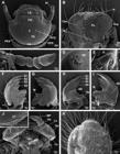
Detailed larval characters of G. jucunda. (A) Head; (B) Epipharynx; (C) Antenna lateral view; (D) Sensilla on the apex of antenna; (E) Magnification of nesium; (F) Left mandible, dorsal surface; (G) Right mandible, dorsal surface; (H) Right mandible, ventral surface; (I) Left mandible, ventral surface; (J) Maxillae and labia, ventral surface, insert showing the magnification of the stridulatory teeth; (K) Raster. Acp, acanthoparia; At, antenna; Clp, clypeus; Ca, cardo; Cpa, chaetoparia; CS, coronal suture; DES, dorsoepicranial seta; F, frons; FS, frontal suture; Ga, galea; Gl, glossa; H, haptomerum; La, lacinia; Lb, labrum; LP, labial palp; M, molar area; MP, maxillary palp; N, nesium; P, palidia; Pe, pedium; PES, posteroepicranial seta; PFS, posterofrontal seta; S, sensillum; S1–4, scissorial teeth; SB, sensillum basiconicum; Sp, stipes; ST, stridulatory teeth.
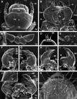
Detailed larval characters of P. brevitarsis. (A) Head; (B) Epipharynx; (C) Antenna lateral view; (D) Sensilla on the apex of antenna; (E) Magnification of nesium; (F) Left mandible, dorsal surface; (G) Right mandible, dorsal surface; (H) Right mandible, ventral surface; (I) Left mandible, ventral surface; (J) Maxillae and labia, ventral surface, insert showing the magnification of the stridulatory teeth; (K) Raster. Acp, acanthoparia; At, antenna; Clp, clypeus; Ca, cardo; Cpa, chaetoparia; CS, coronal suture; DES, dorsoepicranial seta; Es, eyespot; F, frons; FS, frontal suture; Ga, galea; Gl, glossa; H, haptomerum; La, lacinia; Lb, labrum; LP, labial palp; M, molar area; MP, maxillary palp; N, nesium; P, palidia; Pe, pedium; PES, posteroepicranial seta; PFS, posterofrontal seta; S, sensillum; S1–4, scissorial teeth; SB, sensillum basiconicum; Sp, stipes; ST, stridulatory teeth.
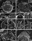
Detailed larval characters of L. succinctus. (A) Head; (B) Epipharynx, insert showing the magnification of the haptomerum; (C) Antenna lateral view; (D) Sensilla on the apex of antenna; (E) Magnification of nesium; (F) Left mandible, dorsal surface; (G) Right mandible, dorsal surface; (H) Right mandible, ventral surface; (I) Left mandible, ventral surface; (J) Maxillae and labia, ventral surface, insert showing the magnification of the stridulatory teeth; (K) Raster. Acp, acanthoparia; At, antenna; Clp, clypeus; Ca, cardo; Cpa, chaetoparia; CS, coronal suture; DES, dorsoepicranial seta; Es, eyespot; F, frons; FS, frontal suture; Ga, galea; Gl, glossa; Gp, gymnoparia; H, haptomerum; Lb, labrum; LP, labial palp; M, molar area; MP, maxillary palp; N, nesium; P, palidia; Pe, pedium; PES, posteroepicranial seta; PFS, posterofrontal seta; S, sensillum; S1–3, scissorial teeth; SB, sensillum basiconicum; Sp, stipes; ST, stridulatory teeth.
Paired mandibles are heavily sclerotized, asymmetric, differentiated into an apical incisor region and a basal molar region. The mandibles are generally triangular, bearing a notched precoila and a knob-like postcoila on the dorsal and ventral corners, respectively. Dorsal surface of mandibles with two setae near the proximal end of the scissorial area. The mandibles are equipped with elongate-oval stridulatory area on ventral surface, featuring stridulatory ridges.
Paired maxillae are symmetric, each consists of a cardo, a stipes, a fused galea and lacinia forming mala, and a three-segmented maxillary palp with the distal segment longest, bearing a cluster of sensilla. Maxillary stipes is furnished with stridulatory teeth on the dorsal surface and an additionally globose process.
Labium consists of a basal submentum, a mentum and an apical prementum, a pair of two-segmented labial palps. Hypopharynx is specialized into a hypopharyngeal sclerome with right lobe more prominent. The glossa bears two tufts of setae laterally and also two tufts of setae below the margin of lateral lobe on the dorsal surface.
Respiratory system is of the peripneustic type, bearing a pair of thoracic spiracles on prothorax, and eight pairs of abdominal spiracles (Fig. 1DG). All the spiracles are C-shaped, composed of numerous minute openings surrounding the oval bulla and a C-shaped spiracular slit. The openings of the C-shaped spiracles are different, with the prothoracic ones toward caudally, and the abdominal pairs toward cephalically. The prothoracic spiracles are slightly larger than the abdominal ones. The abdominal spiracles are similar in size.
The anal slit is transverse. The raster is situated at the sternite of the distal abdominal segment, well-developed and subdivided into various slender setae on the caudal margin.
Larval morphology of Cetonia magnifica Ballion, 1871
Dorsal body lengths are 3.85 ± 0.05 mm on average (Fig. 1D).
Head capsules are 1.39 ± 0.03 mm in width (Fig. 2A). Head is symmetrically furnished with 12 pairs of setae: two pairs on clypeus, one pair of posterofrontal setae (PFS), three pairs on the area surrounding the eyespot, two pairs of long dorsoepicranial setae (DES), four pairs short posteroepicranial setae on lateral along the coronal suture (PES). Cranium bears a pair of eyespots (Fig. 2A).
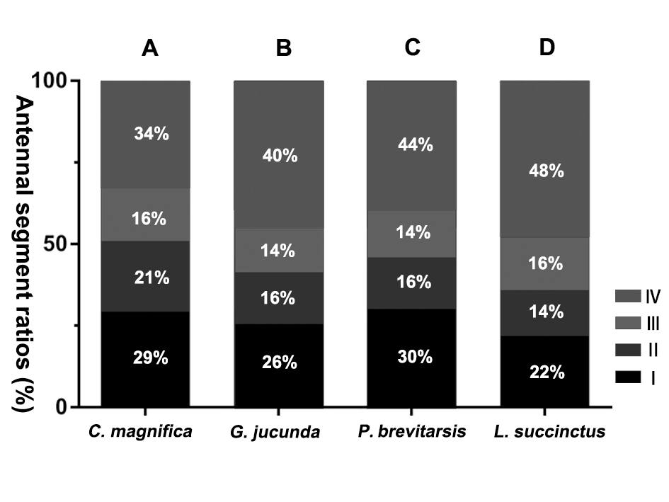
Antennomere IV is the longest with two dorsal and three ventral sensory spots, but longer than antennomeres II and III together (Figs. 2 C and 6A). The antennomere IV bears seven sensilla basiconica at apex (Fig. 2D).
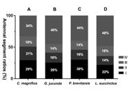
Segment ratios of larval Antennae. (A) C. magnifica; (B) G. jucunda; (C) P. brevitarsis; (D) L. succinctus. I–IV are the number of antennal segments.
The epipharynx is tri-lobed with a smooth haptomerum. The haptomerum possesses 15 setae arranged in an arc and raised irregular arrangement of four setae. The acanthopariae are equipped with six setae on the left side, and eight setae on the right. The nesium is coniform with four sensilla (Fig. 2B, E).
Paired mandibles usually possess a blunt bulge on the proximal lateral end of the scissorial area with two long setae ventrally (Fig. 2F, G). Incisors are sharp triangle-shaped, bearing four scissorial teeth on the left (Fig. 2F, I), and three scissorial teeth II–IV with an indistinctly tooth I on the right (Fig. 2G, H). Molar regions possess a larger blunt ventral protuberance and two molar teeth on left, and a smaller blunt ventral protuberance and three molar teeth on right. Stridulatory area bears 13 or 15 narrow ridges (Fig. 2H, I). Each mandible bears a tuft of molar setae on mesal surface of stridulatory area dorsally and ventrally. Lateral margins of mandibles usually bear four setae on each molar region (Fig. 2FI).
Paired maxillae each possess a cardo with 7 or 8 setae and a stipes with 19–22 setae dorsally. The stipes is notably equipped with six or seven acute stridulatory teeth and an additionally globose process dorsally. Mala has a large uncus at the apex and two subterminal unci fused at base. The stridulatory teeth are various in length, with the median setae comparative more prominent (Fig. 2J).
The raster is furnished with paired elongated palidia opened posteriorly and indistinctly closed anteriorly. Each palidium consisting of 17 or 18 short, acute pali. The anal slit is furnished with setae along the margin (Fig 2K).
Larval morphology of Gametis jucunda (Faldermann, 1835)
Dorsal body lengths are 3.92 ± 0.04 mm on average (Fig. 1E).
Head capsules are 1.17 ± 0.02 mm in width (Fig. 3A). Head is symmetrically furnished with 10 pairs of setae: two pairs on clypeus, one pair of posterofrontal setae (PFS), three pairs on the area surrounding the antennifer, one pair of long dorsoepicranial setae (DES), three pairs of short posteroepicranial setae on lateral along the coronal suture (PES). Eyespots are absent (Fig. 3A).
Antennomere IV is longest with two dorsal and three ventral sensory spots, but shorter than antennomeres II and III together (Figs. 3 C and 6B). The antennomere IV bears seven sensilla basiconica at apex (Fig. 3D).
The epipharynx is tri-lobed with a smooth haptomerum. The haptomerum possesses 18 setae arranged in an arc and raised irregular arrangement of seven setae. The acanthopariae are equipped with eight setae on the left side, and seven setae on the right. The nesium is coniform with four sensilla (Fig. 3B, E).
Paired mandibles usually possess a blunt bulge on the proximal lateral end of the scissorial area with two long setae ventrally (Fig. 3F, G). Incisors are sharp triangle-shaped, bearing four scissorial teeth on the left (Fig. 3F, I), and three scissorial teeth II–IV with an indistinctly tooth I on the right (Fig. 3G, H). Molar regions possess a larger blunt ventral protuberance and two molar teeth on left, and a smaller blunt ventral protuberance and three molar teeth on right. Stridulatory area bears 10 or 11 narrow ridges (Fig. 3H, I). Each mandible bears a tuft of molar setae on mesal surface of stridulatory area dorsally and ventrally. Lateral margins of mandibles usually bear five or six setae on each molar region (Fig. 3FI).
Paired maxillae each possess a cardo with 7 or 8 setae and a stipes with 14 or 15 setae dorsally. The stipes is notably equipped with seven or eight acute stridulatory teeth and an additionally globose process dorsally. Mala has large uncus at apex and two subterminal unci fused at base. The stridulatory teeth are various in length, with the median setae comparative more prominent (Fig. 3J).
The raster is furnished with elongated paired palidia opened posteriorly and indistinctly closed anteriorly. Each palidium consisting of 12 or 13 short, acute pali. The anal slit is furnished with setae along the margin (Fig. 3K).
Larval morphology of Protaetia brevitarsis (Lewis, 1879)
Dorsal body lengths are 4.56 ± 0.03 mm on average (Fig. 1F).
Head capsules are 1.21 ± 0.02 mm in width (Fig. 4A). Head is symmetrically furnished with 11 pairs of setae: two pairs on clypeus, one pair of posterofrontal setae (PFS), three pairs on the area surrounding the antennifer, one pair of long dorsoepicranial setae (DES), four pairs short posteroepicranial setae on lateral along the coronal suture (PES). Eyespots are absent (Fig. 4A).
Antennomere IV is longest with two dorsal and three ventral sensory spots, but longer than antennomeres II and III together (Figs. 4 C and 6C). The antennomere IV bears nine sensilla basiconica at apex (Fig. 4D).
The epipharynx is tri-lobed with a smooth haptomerum. The haptomerum possesses 16 setae arranged in an arc and raised irregular arrangement of seven setae. The acanthopariae are equipped with seven setae on the left side, and five setae on the right. The nesium is coniform with four sensilla (Fig. 4B, E).
Paired mandibles usually possess a blunt bulge on the proximal lateral end of the scissorial area with two long setae ventrally (Fig. 4F, G). Incisor regions are equipped with four apical scissorial teeth on left mandible, and three teeth on right mandible (Fig. 4FI). Molar regions possess a larger blunt ventral protuberance and two molar teeth on left, and a smaller blunt ventral protuberance and three molar teeth on right. Stridulatory area bears 20 or 21 narrow ridges (Fig. 4H, I). Each mandible bears a tuft of molar setae on mesal surface of stridulatory area dorsally and ventrally. Lateral margins of mandibles usually bear five or six setae on each molar region (Fig. 4FI).
Paired maxillae each possess a cardo with 9 or 10 setae and a stipes with 22 or 23 setae dorsally. The stipes is notably equipped with four to seven acute stridulatory teeth and an additionally globose process dorsally. Mala has large uncus at apex and two subterminal unci fused at base. The stridulatory teeth are various in length, with the median setae comparative more prominent (Fig. 4J).
The raster is furnished with paired elongated palidia opened posteriorly and indistinctly closed anteriorly. Each palidium consisting of 9–13 short, acute pali. The anal slit is furnished with setae along the margin (Fig. 4K).
Larval morphology of Lasiotrichius succinctus (Pallas, 1781)
Dorsal body lengths are 3.98 ± 0.03 mm on average (Fig. 1G).
Head capsules are 1.17 ± 0.03 mm in width (Fig. 5A). Head is symmetrically furnished with 13 pairs of setae: two pairs on clypeus, one pair of anterofrontal setae (AFS), two pairs of posterofrontal setae (PFS), five pairs on the area surrounding the eyespot, two pairs of long dorsoepicranial setae (DES), one pair posteroepicranial setae on lateral along the head capsule (PES). Cranium bears a pair of eyespots (Fig. 5A).
Antennomere IV is longest with one dorsal and two ventral sensory spots, but longer than antennomeres II and III together (Figs. 5 C and 6D). The antennomere IV bears seven sensilla basiconica at apex (Figs. 5D). The epipharynx is fan-shaped with a bulgy haptomerum. The haptomerum possesses four sensilla. The acanthopariae are equipped with 11 setae on the left side, and 13 setae on the right. The nesium is coniform with five sensilla (Fig. 5B, E).
Paired mandibles are generally triangular with two long setae on the proximal lateral end of the scissorial area ventrally (Fig. 5F, G). Incisor regions are equipped with three apical scissorial teeth on left mandible, and two teeth on right mandible (Fig. 5FI). Molar regions possess a larger blunt ventral protuberance and two molar teeth on left, and a smaller blunt ventral protuberance and three molar teeth on right. Stridulatory area bears 19 to 21 narrow ridges (Fig. 5H, I). Each mandible bears a tuft of molar setae on mesal surface of stridulatory area dorsally and ventrally. Lateral margins of mandibles usually bear two or three setae on each molar region (Fig. 5FI).
Paired maxillae each possess a cardo with 2 or 3 setae and a stipes with 4 or 5 setae dorsally. The stipes is notably equipped with three or four acute stridulatory teeth and an additionally globose process dorsally. Mala has a large uncus at apex and two subterminal unci fused at base. The stridulatory teeth are various in length, with the median one comparative more prominent (Fig. 5J).
The raster is furnished with 49–53 short setae and 28–32 long setae. palidium is absent. The anal slit is furnished with sparse setae along the margin (Fig. 5K).
Discussions
This study provided the first morphological comparison between the first instar larvae of C. magnifica, G. jucunda, P. brevitarsis and L. succinctus using SEM. The larvae of four species share the following features: i) the antennomere IV is the longest; ii) the nesium possesses minute sensilla anterior; iii) the mandibles are equipped with elongate-oval stridulatory area with ridges; iv) the maxillary stipes is furnished with stridulatory teeth on the dorsal surface with an additionally globose process. On the other hand, the larvae of the four species exhibit distinctly morphological differences, such as the eyespot of cranium, the number of sensilla on nesium, the prominent process on haptomerum, the scissorial teeth, and the palidia on raster (Table 2).
Haptomerum is a critical character for larval taxonomy in Scarabaeidae (Hayes, 1928; Böving, 1936; Ritcher, 1966; Zhang, 1984). The haptomerum of saprophagous larvae in Cetoniinae and Euchirinae are typically smooth with some setae (Šípek et al., 2011; Wen et al., 2024); phytophagous larvae in Sericinae, Melolonthinae and part of Rutelinae are usually protuberant with varying numbers of heli (Jia et al., 2023; Cao et al., 2024; Sampaio et al., 2024). In addition, the coprophagous larvae in Aphodiinae and Scarabaeinae are also smooth, but featuring diverse forms of phobae (Edmonds and Halffter, 2008; Li et al., 2019). In this study, the first instar larvae of L. succinctus exhibits a prominent process and other three Cetoniini larvae are smooth on haptomerum, consistent with their third instar larvae described by (Zhang, 1984; Sawada, 1991; Shabalin and Kalinina, 2006; Fang et al., 2000; Dong et al., 2021). The prominent process without heli is a suitable character for determining Trichiini larvae in Cetoniinae.
Paired mandibles are prominent chewing structures for larval development that are morphologically related to feeding habits (Krings et al., 2024; Hummig-Neto et al., 2025). The mandibular incisors typically varies: blade-like and specialized to forma cutting edge in some of phytophagous Rutelinae, Sericinae and Melolonthinae (Ernesto and Carrillo, 2010; Šípek and Ahrens, 2011; Neita-Moreno and Morón, 2017), but curved, dentate and asymmetrical in other saproxylic, coprophagous and saprophagous species of Scarabaeidae (Ratcliffe and Skelley, 2011; Qu et al., 2019; Sun et al., 2024). Within Cetoniinae, the mandibular incisors are typically asymmetrical incisors with counts of 4:4, 4:3, or 3:2 (Micó and Galante, 2003; Grebennikov and Scholtz, 2004; Sousa et al., 2018). In this study, the first instar larval incisors of all four Cetoniinae are consistent with some previously published descriptions of final-instar larvae (Zhang, 1984; Sawada, 1991; Dong et al., 2021). The number of incisors is not a reliable character for larval identification within Cetoniinae.
The stridulatory teeth on maxillae have been extensively described in Scarabaeidae larvae (Hayes, 1929; Zhang, 1984). However, additionally processes arranged with the stridulatory teeth are typically found only in Cetoniinae, Rutelinae and Dynastinae (Orozco and Pardo-Locarno, 2004; Pardo et al., 2021; Long et al., 2024). These additional processes vary in shape: Cetoniinae larvae are globose (Vendl et al., 2014; Shabalin, 2019), larvae in Rutelinae and Dynastinae are truncate (Micó et al., 2003; Sun et al., 2024; Jiang et al., 2025). Our study found that the first instar larvae of all four Cetoniinae exhibit the additionally globose processes, consistent with previous descriptions of some third-instar larvae (Zhang, 1984; Sawada, 1991, Dong et al., 2021), indicating that the additional process may not change with the larval instars. The additional globose process can therefore serve as an additional criterion for Cetoniinae larval identification.
Raster of the white grubs exhibits diverse morphological features, primarily in the arrangement of palidia, hamate setae, and barbula (Böving, 1936; Medvedev, 1952; Zhang, 1984). The arrangements of palidia are relatively consistent at the tribal or subfamily level, appearing as curved transverse combs in Sericinae larvae (Šípek and Ahrens, 2011), paired mustache-like diverging posteriorly in some species of Melolonthinae larvae (Jia et al., 2021; Zhang et al., 2024), and part of Cetoniinae and Rutelinae larvae (Vondráček et al., 2018; Sun et al., 2024). The three first-instar larvae of Cetoniini, C. magnifica, G. jucunda and P. brevitarsis both exhibit paired elongated palidia opened posteriorly and indistinctly closed anteriorly, Trichiini larvae L. succinctus lack palidia and possess barbula, consistent with previous descriptions of the final instar larvae, though palidia of other three final instar larvae has distinctly closed anteriorly (Zhang, 1984; Fang et al., 2000; Shabalin and Kalinina, 2006; Dong et al., 2021). The raster can serve as a determining criterion in some Cetoniini and Trichiini larvae.
Most scarab larvae are usually devoid of eyespots (Ritcher, 1966; Grebennikov and Scholtz, 2004; Dong et al., 2021), but the eyespots are only present in some species, such as Adoretus tenuimaculatus (Adoretini), and some other Cetoniinae and Rutelinae (Šípek et al., 2009; Vendl et al., 2014; Fang et al., 2018; Carvalho et al., 2019). Notably, the eyespots are also observed in the first-instar Cetoniini larvae of C. magnifica that absent in third-instar larvae (Fang et al., 2000), but the first and third instar larvae L. succinctus all possess eyespots (Dong et al., 2021). The eyespots of first-instar G. jucunda and P. brevitarsis larvae are also absent, consistent with previous descriptions of their final instar larvae (Zhang, 1984; Shabalin and Kalinina, 2006). Therefore, the eyespots are unstable for determining tribal larvae in Cetoniinae.
In this study, SEM was used to better characterize the tiny features of first-instar larvae in Cetoniinae, particularly the sensilla of nesium, which have been inadequately described using LM (Vendl and Šípek, 2016; Sousa and Fuhrmann, 2020). Moreover, based on a systematic comparison of the ultrastructure of first-instar larvae from four species of flower chafers, certain common and distinctive characteristics among these Cetoniinae species can serve as important foundational data for the identification of Cetoniinae larvae.
Acknowledgments
This research was financially supported by National Natural Science Foundation of China (grant nos. 32370470 and 31702036), Scientific Research Fund of Liaoning Provincial Education Department (JYTQN2024002), and Scientific Research Foundation for the Introduced Talent of Shenyang Agricultural University (grant no. 880417008).
Data statement
The entire dataset supporting the results of this study was published in the article itself.
References
-
Alpert, G. D., Ritcher, P., 1975. Notes on the life cycle and myrmecophilous adaptations of Cremastocheilus armatus (Coleoptera: scarabaeidae). Psyche (Camb., Mass.) 82 (3-4), 283-291. https://doi.org/10.1155/1975/79470
» https://doi.org/10.1155/1975/79470 - Ballion, E. V., 1871. Eine Centurie neuer Käfer aus der Fauna des russischen Reiches. Bull. Soc. Imp. Naturalistes Moscou 43 (4), 320-353.
- Böving, A. G., 1936. Description of the larva of Plectris aliena Chapin and explanation of new terms applied to the epipharynx and raster. Proc. Entomol. Soc. Wash. 38 (8), 169-185.
-
Cao, W. J., Chen, Y. N., Zhou, R. J., Jiang, L., 2024. Cross-instar larval identification leads divergent decision on pest management: an example from white grubs on peanuts. J. Asia Pac. Entomol. 27 (3), 102283. https://doi.org/10.1016/j.aspen.2024.102283
» https://doi.org/10.1016/j.aspen.2024.102283 -
Carvalho, T., Duarte, P., Fuhrmann, J., Grossi, P., 2019. Description of the last larval instar and pupa of Chlorota paulistana Ohaus, 1912 (Coleoptera: Melolonthidae: Rutelinae: Rutelini). Rev. Bras. Entomol. 63 (3), 245-249. https://doi.org/10.1016/j.rbe.2019.05.002
» https://doi.org/10.1016/j.rbe.2019.05.002 -
Cazier, M. A., Statham, M., 1962. The behavior and habits of the myrmecophilous scarab Cremastocheilus Stathamae Cazier with notes on other species (Coleoptera: scarabaeidae). J. N.Y. Entomol. Soc. 70 (3), 125-149. Available in: http://www.jstor.org/stable/25005817 (accessed 01 October 2025).
» http://www.jstor.org/stable/25005817 -
Chen, Y. N., Ying, M., Lu, C., Jiang, L., 2025. Consumer or decomposer? Behavioral and morphological diagnosis of white grubs. Ecol. Evol. 15 (8), e71925. https://doi.org/10.1002/ece3.71925
» https://doi.org/10.1002/ece3.71925 -
Dent, D., Binks, R. H., 2020. Insect Pest Management, 3rd ed. Cabi, Wallingford.. https://doi.org/10.1079/9781789241051.0000
» https://doi.org/10.1079/9781789241051.0000 -
Dong, X. M., Jia, Z. C., Jiang, L., 2021. Morphological description of the third instar larva of Lasiotrichius succinctus hananoi (Sawada) (Coleoptera: Scarabaeidae: Cetoniinae: Trichiini), using scanning electron microscopy. Microsc. Res. Tech. 84 (2), 368-375. https://doi.org/10.1002/jemt.23594
» https://doi.org/10.1002/jemt.23594 - Edmonds, W., Halffter, G., 2008. Taxonomic review of immature dung beetles of the subfamily Scarabaeinae (Coleoptera: scarabaeidae). Syst. Entomol. 3 (4), 307-331.
-
Ernesto, C., Carrillo, R., 2010. Description of the Larvae of Phytholaema herrmanni Germain and Phytholaema dilutipes (Fairmaire and Germain) (Coleoptera: Scarabaeidae: Melolonthinae). Coleopt. Bull. 64 (2), 141-147. https://doi.org/10.1649/072.064.0210
» https://doi.org/10.1649/072.064.0210 - Faldermann, F., 1835. Coleopterorum ab Illustrissimo Bungio in China boreali, Mongolia et Montibus Altaicis Collectorum, nec non ab Illustrissimo Turczaninoffio et Stschukino e Provinci Irkutzk Missorum Illustrationes. Caesareae Academiae Scientiarum Impress, Petrópolis.
-
Fang, H., Li, C., Jiang, L., 2018. Morphology of the immature stages of Adoretus tenuimaculatus Waterhouse, 1875 (Coleoptera: Scarabaeidae: Rutelinae: Adoretini). J. Asia Pac. Entomol. 21 (4), 1159-1164. https://doi.org/10.1016/j.aspen.2018.09.001
» https://doi.org/10.1016/j.aspen.2018.09.001 - Fang, H., Zhang, Z. L., Wang, Y., 2000. Study on species of scarabs larvae IV. Shenyang Nongye Daxue Xuebao (05), 511-514.
-
Gasca-Álvarez, H. J., Woodruff, R. E., López, C. D., 2019. Description of the larvae of Cotinis aliena Woodruff, 2008 (Coleoptera: Scarabaeidae: Cetoniinae: Gymnetini), with a key to the known larvae of New World Gymnetini. Zootaxa 4585 (1), 142-150. https://doi.org/10.11646/zootaxa.4585.1.8
» https://doi.org/10.11646/zootaxa.4585.1.8 -
Grebennikov, V. V., Scholtz, C. H., 2004. The basal phylogeny of Scarabaeoidea (Insecta: Coleoptera) inferred from larval morphology. Invertebr. Syst. 18 (3), 321-348. https://doi.org/10.1071/IS03013
» https://doi.org/10.1071/IS03013 -
Hayes, W. P., 1928. The epipharynx of lamellicorn larvae (Coleop.), with a key to common genera. Ann. Entomol. Soc. Am. 21 (2), 282-306. https://doi.org/10.1093/aesa/21.2.282
» https://doi.org/10.1093/aesa/21.2.282 - Hayes, W. P., 1929. Morphology, Taxonomy, and Biology of Larval Scarabaeoidea. University of Illinois, Urbana, IL.
-
Hummig-Neto, E., Rafael, J., Barros, L., Ferreira-Keppler, R., 2025. Taxonomy of the immature stages of Oxelytrum cayennense (Sturm, 1826) (Coleoptera: Staphylinidae: Silphinae) of forensic importance from Central Amazon, Amazonas, Brazil. Rev. Bras. Entomol. 69 (3), e20250024. https://doi.org/10.1590/1806-9665-rbent-2025-0014
» https://doi.org/10.1590/1806-9665-rbent-2025-0014 -
Ibarra-Polesel, M. G., Neita-Moreno, J. C., Larrea, D. D., Damborsky, M. P., 2017. Description of the larva and pupa of Neocorvicoana reticulata (Kirby, 1819)(Coleoptera: Scarabaeidae: Cetoniinae: Gymnetini). Zootaxa 4358 (3), 430-440. https://doi.org/10.11646/zootaxa.4358.3.2
» https://doi.org/10.11646/zootaxa.4358.3.2 -
Ibarra-Polesel, M. G., Valle, N. G., Cave, R. D., Damborsky, M. P., 2022. Descriptions of the larva and pupa of Gymnetis pudibunda Burmeister, 1866 (Coleoptera: Scarabaeidae: Cetoniinae: Gymnetini), with notes on natural history and a key to the known larvae of New World Gymnetini. J. Nat. Hist. 56 (13-16), 969-987. https://doi.org/10.1080/00222933.2022.2080607
» https://doi.org/10.1080/00222933.2022.2080607 -
Jia, Z. C., Dong, H., Li, Y. T., Zhao, X. X., Jiang, L., 2023. Morphology of a grass‐feeding white grub Apogonia cupreoviridis (Coleoptera: Scarabaeidae: Melolonthinae: Diplotaxini) using scanning electron microscopy. Acta Zool. 105 (2), 225-233. https://doi.org/10.1111/azo.12460
» https://doi.org/10.1111/azo.12460 -
Jia, Z. C., Fang, H., Jiang, L., 2021. Morphological description of the white grub Melolontha incana (Coleoptera: Scarabaeidae: Melolonthinae Melolonthini). Microsc. Res. Tech. 84 (5), 921-928. https://doi.org/10.1002/jemt.23653
» https://doi.org/10.1002/jemt.23653 -
Jiang, L., Hua, B. Z., 2015. Morphological comparison of the larvae of Panorpa obtusa Cheng and Neopanorpa lui Chou & Ran (Mecoptera: panorpidae). Zool. Anz. 225, 62-70. https://doi.org/10.1016/j.jcz.2015.02.004
» https://doi.org/10.1016/j.jcz.2015.02.004 -
Jiang, L., Wang, Y. L., Wu, Y. H., Zhang, Z. H., 2025. Larval morphology of Eophileurus chinensis (Coleoptera: Scarabaeidae: Dynastinae), with discussions on their living strategies. Acta Zool. azo.12547. https://doi.org/10.1111/azo.12547
» https://doi.org/10.1111/azo.12547 - Kouklík, O., 2017. Larvální Morfologie Tribu Goliathini (Coleoptera: Cetoniinae) a Její Využití Při Studiu Fylogeneze Zlatohlávků. Univerzita Karlova, Prague.
-
Krings, W., Below, P., Gorb, S. N., 2024. Mandible mechanical properties and composition of the larval Glossosoma boltoni (Trichoptera, Insecta). Sci. Rep. 14 (1), 4695. https://doi.org/10.1038/s41598-024-55211-5
» https://doi.org/10.1038/s41598-024-55211-5 -
Lawrence, J. F., Ślipiński, A., Seago, A. E., Thayer, M. K., Newton, A. F., Marvaldi, A. E., 2011. Phylogeny of the Coleoptera based on morphological characters of adults and larvae. Ann. Zool. 61 (1), 1-217. https://doi.org/10.3161/000345411X576725
» https://doi.org/10.3161/000345411X576725 -
Lewis, G., 1879. LIII.—On certain new species of Coleoptera from Japan. Annu. Mag. Nat. Hist. 4 (24), 459-467. https://doi.org/10.1080/00222937908679867
» https://doi.org/10.1080/00222937908679867 -
Li, C., Lu, Y., Fang, H., Jiang, L., 2019. Morphology of the third instar larva of Colobopterus quadratus (Coleoptera: Scarabaeidae: Aphodiinae) using scanning electron microscopy. Microsc. Res. Tech. 82 (8), 1372-1379. https://doi.org/10.1002/jemt.23289
» https://doi.org/10.1002/jemt.23289 -
Liu, J., Wang, Z., Shi, C., Lu, W., Pang, J., Ma, D., Bai, M., Li, J., 2025. How did the white-spotted flower chafer (Protaetia brevitarsis) come to Xinjiang? J. Pest Sci. 98 (4), 2217-2234. https://doi.org/10.1007/s10340-025-01934-x
» https://doi.org/10.1007/s10340-025-01934-x -
Long, T. T., Jiang, R., Zhu, W., Long, J., Chen, X., 2024. Biology of Callistethus plagiicollis (Coleoptera: Scarabaeoidea: Rutelinae) and comparative morphology of its larvae. Microsc. Res. Tech. 87 (8), 1693-1703. https://doi.org/10.1002/jemt.24555
» https://doi.org/10.1002/jemt.24555 - Medvedev, S. I., 1952. Larvae of the Scarabaeoidea (Coleoptera) of the Soviet Union. USSR Academy of Sciens, Moscow. Keys to the Identification of the Fauna of USSR 47.
-
Mendoza, A. E. G., Cobián, C. V., Ratcliffe, B. C., 2024. Description of the immature stages of Gymnetis merops Ratcliffe (Coleoptera: Scarabaeidae: Cetoniinae: Gymnetini). Coleopt. Bull. 78 (1), 99-107. https://doi.org/10.1649/0010-065X-78.1.99
» https://doi.org/10.1649/0010-065X-78.1.99 -
Micó, E., Galante, E., 2003. Biology and new larval descriptions for three Cetoniine beetles (Coleoptera: Scarabaeidae: Cetoniinae: Cetoniini: Cetoniina, Leucocelina). Ann. Entomol. Soc. Am. 96 (2), 95-106. https://doi.org/10.1603/0013-8746(2003)096[0095:BANLDF]2.0.CO;2
» https://doi.org/10.1603/0013-8746(2003)096[0095:BANLDF]2.0.CO;2 -
Micó, E., Morón, M., Galante, E., 2003. New larval descriptions and biology of some New World Anomalini beetles (Scarabaeidae: rutelinae). Ann. Entomol. Soc. Am. 96 (5), 597-614. https://doi.org/10.1603/0013-8746(2003)096[0597:NLDABO]2.0.CO;2
» https://doi.org/10.1603/0013-8746(2003)096[0597:NLDABO]2.0.CO;2 -
Micó, E., Moron, M. A., Šípek, P., Galante, E., 2008. Larval morphology enhances phylogenetic reconstruction in Cetoniidae (Coleoptera: Scarabaeoidea) and allows the interpretation of the evolution of larval feeding habits. Syst. Entomol. 33 (1), 128-144. https://doi.org/10.1111/j.1365-3113.2007.00399.x
» https://doi.org/10.1111/j.1365-3113.2007.00399.x -
Micó, E., Smith, A. B. T., Morón, M. A., 2000. New larval descriptions for two species of Euphoria Burmeister (Coleoptera: Scarabaeidae: Cetoniinae: Cetoniini: Euphoriina) with a key to the known larvae and a review of the larval biology. Ann. Entomol. Soc. Am. 93 (4), 795-801. https://doi.org/10.1603/0013-8746(2000)093[0795:NLDFTS]2.0.CO;2
» https://doi.org/10.1603/0013-8746(2000)093[0795:NLDFTS]2.0.CO;2 -
Neita-Moreno, J., Morón, M., 2017. Description of immature stages of Platycoelia valida Burmeister, 1844 (Coleoptera: Melolonthidae: Rutelinae: Anoplognathini). Rev. Bras. Entomol. 61 (4), 359-364. https://doi.org/10.1016/j.rbe.2017.07.002
» https://doi.org/10.1016/j.rbe.2017.07.002 -
Nogueira, G., Morón, M., Fierros-López, H., Navarrete-Heredia, J., 2009. The immature stages of Neoscelis Dohrni (Westwood) (Coleoptera: Scarabaeidae: Cetoniinae: Goliathini) with notes on adult behavior. Coleopt. Bull. 58 (2), 171-183. https://doi.org/10.1649/604
» https://doi.org/10.1649/604 -
Orozco, J., Pardo-Locarno, L. C., 2004. Description of immature stages of three species of American Cetoniinae (Coleoptera: Scarabaeidae: Cetoniinae). Zootaxa 769 (1), 1-14. https://doi.org/10.11646/zootaxa.769.1.1
» https://doi.org/10.11646/zootaxa.769.1.1 - Pallas, P. S., 1781. Icones Insectorvm: Praesertim Rossiae Sibiriaeqve Pecvliarivm. Svmptv Wolfgangi Waltheri, Erlangen.
-
Pardo, L., Fonseca, L., Serna, A., 2021. Morfología larval, clave para inmaduros y ciclos de vida de Dynastes hercules L (D. h. lichyi Lachaume, D. h. occidentalis Lachaume) y de Dynastes neptunus Quenzel (Coleoptera: Melolonthidae: Dynastini) en Colombia. Rev. Novedades Colomb. 16, 9-44. https://doi.org/10.47374/novcol.2021.v16.2006
» https://doi.org/10.47374/novcol.2021.v16.2006 -
Perissinotto, R., Clennell, L., 2021. Census of the fruit and flower chafers (Coleoptera, Scarabaeidae, Cetoniinae) of the Macau SAR, China. ZooKeys 1026, 17-43. https://doi.org/10.3897/zookeys.1026.60036
» https://doi.org/10.3897/zookeys.1026.60036 -
Perissinotto, R., Orozco, J., 2013. Eudicella trimeni Janson, 1884 (Coleoptera: Scarabaeidae: Cetoniinae: Goliathini): description of larva with notes on conservation status, biology and taxonomy: Insecta: Coleoptera. Afr. Invertebr. 54 (2), 417-426. https://doi.org/10.5733/afin.054.0222
» https://doi.org/10.5733/afin.054.0222 -
Perissinotto, R., Sípek, P., Ball, J. B., 2014. Description of adult and third instar larva of Trichostetha curlei sp. n. (Coleoptera, Scarabaeidae, Cetoniinae) from the Cape region of South Africa. ZooKeys 428, 41-56. https://doi.org/10.3897/zookeys.428.7855
» https://doi.org/10.3897/zookeys.428.7855 -
Puker, A., Ad’vincula Miranda, H., Korasaki, V., Ferreira, F. N., Orozco, J., 2014. Biodiversity of Cetoniinae beetles (Coleoptera: Scarabaeidae) in introduced and native habitats in the Brazilian Atlantic Forest. Entomol. Sci. 17 (3), 309-315. https://doi.org/10.1111/ens.12069
» https://doi.org/10.1111/ens.12069 -
Puker, A., Rosa, C. S., Orozco, J., Solar, R. R., Feitosa, R. M., 2015. Insights on the association of American Cetoniinae beetles with ants. Entomol. Sci. 18 (1), 21-30. https://doi.org/10.1111/ens.12085
» https://doi.org/10.1111/ens.12085 -
Qu, Z. F., Jia, Z. C., Jiang, L., 2019. Description of the third instar larva of the stag beetle Prismognathus dauricus Motschulsky, 1860 (Coleoptera: Scarabaeoidea: Lucanidae) using electron microscopy. Micron 120, 10-16. https://doi.org/10.1016/j.micron.2019.02.001
» https://doi.org/10.1016/j.micron.2019.02.001 -
Ratcliffe, B., Skelley, P., 2011. Description of the larva of Homophileurus integer (Burmeister, 1847) (Coleoptera: Scarabaeidae: Dynastinae: Phileurini), with notes on biology and a key to the known larvae of New World Phileurini. Coleopt. Bull. 65 (3), 297-304. https://doi.org/10.1649/072.065.0311
» https://doi.org/10.1649/072.065.0311 - Ritcher, P. O., 1966. White Grubs and Their Allies. A Study of North American Scarabaeoid Larvae. Oregon State University Press, Corvallis.
-
Rodrigues, S. R., Garcia, F. P., Falco, J. S., Morón, M. A., 2016. Biology and description of immature stages of Gymnetis rufilateris (Illiger, 1800)(Coleoptera: Cetoniidae: Cetoniinae). Biota Neotrop. 16 (3), e20140176. https://doi.org/10.1590/1676-0611-BN-2014-0176
» https://doi.org/10.1590/1676-0611-BN-2014-0176 -
Sampaio, A., Bento, M., Da Fonseca, C. R. V., Fuhrmann, J., 2024. Description of the third instar larva and pupa of Cnemida leprieuri Arrow, 1899 and Cnemida retusa (Fabricius, 1801)(Coleoptera: Scarabaeidae: Rutelinae). Ann. Zool. 74 (4), 603-625. https://doi.org/10.3161/00034541ANZ2024.74.4.006
» https://doi.org/10.3161/00034541ANZ2024.74.4.006 - Sawada, H., 1991. Morphological and Phylogenetical Study on the Larvae of Pleurostict Lamellicornia in Japan. Tokyo University of Agriculture Press, Tokyo.
-
Shabalin, S., 2019. The first description of the larva in the genus Clinterocera Motschulsky (Coleoptera, Scarabaeidae: cetoniinae). Entomol. Rev. (Engl. Transl.) 99 (3), 403-406. https://doi.org/10.1134/S0013873819030126
» https://doi.org/10.1134/S0013873819030126 -
Shabalin, S. A., Bezborodov, V. G., 2009. Larvae of scarab beetles of the subfamily Trichiinae (Coleoptera, Scarabaeidae) from the Russian Far East. Entomol. Rev. (Engl. Transl.) 89 (9), 1131-1136. https://doi.org/10.1134/S0013873809090139
» https://doi.org/10.1134/S0013873809090139 - Shabalin, S. A., Kalinina, O. I., 2006. On the ecology and biology of the Far Eastern red deer – Oxycetonia jucunda (Faldermann, 1835) (Coleoptera, Scarabaeidae: Cetoniinae) in Primorsky Krai.Readings in Memory of A. I. Kurentsov (17), 89-93.
-
Silva, R. S., Amaral, E. A., Fuhrmann, J., Rodrigues, S. R., 2023. Antennal sensilla of two species of Gymnetis MacLeay, 1819 (Coleoptera: Scarabaeidae: Cetoniinae). Rev. Bras. Entomol. 67 (1), 1806-9665. https://doi.org/10.1590/1806-9665-rbent-2022-0077
» https://doi.org/10.1590/1806-9665-rbent-2022-0077 -
Sims, S., Shapiro, A., 2014. Interspecific variation in size, diapause intensity, and moisture responses of first-instar Speyeria (Lepidoptera: Nymphalidae) larvae. Ann. Entomol. Soc. Am. 107 (1), 163-169. https://doi.org/10.1603/AN13099
» https://doi.org/10.1603/AN13099 - Šípek, P., 2010. Immature Stages of the Pleurostict Scarab Beetles (Coleoptera: Scarabaeidae: Pleurosticti): Morphology, Biology and Phylogenetic Implication. Charles University, Prague.
-
Šípek, P., Ahrens, D., 2011. Inferring larval taxonomy and morphology in Maladera species (Coleoptera: Scarabaeidae: Sericini) using DNA taxonomy tools. Syst. Entomol. 36 (4), 628-643. https://doi.org/10.1111/j.1365-3113.2011.00582.x
» https://doi.org/10.1111/j.1365-3113.2011.00582.x -
Šípek, P., Gill, B., Grebennikov, V., 2009. Afromontane Coelocorynus (Coleoptera: Scarabaeidae: Cetoniinae): Larval descriptions, biological notes and phylogenetic placement. Eur. J. Entomol. 106 (1), 95-106. https://doi.org/10.14411/eje.2009.014
» https://doi.org/10.14411/eje.2009.014 -
Šípek, P., Jansta, P., Král, D., 2011. Immature stages of Euchirinae (Coleoptera Cheirotonus and Propomacrus: Scarabaeoidea): genera with comments on their phylogeny based on larval and adult characters. Invertebr. Syst. 25 (4), 282-302. https://doi.org/10.1071/IS11028
» https://doi.org/10.1071/IS11028 -
Šípek, P., Král, D., 2012. Immature stages of the rose chafers (Coleoptera: Scarabaeidae: Cetoniinae): a historical overview. Zootaxa 3323 (1), 1-26. https://doi.org/10.11646/zootaxa.3323.1.1
» https://doi.org/10.11646/zootaxa.3323.1.1 -
Šípek, P., Ricchiardi, E., Perissinotto, R., 2012. Immature stages and ecology of two species of the South African genus Stripsipher Gory & Percheron, 1833 (Coleoptera, Scarabaeidae, Cetoniinae, Trichiini). ZooKeys 180, 19-40. https://doi.org/10.3897/zookeys.180.2315
» https://doi.org/10.3897/zookeys.180.2315 -
Sousa, R., Fuhrmann, J., 2020. Morphology of immature stages of Paraegidium costalimai (Coleoptera: Scarabaeidae: Orphninae) and remarks on egg-busters in Scarabaeidae first-instar larvae. Pap. Avulsos Zool. 60, 1-16. https://doi.org/10.11606/1807-0205/2020.60.special-issue.13
» https://doi.org/10.11606/1807-0205/2020.60.special-issue.13 -
Sousa, R., Fuhrmann, J., Kouklík, O., Šípek, P., 2018. Immature stages of three species of Inca LePeletier & Serville, 1828 (Coleoptera: Scarabaeidae: Cetoniinae) and morphology of phytophagous scarab beetle pupa. Zootaxa 4434 (1), 65-88. https://doi.org/10.11646/zootaxa.4434.1.4
» https://doi.org/10.11646/zootaxa.4434.1.4 -
Sun, X. Y., Dong, X. M., Jiang, L., 2024. Larval morphology of a Palearctic Rutelini, Parastasia ferrieri (Coleoptera, Scarabaeidae), with discussions on their feeding habits. Dtsch. Entomol. Z. 71 (1), 185-192. https://doi.org/10.3897/dez.71.113727
» https://doi.org/10.3897/dez.71.113727 -
Truman, J. W., 2019. The evolution of insect metamorphosis. Curr. Biol. 29 (23), R1252-R1268. https://doi.org/10.1016/j.cub.2019.10.009
» https://doi.org/10.1016/j.cub.2019.10.009 -
Uliana, M., Vas’ko, B., Polat, A., 2019. Protaetia (Potosia) jelineki (Coleoptera: Scarabaeidae: Cetoniinae: Cetoniini): preimaginal stages, biology, intrapopulation variability and taxonomy. Zootaxa 4565 (1), 89-107. https://doi.org/10.11646/zootaxa.4565.1.7
» https://doi.org/10.11646/zootaxa.4565.1.7 - Vanin, S. A., Costa, C., 1984. Larvae of neotropical Coleoptera. IX: Scarabaeidae, Cetoniinae, Gymnetini (1984). Rev. Bras. Entomol. 28 (3), 329-335.
-
Vendl, T., Šípek, P., 2016. Immature stages of giants: morphology and growth characteristics of Goliathus Lamarck, 1801 larvae indicate a predatory way of life (Coleoptera, Scarabaeidae, Cetoniinae). ZooKeys 619, 25-44. https://doi.org/10.3897/zookeys.619.8145
» https://doi.org/10.3897/zookeys.619.8145 - Vendl, T., Vondráček, D., Kubáň, V., Šípek, P., 2014. Immature stages of Taenioderini (Coleoptera: Scarabaeidae: Cetoniinae): a report of hidden morphological diversity. Acta Entomol. Mus. Natl. Pragae 54 (2), 571-604.
-
Vondráček, D., Hadjiconstantis, M., Šípek, P., 2018. Immature stages of the genus Oxythyrea (Coleoptera: Scarabaeidae: Cetoniinae) with a key to third instar larvae, and notes on the biology of the genus. Zootaxa 4486 (4), 401-434. https://doi.org/10.11646/zootaxa.4486.4.1
» https://doi.org/10.11646/zootaxa.4486.4.1 -
Wen, H., Zhang, X. Y., Qiu, J. Y., 2024. Immature stages of Pleuronota rufosquamosa (Fairmaire, 1893)(Coleoptera: Scarabaeidae: Cetoniinae), with notes on its biology. Zootaxa 5528 (1), 311-323. https://doi.org/10.11646/zootaxa.5528.1.22
» https://doi.org/10.11646/zootaxa.5528.1.22 -
Zalucki, M. P., Clarke, A. R., Malcolm, S. B., 2002. Ecology and behavior of first instar larval Lepidoptera. Annu. Rev. Entomol. 47 (1), 361-393. https://doi.org/10.1146/annurev.ento.47.091201.145220
» https://doi.org/10.1146/annurev.ento.47.091201.145220 -
Zhang, G. Z., Wang, X. T., Li, Y. T., Fang, H., Jiang, L., 2024. Ultramorphology of the third instar larvae of Miridiba trichophora (Coleoptera: Melolonthinae: Rhizotrogini). Zoomorphology 143 (3), 681-690. https://doi.org/10.1007/s00435-024-00678-z
» https://doi.org/10.1007/s00435-024-00678-z - Zhang, Z. L., 1984. Economic Insect Fauna of China, Fascicule 28, Coleoptera: Larvae of Scarabaeoidea. Science Press, Beijing.
Edited by
-
Associate Editor:
Marcela Monné