Figure 1
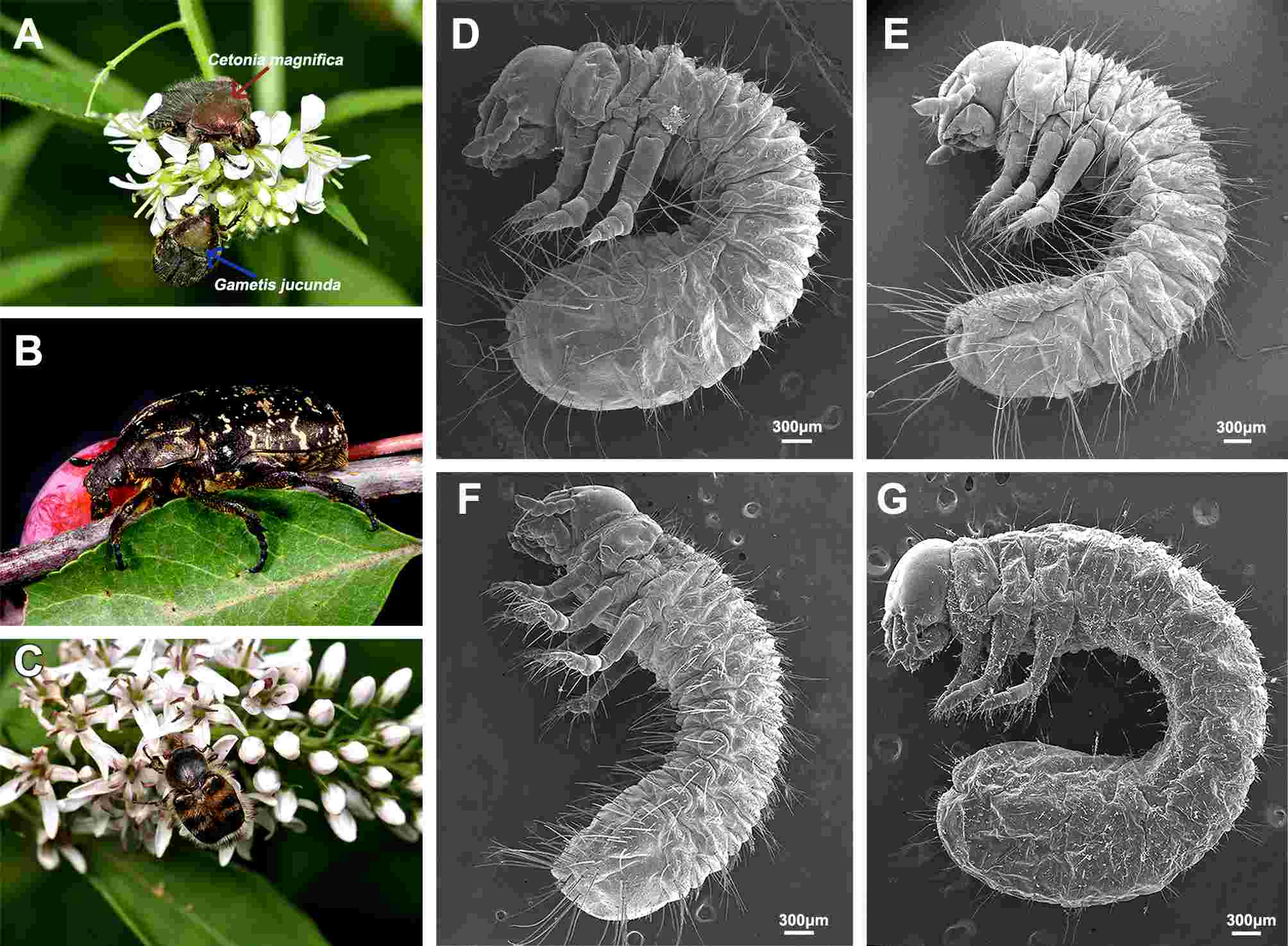
Adult and first instar larvae in habitus. (A) Adults of C. magnifica and G. jucunda in habitus; (B) Adult of P. brevitarsis in habitus; (C) Adult of L. succinctus in habitus; (D) Larva of C. magnifica; (E) Larva of G. jucunda; (F) Larva of P. brevitarsis; (G) Larva of L. succinctus.
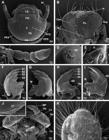
Figure 2
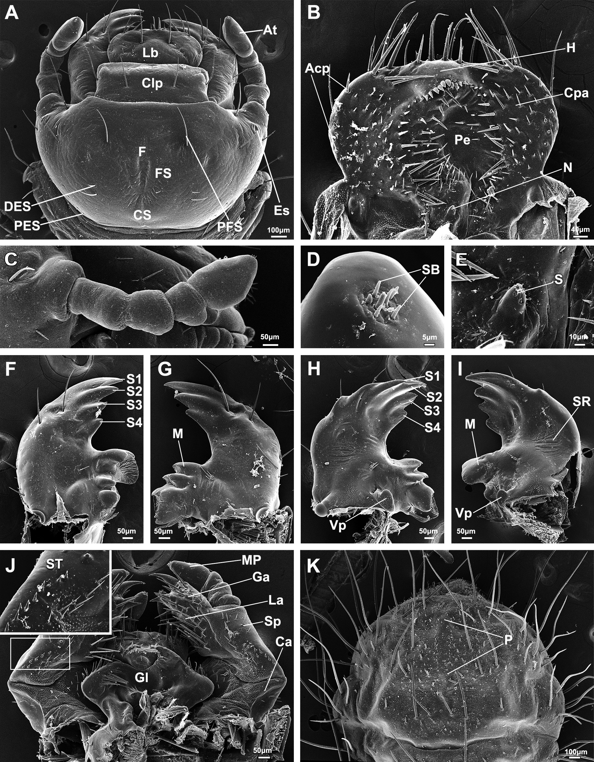
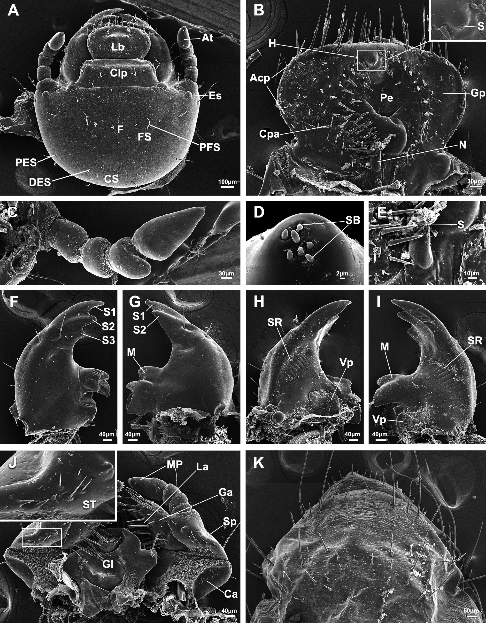
Detailed larval characters of C. magnifica. (A) Head; (B) Epipharynx; (C) Antenna lateral view; (D) Sensilla on the apex of antenna; (E) Magnification of nesium; (F) Left mandible, dorsal surface; (G) Right mandible, dorsal surface; (H) Right mandible, ventral surface; (I) Left mandible, ventral surface; (J) Maxillae and labia, ventral surface, insert showing the magnification of the stridulatory teeth; (K) Raster. Acp, acanthoparia; At, antenna; Clp, clypeus; Ca, cardo; Cpa, chaetoparia; CS, coronal suture; DES, dorsoepicranial seta; Es, eyespot; F, frons; FS, frontal suture; Ga, galea; Gl, glossa; H, haptomerum; La, lacinia; Lb, labrum; LP, labial palp; M, molar area; MP, maxillary palp; N, nesium; P, palidia; Pe, pedium; PES, posteroepicranial seta; PFS, posterofrontal seta; S, sensillum; S1–4, scissorial teeth; SB, sensillum basiconicum; Sp, stipes; ST, stridulatory teeth.
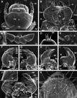
Figure 3
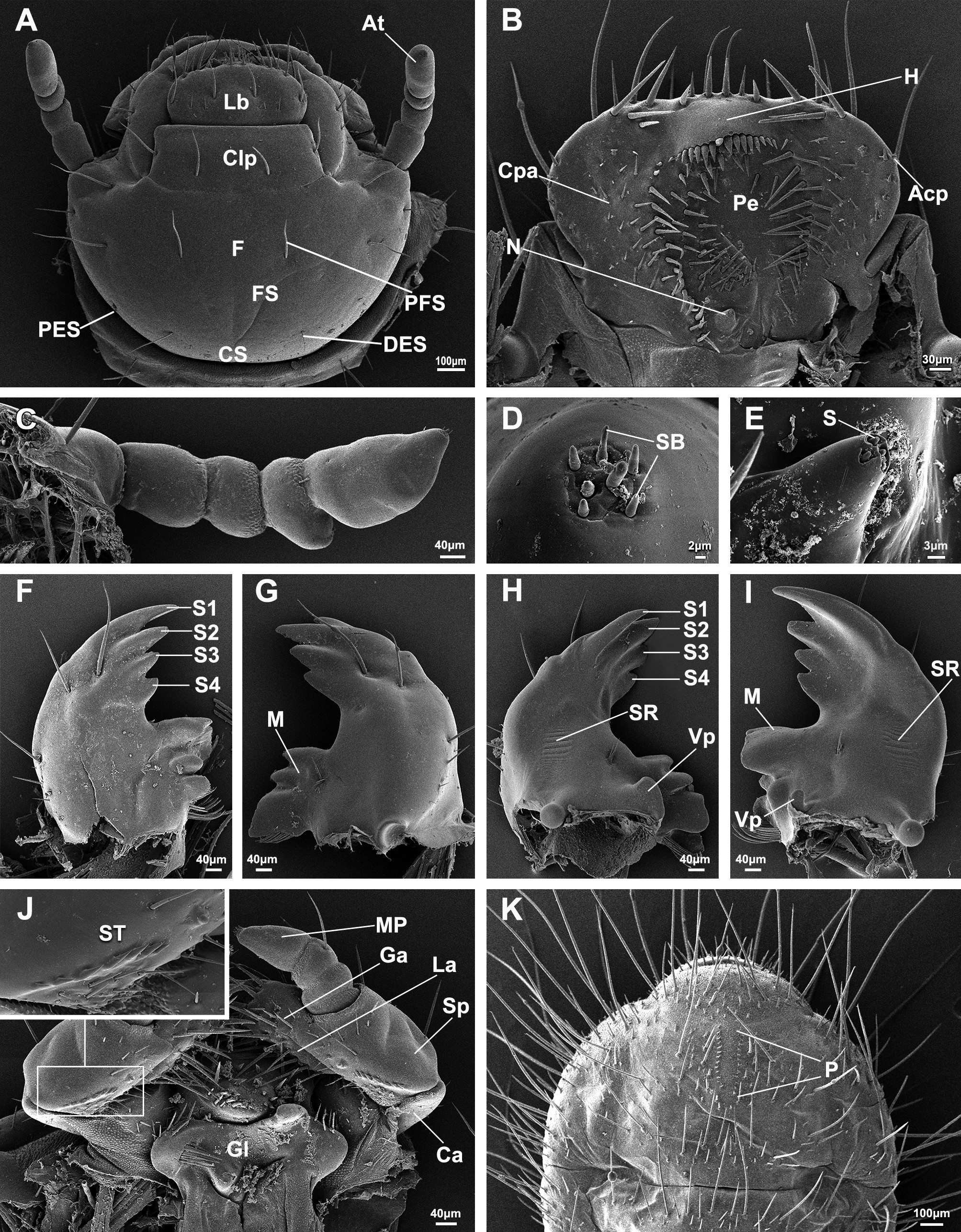
Detailed larval characters of G. jucunda. (A) Head; (B) Epipharynx; (C) Antenna lateral view; (D) Sensilla on the apex of antenna; (E) Magnification of nesium; (F) Left mandible, dorsal surface; (G) Right mandible, dorsal surface; (H) Right mandible, ventral surface; (I) Left mandible, ventral surface; (J) Maxillae and labia, ventral surface, insert showing the magnification of the stridulatory teeth; (K) Raster. Acp, acanthoparia; At, antenna; Clp, clypeus; Ca, cardo; Cpa, chaetoparia; CS, coronal suture; DES, dorsoepicranial seta; F, frons; FS, frontal suture; Ga, galea; Gl, glossa; H, haptomerum; La, lacinia; Lb, labrum; LP, labial palp; M, molar area; MP, maxillary palp; N, nesium; P, palidia; Pe, pedium; PES, posteroepicranial seta; PFS, posterofrontal seta; S, sensillum; S1–4, scissorial teeth; SB, sensillum basiconicum; Sp, stipes; ST, stridulatory teeth.
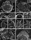
Figure 4
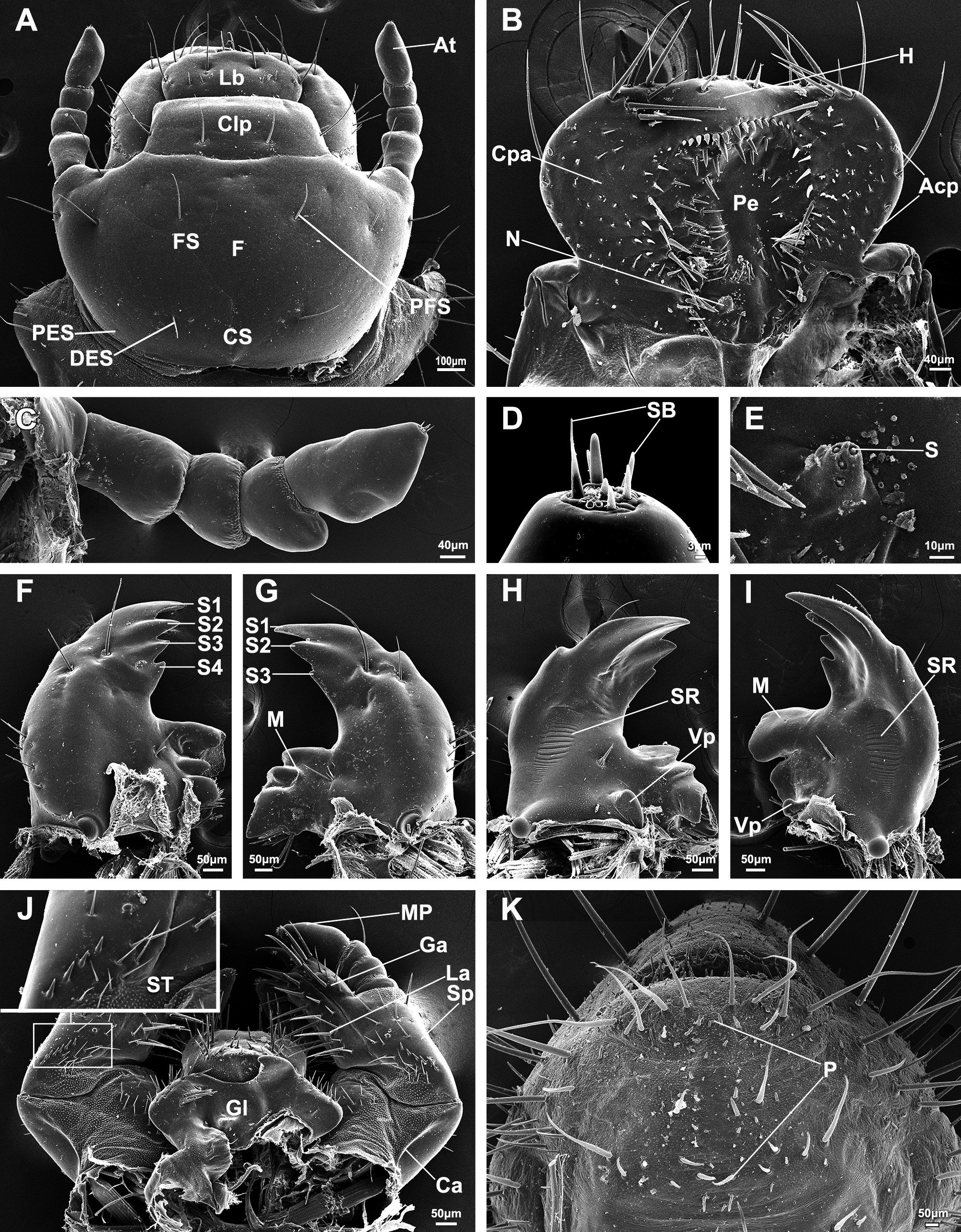
Detailed larval characters of P. brevitarsis. (A) Head; (B) Epipharynx; (C) Antenna lateral view; (D) Sensilla on the apex of antenna; (E) Magnification of nesium; (F) Left mandible, dorsal surface; (G) Right mandible, dorsal surface; (H) Right mandible, ventral surface; (I) Left mandible, ventral surface; (J) Maxillae and labia, ventral surface, insert showing the magnification of the stridulatory teeth; (K) Raster. Acp, acanthoparia; At, antenna; Clp, clypeus; Ca, cardo; Cpa, chaetoparia; CS, coronal suture; DES, dorsoepicranial seta; Es, eyespot; F, frons; FS, frontal suture; Ga, galea; Gl, glossa; H, haptomerum; La, lacinia; Lb, labrum; LP, labial palp; M, molar area; MP, maxillary palp; N, nesium; P, palidia; Pe, pedium; PES, posteroepicranial seta; PFS, posterofrontal seta; S, sensillum; S1–4, scissorial teeth; SB, sensillum basiconicum; Sp, stipes; ST, stridulatory teeth.
Figure 5
Detailed larval characters of L. succinctus. (A) Head; (B) Epipharynx, insert showing the magnification of the haptomerum; (C) Antenna lateral view; (D) Sensilla on the apex of antenna; (E) Magnification of nesium; (F) Left mandible, dorsal surface; (G) Right mandible, dorsal surface; (H) Right mandible, ventral surface; (I) Left mandible, ventral surface; (J) Maxillae and labia, ventral surface, insert showing the magnification of the stridulatory teeth; (K) Raster. Acp, acanthoparia; At, antenna; Clp, clypeus; Ca, cardo; Cpa, chaetoparia; CS, coronal suture; DES, dorsoepicranial seta; Es, eyespot; F, frons; FS, frontal suture; Ga, galea; Gl, glossa; Gp, gymnoparia; H, haptomerum; Lb, labrum; LP, labial palp; M, molar area; MP, maxillary palp; N, nesium; P, palidia; Pe, pedium; PES, posteroepicranial seta; PFS, posterofrontal seta; S, sensillum; S1–3, scissorial teeth; SB, sensillum basiconicum; Sp, stipes; ST, stridulatory teeth.
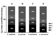
Figure 6
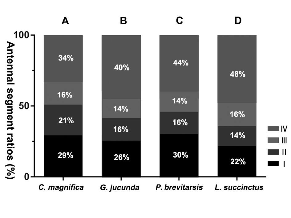
Segment ratios of larval Antennae. (A) C. magnifica; (B) G. jucunda; (C) P. brevitarsis; (D) L. succinctus. I–IV are the number of antennal segments.

 Description of the first instar larvae of four species of Cetoniinae (Coleoptera: Scarabaeidae) in northeastern China
 Thumbnail
 Thumbnail
 Thumbnail
 Thumbnail
 Thumbnail
 Thumbnail