Incredible fast improvement in visual acuity after YAG laser for Valsava retinopathy
ABSTRACT
This study was aimed at reporting a case of Valsava Retinopathy (VR) successfully treated with early YAG laser membraniectomy. This is a case report with analysis of medical records, performance, and recording of complementary exams, performing the laser procedure, assessment of immediate visual acuity, 7 and 30 days after the laser procedure. The patient demonstrated full recovery of visual acuity within 1 week. YAG laser membranectomy has proven effective in dispersing bleeding, accelerating visual recovery faster and more accessible than a vitreoretinal surgery in this case.
Keywords:
Laser therapy; Retinal hemorrhage; Valsava maneuver
RESUMO
O objetivo deste estudo foi relatar um caso de retinopatia de Valsalva tratada de maneira imediata com sucesso pela membraniectomia com YAG laser. Trata-se de um relato de caso com análise de prontuário; realização e interpretação de exames complementares; realização do procedimento a laser; registro pré e pós-laser; e avaliação da acuidade visual imediata, 7 e 30 dias após o procedimento. O paciente demonstrou recuperação total da acuidade visual em 1 semana. A membraniectomia com YAG laser provou-se eficaz em dispersar a hemorragia para a cavidade vítrea, acelerando a recuperação visual de forma rápida e mais acessível que uma cirurgia vitreorretiniana neste caso.
Descritores:
Terapia a laser; Hemorragia retiniana; Manobra de Valsava
INTRODUCTION
Valsalva retinopathy (VR) is a retinal hemorrhagic condition caused by rupture of superficial capillaries due to a sudden increase in intrathoracic or intra-abdominal pressure. Blood extravasation typically occurs between the internal limiting membrane (ILM) and the posterior hyaloid, potentially leading to significant visual impairment. Clinically, it presents as a sudden, painless central scotoma, making it essential to differentiate from other retinal hemorrhages, such as macroaneurysms and Terson syndrome.(1)
The management of VR with small hemorrhages is usually conservative with spontaneous hemorrhage resorption occurring within 6 to 9 months.(2) However, it should be highlighted that in cases of extensive hemorrhage or patients requiring rapid visual recovery, intervention may be considered.(3) YAG laser membranectomy is an effective alternative, promoting blood dispersion into the vitreous cavity and accelerating visual improvement, thereby avoiding the need for vitreoretinal surgery.(4)
In this case report, we present a young patient with VR secondary to an episode of vomiting. The patient was treated with YAG laser membranectomy, resulting in significant visual recovery within a few weeks. This case reinforces the importance of quick individualized treatment and the potential benefits of laser therapy in selected cases. This study was approved by the Ethics Committee (CAAE: 77888423.1.0000.0093).
CASE REPORT
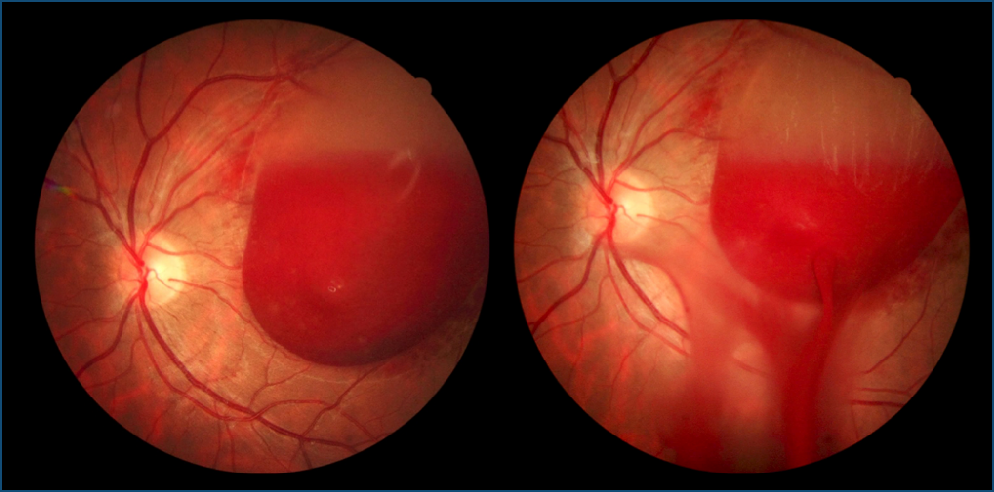
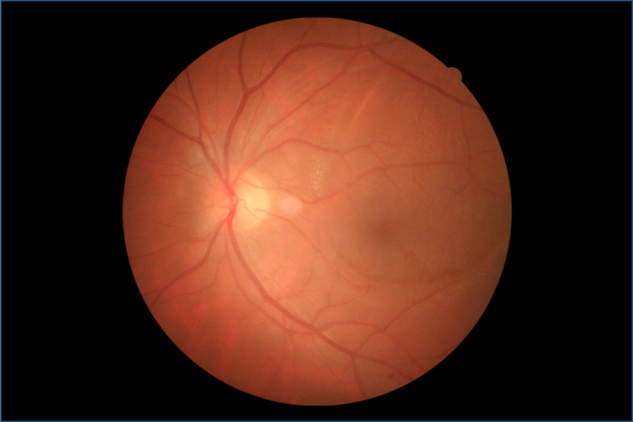
A 26-year-old male presented to the emergency department with a sudden painless central scotoma in OS, after an episode of vomiting. His BCVA was counting fingers at 1 m in OS, with no afferent pupillary defect. Dilated fundus examination showed circumscribed hemorrhage in the pre-macular region (Figure 1). Given the need for rapid visual rehabilitation, immediate YAG laser membranectomy was performed in OS. Five ampere laser shots were applied to the inferior portion of hemorrhage, targeting the hyaloid membrane and dispersing blood into the vitreous cavity. One week post-procedure, the patient was asymptomatic with BCVA of 20/60 in OS. Fundus evaluation showed vitreous hemorrhage in partial resolution in the inferior posterior pole and no pre-macular hemorrhage (Figure 2). After one month, BCVA improved to 20/25 in OS with complete hemorrhage absorption and a physiological foveal reflex (Figure 3).
Retinography showing circumscribed hemorrhage in the pre-macular region immediately after YAG laser membranotomy.
Retinography after 1 week showing vitreous hemorrhage in partial resolution in the inferior posterior pole and no pre-macular hemorrhage.
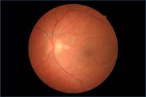
Retinography after 1 month with complete hemorrhage absorption and a physiological foveal reflex.
DISCUSSION
Valsalva retinopathy is caused by rupture of superficial retinal capillaries due to a sudden increase in intrathoracic or intra-abdominal pressure.(4) In the presented case, the Valsava maneuver was secondary to episodes of vomiting. These pre-retinal hemorrhages occur between the posterior hyaloid and the ILM or – less frequently – between the ILM and the retinal nerve fiber layer (RNFL).(4)
Valsalva retinopathy can manifest with floaters, central scotoma, and – in severe cases – reduced visual acuity due to hemorrhagic detachment of the ILM.(4) The patient in the case had BCVA of counting fingers at 1 m in OS alone, with no other symptoms. The diagnosis is clinical, with fundoscopic examination being essential to differentiate from ruptured macroaneurysm, hemorrhagic detachment, and Terson syndrome.(5) The optical coherence tomography (OCT) is fundamental to locate the pre-macular hemorrhage, since the biomicroscopy is not accurate to determinate the exact location between layers of the retina.(6,7) Despite that, OCT may be limited due to the high reflectivity of recent blood.(8)
Management is primarily conservative with observation. Focal opening of the posterior hyaloid or ILM with YAG laser has been described as a non-invasive alternative to surgical approach, achieving rapid intravitreal drainage.(9) This method offers an effective treatment to restoring vision in a short period.(10) The patient of the case had improved best-corrected visual acuity (BVCA) from counting on fingers 1 m to 20/60 in only one week and 20/25 after a month. In these cases, YAG laser membranectomy has proven effective in dispersing the blood, accelerating visual recovery, and preventing a vitreoretinal surgery.(11,12)
Extensive or persistent cases may cause permanent damage to the photoreceptors. There is no consensus on the effects of long contact between blood and retina, but the formation of epiretinal membrane and toxic effect on the retina after dissolving hemoglobin was observed.(13,14) In some cases, a pars plana vitrectomy for membrane removal is necessary after YAG.(15)
Data Availability Statement:
The datasets generated and/or analyzed during the current study are included in the manuscript.
REFERENCES
- 1 Kuruvilla O, Munie M, Shah M, Desai U, Miller JA, Ober MD. Nd: YAG membranotomy for preretinal hemorrhage secondary to Valsalva retinopathy. Saudi J Ophthalmol. 2014;28(2):145-51.
- 2 Kirwan RP, Cahill MT. Nd:YAG laser hyaloidotomy for Valsalva pre-macular haemorrhage. Ir J Med Sci. 2011;180:749-52.
- 3 Moreira Jr CA, Vianello SM, Cardillo JA. Retinopatia de Valsalva: intervir ou não? Relato de caso com avaliação por OCT. e-Oftalmo.CBO: Rev Dig Oftalmol. 2015;1(1):1-2.
-
4 Lam L. Valsalva Retinopathy. American Academy of Ophtalmology. 2023 [cited 2026 Jan 7]. Available from: https://eyewiki.org/Valsalva_Retinopathy
» https://eyewiki.org/Valsalva_Retinopathy - 5 Lavezzo MM, Zacharias LC, Takahashi WY. Hemorragia submembrana limitante interna em paciente após Valsalva: relato de caso. Arq Bras Oftalmol. 2012;75(6)436-8.
- 6 Shukla D, Naresh KB, Kim R. Optical coherence tomography findings in Valsalva retinopathy. Am J Ophthalmol. 2005;140(1):134-6.
- 7 Pérez-Rico C, Montes-Mollón Á, Castro-Rebollo M, Pareja-Esteban J, Benítez-Herreros J. Optical coherence tomography features of sub-internal limiting membrane hemorrhage and temporary premacular cavity following Nd-YAG laser membranotomy in Valsalva retinopathy. Jpn J Ophthalmol. 2008 Nov-Dec;52(6):513-515.
- 8 Pollack AL, McDonald HR, Ai E, Johnson RN, Dugel PU, Folk J, Grand MG, Lambert HM, Schwartz S, Miller RD. Massive suprachoroidal hemorrhage during pars plana vitrectomy associated with Valsalva maneuver. Am J Ophthalmol. 2001 Sep;132(3):383-7.
- 9 Meyer CH, Mennel S, Rodrigues EB, Schmidt JC. Persistent premacular cavity after membranotomy in Valsalva retinopathy evident by optical coherence tomography. Retina. 2006;26(1):116-8. Erratum in: Retina. 2006;26(3):377.
- 10 Lim JI, Drews-Botsch C, Sternberg P Jr, Capone A Jr, Aaberg TM Sr. Submacular hemorrhage removal. Ophthalmology. 1995;102(9):1393-9.
- 11 Meyer CH, Mennel S, Rodrigues EB, Schmidt JC. Persistent premacular cavity after membranotomy in Valsalva retinopathy evident by optical coherence tomography. Retina. 2006;26(1):116-8. Erratum in: Retina. 2006;26(3):377.
- 12 Khan MT, Saeed MU, Shehzad MS, Qazi ZA. Nd:YAG laser treatment for Valsalva premacular hemorrhages: 6 month follow up : alternative management options for preretinal premacular hemorrhages in Valsalva retinopathy. Int Ophthalmol. 2008;28(5):325-7.
- 13 Celebi S, Kükner AS. Photodisruptive Nd:YAG laser in the management of premacular subhyaloid hemorrhage. Eur J Ophthalmol. 2001;11(3):281-6.
- 14 Liu Z, Pan X, Bi H. Treatment of Valsalva retinopathy. Optom Vis Sci. 2014;91(11):e278-81.
- 15 Kwok AK, Lai TY, Chan NR. Epiretinal membrane formation with internal limiting membrane wrinkling after Nd:YAG laser membranotomy in Valsalva retinopathy. Am J Ophthalmol. 2003;136(4):763-6.
Edited by
-
Associate editor:
Ricardo Augusto Paletta Guedes Universidade Federal de Juiz de Fora, Juiz de Fora, MG, Brazil https://orcid.org/0000-0002-9451-738X