Evaluating peripheral retina: a cautionary note on miotics
ABSTRACT
Objective: To underscore the critical importance of assessing the peripheral retina in all patients, especially those planning to use miotics.
Methods: A retrospective and observational study was conducted at a single ophthalmological medical center, using records from the International Classification of Diseases (ICD-10) for closed-angle glaucoma and lesions predisposing to detachment. Participants in this study underwent anterior chamber assessment using the Goldman 3-Mirror lens and the Volk G-4 Gonioscopy lens. Peripheral retinal evaluation was conducted with indirect binocular ophthalmoscopy and posterior biomicroscopy, also utilizing the Goldman 3-Mirror lens. The medical records were analyzed, and all information was recorded, including procedures.
Results: Among 9,854 patients assessed, 1,144 (11.6%) were classified with primary angle closure suspect, primary angle closure, or primary angle closure glaucoma. Additionally, 1,032 patients exhibited lesions predisposing to retinal detachment, constituting 10.4% of the cohort. Notably, 71 eyes from 56 (0.56%) patients demonstrated an association between primary angle closure suspect, primary angle closure, or primary angle closure glaucoma and these predisposing lesions. Within the primary angle closure suspect, primary angle closure, or primary angle closure glaucoma group, 56 (4.89%) presented with retinal lattice degeneration, holes, and/or tears. The majority were female, over 40 years old, hyperopic, with unilateral involvement and symptomatic. The average follow-up duration was 6.4 years.
Conclusion: Peripheral degenerative lesions that may lead to retinal detachment are relatively common and can be found even in selected cases of angular narrowing. Peripheral retinal evaluation may be advisable in angle-closure eyes, particularly when miotics are planned; prospective studies are needed to confirm risk.
Keywords:
Pilocarpine; Presbyopia; Glaucoma; Glaucoma,angle-closure; Retinal perforations; Retinal detachment
RESUMO
Objetivo: Ressaltar a importância da avaliação da retina periférica em todos os pacientes, especialmente naqueles que planejam usar mióticos.
Métodos: Estudo retrospectivo e observacional, realizado em um único centro médico oftalmológico, utilizando registros da Classificação Internacional de Doenças (CID-10) para glaucoma de ângulo fechado e lesões predisponentes ao descolamento. Os participantes deste estudo foram submetidos à avaliação da câmara anterior utilizando a lente Goldman 3-espelhos e a lente Volk G-4 de Gonioscopia. A avaliação da retina periférica foi realizada com oftalmoscopia binocular indireta e lente de Goldman 3-espelhos. Os prontuários médicos foram analisados e todas as informações foram registradas.
Resultados: Entre os 9.854 pacientes avaliados, 1.144 (11,6%) foram classificados com suspeita de fechamento angular primário, fechamento angular primário ou glaucoma primário de ângulo fechado. Adicionalmente, 1.032 pacientes apresentaram lesões predisponentes de descolamento de retina, representando 10,4% da coorte. 71 olhos de 56 (0,56%) pacientes demonstraram uma associação entre suspeita de fechamento angular primário, fechamento angular primário ou glaucoma primário de ângulo fechado e lesões predisponentes. Dentro do grupo de suspeita de fechamento angular primário, fechamento angular primário ou glaucoma primário de angular fechado, 56 (4,89%) apresentaram degeneração em treliça, buracos e/ou rupturas de retina. A maioria era do sexo feminino, com mais de 40 anos, hipermétrope, com envolvimento unilateral e sintomáticos. A duração média do acompanhamento foi de 6,4 anos.
Conclusão: Lesões degenerativas periféricas que podem levar ao descolamento de retina são relativamente comuns e podem ser encontradas mesmo em casos de estreitamento angular. A avaliação da retina periférica é aconselhável em olhos com fechamento angular, particularmente quando mióticos são planejados. Estudos prospectivos são necessários para confirmar o risco.
Descritores:
Pilocarpina; Presbiopia; Glaucoma; Glaucoma de ângulo fechado; Perfurações; Descolamento retiniano
INTRODUCTION
Pilocarpine, a topical parasympathomimetic agent that acts on postganglionic muscarinic receptors, was used in the treatment of glaucoma.(1) By contracting the ciliary muscle and repositioning the scleral spur posteriorly and internally, pilocarpine facilitates the widening of the spaces between the corneoscleral trabeculae and enhances the distension of the endothelial meshwork.(1,2) This action decreases resistance to aqueous humor outflow, making pilocarpine a common choice for managing both primary angle-closure glaucoma and open-angle glaucoma.(1,3–5) However, its use is associated with various ocular adverse effects, including miosis, accommodative spasm, blurred vision, frontal headache, eye pain, eyelid tremors, conjunctival and ciliary injection, cataracts, iris cysts, and notably, retinal detachment.(6–8) Consequently, in the context of open-angle glaucoma, pilocarpine has been increasingly supplanted by alternative medications that offer fewer side effects and improved efficacy in lowering intraocular pressure (IOP). These alternatives include β-blockers, α-agonists, carbonic anhydrase inhibitors, and prostaglandin analogues.(9,10) For primary angle-closure glaucoma, minimally invasive procedures such as peripheral iridectomy have emerged as successful replacements for pilocarpine, utilizing techniques such as argon laser and neodymium:yttrium-aluminum-garnet (Nd:YAG) lasers.(11–15) However, pilocarpine hydrochloride ophthalmic solution 1.25% has recently received approval from the US Food and Drug Administration (FDA), now for the correction of presbyopia.(7,8,16–18)
Age-related changes in the eye, such as decreased ciliary muscle efficiency, sclerosis of lens fibers, alterations in the lens capsule, and increased lens thickness, contribute to accommodation insufficiency, hindering the ability to focus on nearby objects.(19,20) The resulting decline in near vision can significantly impact the quality of life for many individuals.(21) Although non-invasive alternatives, like contact lenses, have been employed to address this issue, they can be difficult to maintain due to the development of dry eye and decreased manual dexterity with age.(21,22) Invasive options, such as refractive lens exchange (RLE), involve replacing the natural crystalline lens with an intraocular lens (IOL), but these procedures are not without risks.(19,23–27) Pilocarpine, with its reversible effects, low cost, and relative safety, presents a viable option for presbyopia management. By inducing contraction of the ciliary muscle and causing pupillary miosis, pilocarpine can trigger accommodative spasm, thereby enhancing visual acuity in ametropic patients.(28) This accommodative spasm also makes the anterior chamber shallower, as it displaces the lens anteriorly, reducing its radius of curvature, causing a myopic effect and consequent improvement in near vision.(28–31) Furthermore, the pinhole effect induced by pilocarpine may also contribute to enhanced visual acuity.(32)
Despite these benefits, there are growing concerns regarding the association between pilocarpine use and retinal detachment, as highlighted in several recent reports.(33–36) Dynamic activities that mobilize the anterior vitreous, such as the advancement of 0.05 mm of the ora serrata and underlying choroid for each diopter of accommodation, and the anterior displacement of the posterior surface of the lens caused by accommodation,(28,29,37) can reverberate in abnormal vitreoretinal adhesions such as retinal lattices, holes and/or tears, thus triggering retinal detachment.(38) Additionally, pharmacological accommodation may significantly increase subfoveal choroidal thickness and axial length, which may aggravate this process.(24,39)
Historically, a survey among ophthalmologists from the retina society indicated that topical ocular miotics, including pilocarpine, were implicated in precipitating retinal detachment, particularly in eyes with preexisting pathological conditions.(40) Therefore, the present study aims to analyze the medical records of patients who present narrowing of the anterior chamber angle and lesions predisposing to retinal detachment, reaffirming the need for careful peripheral retinal evaluation, especially in those undergoing treatment for presbyopia with miotics.
METHODS
This study received approval from the Medical Ethics Committee of Hospital Angelina Caron, located in Campina Grande do Sul, PR, Brazil (CAAE: 87106025.6.0000.5226).
We conducted a retrospective and observational study at the Ophthalmology Center of Curitiba, Paraná, from December 1, 2015, to September 1, 2023, utilizing patient records categorized under the International Classification of Diseases (ICD-10) codes for closed-angle glaucoma (H40.2) and lesions predisposing to retinal detachment without retinal detachment (H33.3).
Patients included in this study underwent at least the following examinations: static gonioscopy using a Goldman 3-mirror lens, with ×25 magnification, performed with minimal ambient lighting possible and with the eye in the primary gaze position; indentation gonioscopy with the Volk G-4 Gonioscopy lens. These examinations helped detect primary angle closure suspect (PACS), primary angle closure (PAC), or primary angle closure glaucoma (PACG).
Thus, primary angle-suspect was defined as the gonioscopic condition presenting reversible iridotrabecular contact (ITC) of 180° or more, without evidence of permanent obstruction of aqueous flow, without peripheral anterior synechiae (PAS), IOP ≤ 21 mm Hg, and without glaucomatous optic neuropathy.(41) On the other hand, PAC was considered to be a narrow or closed angle, with ITC of 180° or more, with IOP ≥ 21 mmHg or PAS, resulting from long-term iridotrabecular contact.(42)
Primary angle-closure glaucoma was characterized for those participants who, in addition to having closed angles, exhibited glaucomatous damage evidenced by visual field loss, nerve fiber layer damage, or optic nerve damage.(43)
All participants underwent indirect binocular ophthalmoscopy and posterior biomicroscopy with a 3-mirror Goldman lens to assess lesions predisposing for retinal detachment, focusing on retinal lattice degeneration, holes, and tears as the main lesions of interest and amenable to prevention and treatment. Symptoms were documented as floaters alone, floaters with flashes, and flashes alone. Refractive status was classified based on cycloplegic refraction, with emmetropic defined as between −0.50 and +0.50, hyperopic as over +0.50, and myopic as over −0.50. Exclusion criteria encompassed individuals with open-angle glaucoma, secondary glaucoma, congenital glaucoma, retinoschisis, a history of uveitis, ocular trauma, or prior eye surgery. Patients with concurrent PACS, PAC, or PACG and predisposing lesions to retinal detachment in at least one eye were evaluated by experienced subspecialists in glaucoma and retina/vitreous. A thorough analysis of medical records was conducted, capturing essential data such as refraction, biomicroscopy, IOP measurements, stereophotography of the optic disc, retinography, pachymetry, corneal specular microscopy, visual field testing, and optical coherence tomography (OCT) of the optic disc. Treatment procedures, including peripheral iridectomy with neodymium:yttrium-aluminum-garnet (Nd:YAG) and argon green laser photocoagulation aimed at preventing retinal detachment, were meticulously documented.
RESULTS
Among the 9,854 patients evaluated, 1,144 (11.6%) were classified as having PACS, PAC, or PACG. Furthermore, 1,032 patients (10.4%) were diagnosed with lesions predisposing to retinal detachment. An association between PACS, PAC, or PACG and lesions predisposing to retinal detachment was found in 56 patients (4.89%) and 71 eyes (0.36%). The following paragraphs and Table 1 display the main findings in those 56 patients and 71 eyes.
Data for the 56 patients and 71 eyes with association of primary angle closure suspect, primary angle closure, or primary angle closure glaucoma and lesions predisposing to retinal detachment
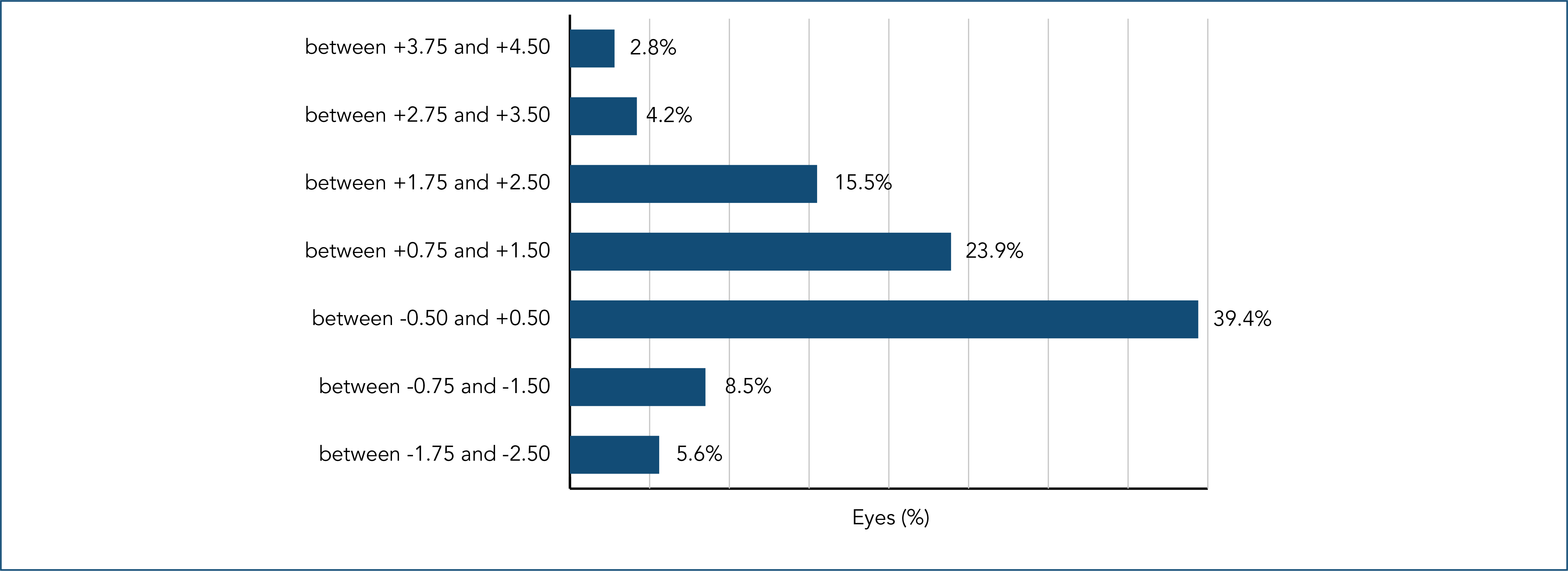
In this cohort of 56 patients, 40 (71.4%) were female, while retinal lesions that predispose to retinal detachment were unilateral in 41 patients (73,2%) and symptoms (flashes and/or floaters) were referred by approximately half of patients. In addition to this, 54 out of the 56 patients (96.5%) were over 40 years of age, and 10 patients (14%) were myopic (Figure 1).
For the cohort of 71 eyes with association of PACS, PAC, or PACG and lesions predisposing to retinal detachment, 25 eyes (35.2%) exhibited tears, 36 eyes (50.7%) had holes, and 28 eyes (39.4%) displayed lattice degeneration. Notably, 15 eyes (15%) showed more than one type of lesion, with the majority located in the inferior temporal periphery.
Ultrasound biomicroscopy (UBM) was performed in 39 out of the 56 patients (69.6%), aiding in diagnostic confirmation. Treatment interventions included peripheral iridectomy in 67 eyes (94.4%) for prevention of primary angle closure and argon laser photocoagulation in 69 eyes (97.2%) to address lesions predisposing to retinal detachment. Phacoemulsification was performed in 21 eyes (29.6%). Comorbidities were prevalent, with cataracts affecting 22 patients (31%), dry age-related macular degeneration in 16 patients (22.5%), and glaucoma in 4 patients (5.6%). Additionally, 13 patients (18.3%) had multiple comorbidities. In symptomatic patients, follow-up was on average 6.1 years, while follow-up time in all patients was on average 6.4 years.
DISCUSSION
The analysis of medical records from a single ophthalmological center, utilizing the International Classification of Diseases codes for PACS, PAC, or PACG (ICD H40-2) and lesions predisposing to retinal detachment without retinal detachment (H33-3), underscores the overlap between eyes susceptible to angle closure and those presenting with retinal detachment risk factors. This finding emphasizes the necessity of thorough peripheral retinal examination for all patients, particularly those undergoing treatment with miotics.
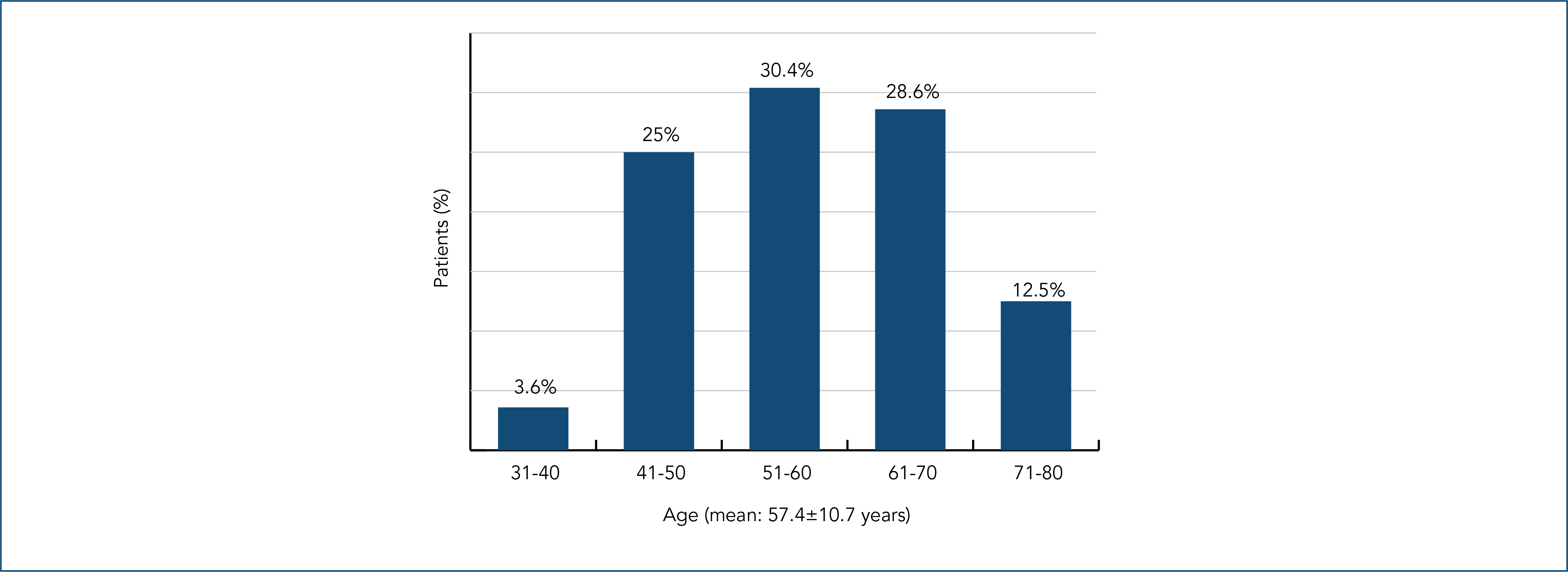
It is noteworthy that the ICD-10 classification does not separately include PACS or PAC; instead, these conditions are subsumed under the ICD H40-2 code, which pertains specifically to PACG. Consequently, this study's methodology involved a careful evaluation of records coded as ICD H40-2 in conjunction with H33-3 to accurately assess the association between these conditions. We excluded retinoschisis from our analysis because when adding ICD H 33-1 (retinoschisis) versus ICD H40-2 (primary angle-closure glaucoma) we found a large number of participants; therefore, deserving a more in-depth reflection on this condition. Our findings showed that 10 eyes had myopia below - 2.50, 28 eyes were emmetropic, 17 eyes had hyperopia up to + 1.50 and 16 eyes had hyperopia above + 1.75. Considering the refractive issue, we can infer that almost 55 (77,4%) of the eyes analyzed in our study would be eligible for presbyopia treatment with miotics, since in studies regarding this type of treatment, 75% of the participants were emmetropic (range of −0.50 D to +0.75 D and/or a cylinder smaller than 0.75 D) and a minority (25%) were non-emmetropic whose best distance correction could vary between -4.00 and +1.00 diopter (D) sphere and cylinder ≤ ±2.00 D.(32) The predominance of unilateral alterations in 41 (73.2%) of patients, along with the notable female representation 40 (71.4%) (Table 1) and age distribution, 54 (96.4%) of participants being over 40 (Figure 2), mirrors trends documented in studies addressing closed-angle glaucoma and retinal tears.(44–47) This age correlation highlights the importance of monitoring older populations for the early detection of these conditions.
The prevalence of lesions predisposing to retinal detachment, identified in 1,032 (10.4%) of patients who were seen at an ophthalmology clinic, is consistent with expectations, particularly as prior studies indicated 12% of myopia surgery candidates had such lesions.(48) Our study covered not only myopic eyes, but mainly emmetropic and hyperopic eyes. Additionally, the 4.89% prevalence in eyes with PACS, PAC, or PACG suggests that these predisposing lesions to retinal detachment, often more common in myopic eyes,(47) can also occur in emmetropic and hyperopic patients, indicating a broader risk profile. This finding highlights the critical need to evaluate retinal health across all refractive errors, including patients with narrow anterior chamber angles. Notably, this group has often been excluded from presbyopia treatment protocols that involve pilocarpine.(32) It is important to note that the miosis induced by pilocarpine instillation moves the peripheral iris away from the anterior chamber angle, alleviating pupillary block and enhancing aqueous flow. Paradoxically, however, a dose-dependent narrowing of the anterior chamber may occur due to anteriorization of the ciliary processes, which has the potential of triggering angle closure in affected eyes.(49)
Historical context highlights that before the introduction of lasers for performing iridectomy, patients at risk for angle closure were often treated with miotic, leading to documented cases of retinal detachment.(40) This highlights the need for careful evaluation of the peripheral retina, which may have been overlooked in cases where narrow angles were present, thus raising concerns about induction of acute glaucoma caused by pupillary dilation. Additionally, studies have shown a connection between predisposing retinal lesions and glaucoma.(50)
Symptoms such as floaters and flashes, which are indicative of posterior vitreous detachment (PVD),(51) are associated with retinal tears in approximately 8% to 15% of cases,(51–55) and can consequently lead to retinal detachment.(40,52–55) In our cohort, 55.3% of patients reported vitreoretinal symptoms, including floaters, flashes, or both. Notably, nearly half of the patients (44.6%) did not exhibit these symptoms, highlighting the critical importance of conducting peripheral retinal evaluations in all individuals, regardless of their symptomatology. It is essential to emphasize that in cases of PVD, follow-up should extend for at least four months after the onset of initial symptoms,(56) as persistent vitreoretinal traction in symptomatic retinal tears can result in retinal detachment in 30% to 50% of cases.(57,58) In this study, all patients with symptoms were closely monitored until they became asymptomatic. Given that subsequent retinal breaks are more commonly observed in myopic individuals,(56) our findings, where significant proportions of participants were emmetropic or hyperopic, further underscore the necessity for vigilant monitoring, particularly if being treated with pilocarpine.
Ultrasound biomicroscopy was performed in 39 patients (69.6%), enhancing our understanding of the anatomical conditions of the anterior chamber that could compromise vision. This evaluation also facilitated patient awareness regarding preventive measures, including peripheral iridectomy with Nd:YAG laser, which was performed in 67 eyes (94.4%). Peripheral iridectomy with Nd:YAG laser is regarded as a safe prophylactic intervention that can effectively prevent progressive angle closure.(59) This procedure is associated with few and rare risks.(11–14) Following iridectomy, pupillary dilation was employed for stereophotography of the optic disc and peripheral retinal evaluation, with 69 eyes (97,2%) receiving preventive treatment via argon green laser photocoagulation for identified retinal lesions, as recommended in the literature.(60–61) Thus, ruptures, holes and lattice degeneration, present in the retinal periphery, were treated with photocoagulation in practically all cases, especially when symptomatic. It is important to highlight that the incidence of ruptures varies with age, being greater in the 4th, 5th and 6th decades of life.(45–47) Therefore, periodic evaluation of the retinal periphery of patients chronically using pilocarpine becomes essential with advancing age. Although there are reports associating vitreoretinal disorders with the use of pilocarpine, including in the reversal of pupillary dilation,(34,62,63) this miotic was used to prevent angle-closure glaucoma in cases of plateau iris, where there was a risk of progressive angle closure despite having undergone peripheral iridectomy. The application of these eye drops remains a topic of debate, as they have been associated with an increased risk of triggering retinal detachment.(48) However, studies investigating the effects of pilocarpine on presbyopia over a one-month period have reported no cases of retinal detachment.(8,16–18) In the current study, we used pilocarpine only once and also observed no occurrence of retinal detachment after pilocarpine instillation, with a follow-up duration of 6.4 years.
Cataracts were a notable comorbidity in our study, affecting 22 eyes (31%), with phacoemulsification performed on 21 eyes, including four with primary closed-angle glaucoma. This reflects the growing recognition of cataract surgery as a viable first-line treatment for PACG.(64) Dry age-related macular degeneration (AMD) was also prevalent, highlighting the need for comprehensive management strategies in this elderly demographic, where conditions like AMD and cataracts are more common.(49,65) Glaucoma was observed in 8 eyes (11.3%), and the association of retinal detachment with glaucoma has been the subject of several publications.(65)
Despite the study's strengths, including its focus on the association between PACS, PAC, or PACG and retinal detachment risk factors, limitations such as its retrospective nature and single-center design must be acknowledged, emphasizing possible selection bias. Furthermore, most cases underwent prophylactic treatment for primary angle-closure glaucoma and/or retinal detachment, and these preventive interventions interfered with the natural histories of the diseases, modifying the causal risk of eventual miotic treatment. Additionally, important parameters, including anterior chamber depth and biometry, were not consistently recorded, which may impact the comprehensiveness of the findings. Future research should aim to address these gaps while continuing to explore the complexities of managing patients with these ocular conditions.
CONCLUSION
In conclusion, peripheral degenerative lesions, which can potentially lead to retinal detachment, are relatively common and may even be present in select cases of angular narrowing, such as primary angle closure suspect, primary angle closure, or primary angle closure glaucoma. Peripheral retinal evaluation may be advisable in angle-closure eyes, particularly when miotics are planned; prospective studies are needed to confirm risk.
Declaração de disponibilidade de dados:
The datasets generated and/or analyzed during the current study are included in the manuscript.
ACKNOWLEDGEMENT
Sandra Zandavalli Avila performed ultrasound biomicroscopy.
REFERENCES
- 1 Duke-Elder S. System of ophthalmology. The Foundations of Ophthalmology. Kimpton: London; 1958. vol. 7.
- 2 Grierson I, Lee WR, Abraham S. Effects of pilocarpine on the morphology of the human outflow apparatus. Br J Ophthalmol. 1978;62(5):302-13.
- 3 Davidorf JM, Bajer ND, Derick R. Treatment of the fellow eye in acute angle-closure glaucoma: a case report and survey of the members of the American Glaucoma Society. J Glaucoma. 1996;5:228-32.
- 4 Abramson DH, Chang S, Coleman J. Pilocarpine therapy in glaucoma: effects on anterior chamber depth and lens thickness in patients receiving long-term therapy. Arch Ophthalmol. 1976;94(6):914-8.
- 5 Drance SM, Bensted M, Schulzer M. Pilocarpine and intraocular pressure. Duration of effectiveness of 4 percent and 8 percent pilocarpine instillation. Arch Ophthalmol. 1974;91(2):104-6.
- 6 Zimmerman TJ, Wheeler TM. Miotics: side effects and ways to avoid them. Ophthalmology. 1982;89(1):76-80.
-
7 Allergan. A phase 3 efficacy study of pilocarpine HCl ophthalmic solution (AGN-190584) in participants with presbyopia (GEMINI 2). 2019 [cited 2026 Mar 12]. Available from: https://classic.clinicaltrials.gov/ct2/show/NCT03857542
» https://classic.clinicaltrials.gov/ct2/show/NCT03857542 - 8 Waring GO 4th, Price FW Jr, Wirta D, McCabe C, Moshirfar M, Guo Q, et al. Safety and efficacy of AGN-190584 in Individuals with presbyopia: the GEMINI 1 Phase 3 randomized clinical trial. JAMA Ophthalmol. 2022;140(4):363-71.
- 9 Boger WP 3rd, Steinert RF, Puliafito CA, Pavan-Langston D. Clinical trial comparing timolol ophthalmic solution to pilocarpine in open-angle glaucoma. Am J Ophthalmol. 1978;86(1):8-18.
- 10 Cohen LP, Pasquale LR. Clinical characteristics and current treatment of glaucoma. Cold Spring Harb Perspect Med. 2014;4(6):a017236.
- 11 Quigley HA. Long-term follow-up of laser iridotomy. Ophthalmology. 1981;88(3):218-24.
- 12 Robin AL, Pollack IP. A comparison of neodymium: YAG and argon laser iridotomies. Ophthalmology. 1984;91(9):1011-6.
- 13 Caronia RM, Liebmann JM, Stegman Z, Sokol J, Ritch R. Increase in iris-lens contact after laser iridotomy for pupillary block angle closure. Am J Ophthalmol. 1996;122(1):53-7.
- 14 Lim LS, Husain R, Gazzard G, Seah SK, Aung T. Cataract progression after prophylactic laser peripheral iridotomy: potential implications for the prevention of glaucoma blindness Ophthalmology. 2005;112(8):1355-9.
- 15 Ng WS, Ang GS, Azuara-Blanco A. Laser peripheral iridoplasty for angle-closure. Cochrane Database Syst Rev. 2012;2012(2):CD006746. Update in: Cochrane Database Syst Rev 2021 Mar 23;3:CD006746.
- 16 Holland E, Karpecki P, Fingeret M, Schaeffer J, Gupta P, Fram N, et al. Efficacy and safety of CSF-1 (0.4% Pilocarpine Hydrochloride) in Presbyopia: pooled results of the NEAR phase 3 randomized, clinical trials. Clin Ther. 2024;46(2):104-13.
- 17 Kannarr S, El-Harazi SM, Moshirfar M, Lievens C, Kim JL, Peace JH, et al. Safety and efficacy of twice-daily pilocarpine hcl in presbyopia: the virgo phase 3, randomized, double-masked, controlled study. Am J Ophthalmol. 2023;253:189-200.
- 18 Lievens CW, Hom MM, McLaurin EB, Yuan J, Safyan E, Liu H. Pilocarpine HCl 1.25% for treatment of presbyopia after laser vision correction: pooled analysis of two phase 3 randomized trials (GEMINI 1 and 2). J Cataract Refract Surg. 2024;50(1):57-63.
- 19 Zuo H, Cheng H, Lin M, Gao X, Xiang Y, Zhang T, et al. The effect of aging on the ciliary muscle and its potential relationship with presbyopia: a literature review. Peer J. 2024;12:e18437.
- 20 Koretz JF, Cook CA, Kaufman PL. Aging of the human lens: Changes in lens shape upon accommodation and with accommodative loss. J Opt Soc Am A Opt Image Sci Vis. 2002;19:144‐51.
- 21 Katz JA, Karpecki PM, Dorca A, Chiva-Razavi S, Floyd H, Barnes E, et al. Presbyopia - a review of current treatment options and emerging therapies. Clin Ophthalmol. 2021;15:2167-78.
- 22 Remón L, Pérez-Merino P, Macedo-de-Araújo RJ, Amorim-de-Sousa AI, González-Méijome JM. Bifocal and multifocal contact lenses for presbyopia and myopia control. J Ophthalmol. 2020;2020:8067657.
- 23 Barraquer C, Cavelier C, Mejia L. Incidence of retinal detachment following clear-lens extraction in myopic patients. Arch Ophthalmol. 1994;112:338-9.
- 24 Sieburth R, Chen M. Intraocular lens correction of presbyopia. Taiwan J Ophthalmol. 2019;9(1):4-17.
- 25 Brito P, Salgado-Borges J, Neves H, Gonzalez-Meijome J, Monteiro M. Light-distortion analysis as a possible indicator of visual quality after refractive lens exchange with diffractive multifocal intraocular lenses. J Cataract Refract Surg. 2015;41(3):613-22.
- 26 Mendicute J, Kapp A, Lévy P, Krommes G, Arias-Puente A, Tomalla M, et al. Evaluation of visual outcomes and patient satisfaction after implantation of a diffractive trifocal intraocular lens. J Cataract Refract Surg. 2016;42(2):203-10
- 27 Fouad YA, Jabbehdari S, Neuhouser A, Soliman MK, Chandra A, Yang YC, et al. Visual outcomes and postoperative complications of eyes with dropped lens fragments during cataract surgery: multicenter database study. J Cataract Refract Surg. 2023;49(5):485-91.
- 28 Abramson DH, Coleman DJ, Forbes M, Franzen LA. Pilocarpine. Effect on the anterior chamber and lens thickness. Arch Ophthalmol. 1972;87(6):615-20.
- 29 Coleman DJ. Unified model for accommodative mechanism. Am J Ophthalmol. 1970;69(6):1063-79.
- 30 Patnaik B. A photographic study of accommodative mechanisms: Changes in the lens nucleus during accommodation. Invest Ophthal. 1967;6:601-11.
- 31 Poinoosawmy D, Nagasubramanian S, Brown NA. Effect of pilocarpine on visual acuity and on the dimensions of the cornea and anterior chamber. Br J Ophthalmol. 1976;60(10):676-79.
- 32 Singh M, Sinha BP, Dutta S, Deokar KK, Mishra D, Goswami K. A Systematic review and meta-analysis on the efficacy and safety of topical pilocarpine 1.25% in presbyopia treatment. J Curr Ophthalmol. 2025;36(2):111-21.
- 33 Al-Khersan H, Flynn HW Jr, Townsend JH. Retinal detachments associated with topical pilocarpine use for presbyopia. Am J Ophthalmol. 2022;242:52-5.
- 34 Eaddy IC, Moushmoush O, Sabbagh O, Barazi MD, Sabbagh O. horseshoe retinal tear minutes after use of a new pilocarpine formulation in a presbyopic, emmetropic man. J Vitreoretin Dis. 2024:24741264241255589.
- 35 Eton EA, Zhao PY, Johnson MW, Rao RC, Huvard MJ. Rhegmatogenous retinal detachment after initiation of pilocarpine hydrochloride ophthalmic solution 1.25% for treatment of presbyopia. Retin Cases Brief Rep. 2024;18(1):98-100.
- 36 Amarikwa L, Michalak SM, Caul S, Mruthyunjaya P, Rahimy E. Vitreofoveal Traction associated with pilocarpine for presbyopia. Ophthalmic Surg Lasers Imaging Retina. 2022;53(7):410-1.
- 37 Abramson DH, Franzen LA, Coleman DJ. Pilocarpine in the presbyope. Arch Ophthalmol. 1973;89:100-5.
- 38 Hogan MJ: The vitreous, its structure, and relation to the ciliary body and retina. Invest Ophthalmol. 1963;2:418-45.
- 39 Bahar A, Pekel G. The effects of pharmacological accommodation and cycloplegia on axial length and choroidal thickness. Arq Bras Oftalmol. 2021;84(2):107-12.
- 40 Beasley H, Fraunfelder FT. Retinal detachments and topical ocular miotics. Ophthalmology. 1979;86(1):95-8.
- 41 American Academy of Ophthalmology. Primary Angle closure (preferred practice pattern). San Francisco: American Academy of Ophthalmology; 2020.
- 42 Gedde SJ, Chen PP, Muir KW. Primary angle-closure disease preferred practice Pattern®. Ophthalmology. 2021;128(1):P30-70.
- 43 Cumba RJ, Nagi KS, Bell NP, Blieden LS, Chuang AZ, Mankiewicz KA, et al. Clinical outcomes of peripheral iridotomy in patients with the spectrum of chronic primary angle closure. ISRN Ophthalmol. 2013;2013:828972.
- 44 Tham YC, Li X, Wong TY, Quigley HA, Aung T, Cheng CY. Global prevalence of glaucoma and projections of glaucoma burden through 2040: a systematic review and meta-analysis. Ophthalmology. 2014;121(11):2081-90.
- 45 Byer NE. The peripheral retina profile: a stereoscopic atlas. Torrance: Criterion Press; 1982.
- 46 Foos RY, Wheelers NC. Vitreoretinal juncture. Synchysis senilis and posterior vitreous detachment. Ophthalmology. 1982;89:1502-12.
- 47 Hyams SW, Neumann E. Peripheral retina in myopia with particular reference to retinal breaks. Br J Ophthalmol. 1969;53:300-6.
- 48 Morales PHA, Farah ME, Höfling-Lima AL, Alleman N, Bonomo PB. Degenerações periféricas da retina em pacientes candidatos à cirurgia refrativa. Arq Bras Oftalmol. 2001;64(1):27-32.
- 49 Abramson DH, Chang S, Coleman DJ, Smith ME. Pilocarpine-induced lens changes. An ultrasonic biometric evaluation of dose response. Arch Ophthalmol. 1974;92(6):464-9
- 50 Griffith JF, Goldberg JL. Prevalence of comorbid retinal disease in patients with glaucoma at an academic medical center. Clin Ophthalmol. 2015;9:1275-84.
- 51 Lindner B. Acute posterior vitreous detachment and its retinal complications: a clinical biomicroscopic study. Acta Ophthamol. 1966;87:1-108.
- 52 Jaffe NS. Complications of acute posterior vitreous detachment. Arch Ophthalmol. 1968;79:568-71.
- 53 Tasman WS. Posterior vitreous detachment and peripheral retinal breaks. Trans Am Acad Ophthalmol Otolaryngol. 1968;72:217-24.
- 54 Novak MA, Welch RB. Complications of acute symptomatic posterior vitreous detachment. Am J Ophthalmol. 1984;97:308-14.
- 55 Gishti O, van den Nieuwenhof R, Verhoekx J, van Overdam K. Symptoms related to posterior vitreous detachment and the risk of developing retinal tears: a systematic review. Acta Ophthalmol. 2019;97(4):347-52.
- 56 Crim N, Esposito E, Monti R, Correa LJ, Serra HM, Urrets-Zavalia JA. Myopia as a risk factor for subsequent retinal tears in the course of a symptomatic posterior vitreous detachment. BMC Ophthalmol. 2017;17(1):226.
- 57 Hikichi T, Trempe CL. Relationship between floaters, light flashes, or both, and complications of posterior vitreous detachment. Am J Ophthalmol. 1994;117:593-8.
- 58 Mastropasqua L, Carpineto P, Ciancaglini M, Falconio G, Gallenga PE. Treatment of retinal tears and lattice degenerations in fellow eyes in high risk patients suffering retinal detachment: a prospective study. Br J Ophthalmol. 1999;83(9):1046-9.
- 59 Nolan WP, Foster PJ, Devereux JG, Uranchimeg D, Johnson GJ, Baasanhu J. YAG laser iridotomy treatment for primary angle closure in east Asian eyes. Br J Ophthalmol. 2000;84(11):1255-9.
- 60 Venkatesh R, James E, Jayadev C. Screening and prophylaxis of retinal degenerations prior to refractive surgery. Indian J Ophthalmol. 2020;68(12):2895-8.
- 61 Curran CD, Adams OE, Vagaggini T, Sodhi GS, Prairie ML, Baker MJ, et al. Prophylactic treatment of lattice degeneration in fellow eyes after repair of uncomplicated primary rhegmatogenous retinal detachment. Retina. 2024;44(1):63-70.
- 62 Rodriguez A, Camacho H. Retinal detachment after refractive surgery for myopia. Retina. 1992;12(3Suppl):46-50.
- 63 Walker JD, Alvarez MM. Vitreofoveal traction associated with the use of pilocarpine to reverse mydriasis. Eye (Lond). 2007;21(11):1430-1.
- 64 Azuara-Blanco A, Burr J, Ramsay C, Cooper D, Foster PJ, Friedman DS, et al.; EAGLE study group. Effectiveness of early lens extraction for the treatment of primary angle-closure glaucoma (EAGLE): a randomised controlled trial. Lancet. 2016;388(10052):1389-97.
- 65 Roa TM, Kanner EM, Netland PA. Glaucoma associated with disorders of the retina, vitreous, and choroid. In: Albert DM, Miller JW, Azar DT, Young LH, editors. Albert and Jakobiec's principles and practice of ophthalmology. Springer; 2022.
Edited by
-
Associate editor:
Bruno Machado Fontes Centro de Microcirurgia e Diagnóstico. Rio de Janeiro, RJ, Brazil. https://orcid.org/0000-0001-5725-1458