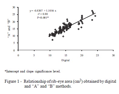
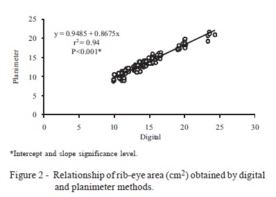
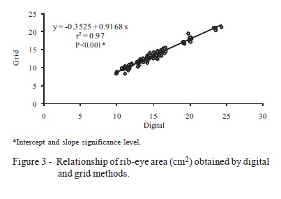
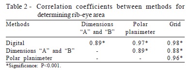
This study evaluated the correlation between measurements of rib-eye areas of sheep carcasses by traditional methods and those obtained by scanned images. Thirty pictures of the longissimus dorsi muscle of sheep carcasses were drawn on tracing paper and analyzed for muscle area (rib-eye) using four methods: scanned images, which utilizes the software DDA -Determinador Digital de Áreas (Digital Area Determiner); measurements "A" and "B" applied to the equation: (A/2 × B/2) × π; Planimeter method and rib-eye grid method. All rib-eye area figures were measured five times by each method, setting up a completely randomized experiment with four treatments and five replicates. Data were submitted to analysis of variance and means were compared by the Tukey test, Pearson correlation and linear regression by the SAS software. Easiness and difficulties perceived by the evaluators in the performance of each method were also recorded. The method of scanned images analyzed by the software DDA showed high correlation with the methods traditionally used, and can be considered feasible to determine carcass rib-eye area, with the advantage of being easy to operate, flexible, and economical.
carcass assessment; image analysis; longissimus dorsi area; software


