Morphology of greater rhea (Rhea americana) blood cells using light and scanning electron microscopy
ABSTRACT
The greater rhea (Rhea americana) is the largest bird native to Brazil and is classified as near threatened with extinction. Hematology is an essential tool for assessing the health status of birds. However, detailed information on the blood cell morphology of this species is still scarce. In this context, this study aimed to describe the morphology of greater rhea blood cells using light microscopy and scanning electron microscopy (SEM). We used 48 healthy adult specimens of both sexes kept in captivity in the Brazilian semiarid. Blood smears were prepared and stained using the Giemsa method for light microscopy analysis, and additional samples were processed for SEM. Elliptical nucleated erythrocytes, heterophiles, eosinophils, basophils, monocytes, lymphocytes, and thrombocytes were identified in light microscopy. Their morphological characteristics were generally compatible with those described for other birds and ratites. The high frequency of heterophiles and the predominantly elliptical shape of thrombocytes, a characteristic described as species-specific, stood out. SEM enabled detailed visualization of the surface architecture of blood cells, revealing irregularities, roughness, granulations, and cytoplasmic projections that are not observable by light microscopy. Greater rhea blood presents all major cell types, including red blood cells, monocytes, lymphocytes, eosinophils, heterophils, basophils, and thrombocytes. Although SEM is not applicable in routine clinical practice, its findings broaden the morphofunctional understanding of blood cells and provide important insights for comparative, physiological, and conservation studies involving other bird species.
Keywords:
Avian Hematology; Blood; Ratite.
RESUMO
A ema (Rhea americana) é a maior ave nativa do Brasil e está classificada como quase ameaçada de extinção. A hematologia constitui uma ferramenta essencial para a avaliação do estado de saúde de aves, porém informações detalhadas sobre a morfologia das células sanguíneas dessa espécie ainda são escassas. Nesse contexto, o presente estudo teve como objetivo descrever a morfologia das células sanguíneas de emas por meio de microscopia de luz e microscopia eletrônica de varredura (MEV). Foram utilizados 48 exemplares adultos, hígidos, de ambos os sexos, mantidos em cativeiro no semiárido brasileiro. Esfregaços sanguíneos foram preparados e corados pelo método de Giemsa para análise em microscopia de luz, enquanto amostras adicionais foram processadas para MEV. Na microscopia de luz, foram identificados eritrócitos nucleados de formato elíptico, além de heterófilos, eosinófilos, basófilos, monócitos, linfócitos e trombócitos, cujas características morfológicas foram, em geral, compatíveis com aquelas descritas para outras aves e ratitas. Destacou-se a elevada frequência de heterófilos e a forma predominantemente elíptica dos trombócitos, característica descrita como particular da espécie. A MEV permitiu a visualização detalhada da arquitetura superficial das células sanguíneas, evidenciando irregularidades, rugosidades, granulações e projeções citoplasmáticas não observáveis pela microscopia de luz. O sangue da ema apresenta todos os principais tipos celulares, incluindo hemácias, monócitos, linfócitos, eosinófilos, heterófilos, basófilos e trombócitos. Embora a MEV não seja aplicável à rotina clínica, seus achados ampliam a compreensão morfofuncional das células sanguíneas e fornecem subsídios importantes para estudos comparativos, fisiológicos e conservacionais envolvendo outras espécies de aves.
Palavras-chave:
Hematologia Aviária; Sangue; Ratita.
INTRODUCTION
Birds are among the most studied vertebrate groups for environmental preservation. Throughout their evolutionary history, they have developed physiological adaptations that are directly or indirectly related to essential ecological processes, such as plant pollination, and play a relevant role in pest control by feeding on various groups of invertebrates. Thus, birds are considered a group of animals of great functional importance, contributing to the balance of the ecosystems they inhabit and, consequently, to environmental sustainability (SANTOS et al., 2023).
The greater rhea (Rhea americana) is the largest bird species native to Brazil. It has conservation relevance, as it is classified as Near Threatened (BIRDLIFE INTERNATIONAL, 2022). Belonging to the order Rheiformes and the family Rheidae, the species is endemic to the American continent and is distributed mainly in open areas of eastern and central Brazil, as well as in Uruguay, Paraguay, and regions of northern and central Argentina (SOUSA et al., 2018; PICASSO; MOSTO, 2016). It is an omnivorous bird that feeds on a wide variety of plant matter, including leaves, fruits, and seeds, as well as invertebrates and small vertebrates (LÈCHE et al., 2021). Given these biological and ecological characteristics, deepening knowledge of the species' physiology, such as blood cell morphology, is fundamental for assessing health status and informing strategies for the management and conservation of this species.
Hematology is a fundamental diagnostic tool for assessing the health status of birds and other animal species, as hematological changes can occur even in the absence of obvious clinical signs, helping to monitor disease progression, evaluate the effectiveness of therapeutic protocols, and establish appropriate prognoses (SAMOUR, 2016). In this context, hematological tests allow the qualitative and quantitative analysis of erythrocytes and leukocytes, as well as the identification of cellular morphological changes that can provide important insights into the pathological mechanisms associated with different diseases (VILA, 2013).
Considering the biological and conservation relevance of rheas, as well as the scarcity of specific information on hematological parameters, especially in breeding conditions in the semiarid, this study aimed to describe the morphology of this species' blood cells to contribute to the advancement of knowledge regarding its blood physiology and subsidize studies aimed at animal health, zootechnical management, and conservation of native fauna.
MATERIAL AND METHODS
The samples were obtained from animals at the Center for the Multiplication of Wild Animals of the Universidade Federal Rural do Semi-Árido (CEMAS/UFERSA), located in the municipality of Mossoró, RN, Brazil, and registered with IBAMA as a scientific breeding facility (Registration No. 478912). This breeding site occupies an area of 20 ha, geographically located at 5°12'49.4" S and 37°18'36.7" W Gr, and 16 m above sea level, with an average annual temperature of about 27.4 °C, reaching 36 °C at certain times of the day, with relative humidity of about 70% (COSTA SARAIVA; VALE; ZANELLA, 2017).
We used 48 healthy adult greater rhea specimens of both sexes, distributed across nine screened paddocks with ample vegetation cover, totaling approximately 0.33 ha. The animals were kept under standardized feeding, receiving specific pelleted feed for rheas, supplemented with fruits, vegetable branches, and water ad libitum, according to the adopted CEMAS protocol. The experimental procedures were conducted in accordance with the ethical standards for the use of animals, as approved by the Ethics Committee for the Use of Animals (CEUA), under protocol CEUA No. 29/2024. Sampling and analysis waived SISBio authorization, in accordance with ICMBio Ordinance No. 748 of September 19, 2022, as they are exclusively ex situ procedures, involving the collection of blood from rheas kept in captivity.
The material was collected between November 2023 and February 2024. The animals were physically contained by the CEMAS keepers, with no more than 10 min per animal. The samples were taken in the morning, between 6:00 and 7:00, to avoid thermal stress. After containment, brachial vein venipuncture was performed, and peripheral blood was collected in sterile tubes containing 10% ethylenediamine tetra-acetic acid (EDTA) (5 mL). The samples were stored in expanded polystyrene boxes containing recyclable ice and taken to the Clinical Pathology Laboratory of the Veterinary Hospital of UFERSA.
Three blood smears from each animal were prepared for morphological analysis of blood cells and stained with the Giemsa method (VALKIÜNAS, 2004). After drying, they were analyzed and photographed under a light microscope (Leica DM 500 HD) at 1000x magnification (immersion oil). The most representative images were photographed using a Leica ICC50 HD equipment, to characterize the morphology of blood cell types.
The protocol described by Choudhary et al. (2021) was used to prepare samples for scanning electron microscopy (SEM) analysis of rheas blood cell types, performed in the Electron Microscopy Laboratory of the Center for Plant Research of the Semi-Arid (CPVSA) of UFERSA. The solution obtained was distributed onto 2x2 cm2 glass slides. After drying, they were stained by the Quick Panoptic method and subsequently subjected to the sputtering gold metallization process (Q150R ES, Quorum Technologies Ltda., Laughton, East Sussex, UK), followed by analysis under a scanning electron microscope, capturing images with a secondary electron detector in SEM (TESCAN VEJA 3 LMU, Tescan, Czech Republic), operating with a 10kV electron beam.
RESULTS AND DISCUSSION
Light Microscopy
In general, the hematology of birds presents similarities with that of other vertebrates, especially humans and other mammals; however, it exhibits relevant specificities, such as the presence of nucleated erythrocytes and the occurrence of thrombocytes and heterophils in peripheral blood, cells functionally equivalent to mammal platelets and neutrophils, respectively (CAMPBELL, 2015).
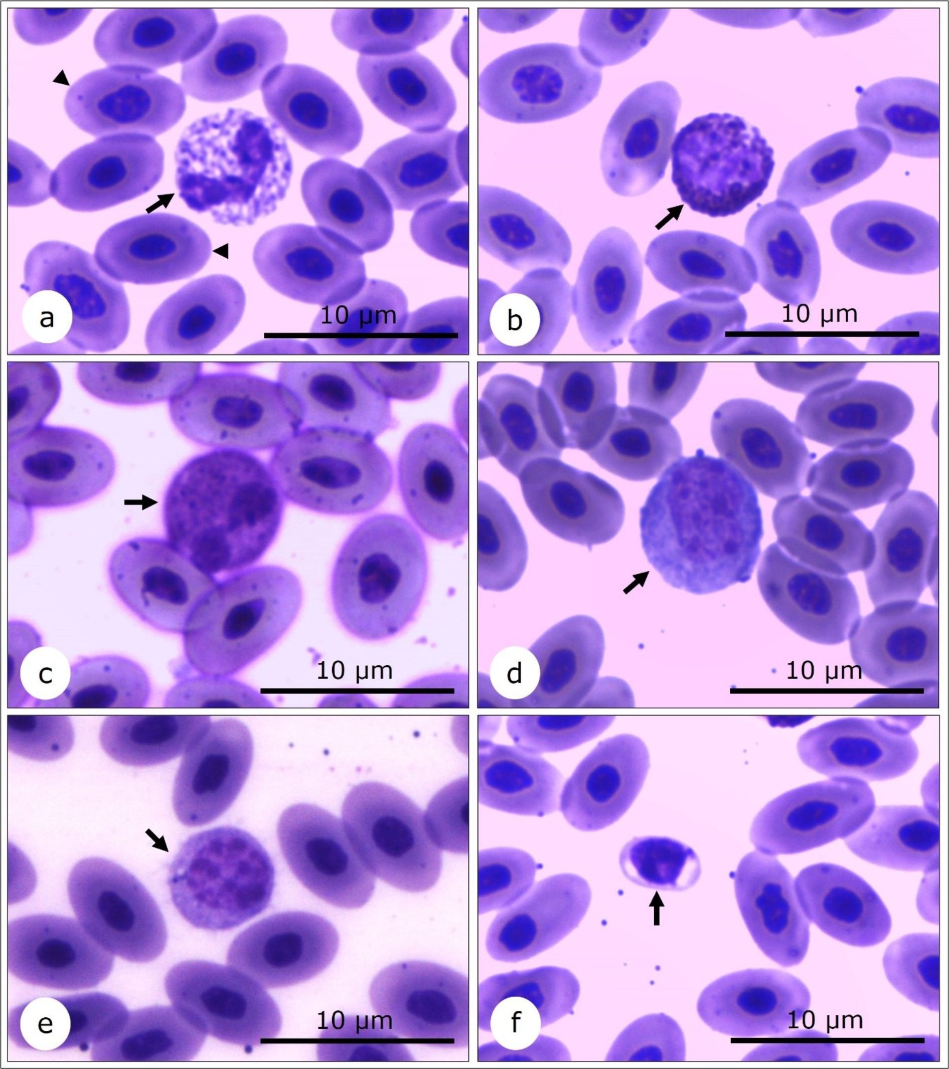
The erythrocyte morphology observed in the analyzed rheas follows the pattern described for most birds, characterized by nucleated cells, oval to elliptical in shape, with a central or slightly eccentric nucleus, also in oval or elliptical shape, and condensed chromatin. This pattern is widely described in the classical avian hematology literature (CAMPBELL, 2015; SAMOUR, 2016; JONES, 2015) and has also been reported in specific studies involving different groups of birds, including chickens, birds of prey, and ratites (CHANIE; HAILE, 2014; PINTO; HOLUM; GONDIM, 2017; CLARK, 2022). In R. americana, the elliptical erythrocyte conformation and the intense cytoplasmic eosinophilia observed in blood smears corroborate previous findings for the species (FORTES et al., 2009; GALLO et al., 2015) (Figure 1a), indicating morphological conservation of these cells among adult individuals maintained under different environmental conditions.
Morphology of greater rhea (Rhea americana) blood cells in blood smears subjected to Giemsa staining. (a) Heterophile (arrow) and mature erythrocytes (arrow heads). (b) Basophil (arrow). (c) Eosinophil (arrow). (d) Monocyte (arrow). (e) Lymphocyte (arrow). (f) Thrombocyte (arrow). 1000x magnification (immersion oil).
Heterophils constituted the most frequent leukocyte type in the blood smears of the rheas analyzed, a pattern compatible with that described for most avian species, in which these cells play a central role in the innate inflammatory response (CAMPBELL, 2015; SAMOUR, 2016). Morphologically, heterophiles presented as large, rounded cells with a high cytoplasm/nucleus ratio. The cytoplasm was clear and abundant, containing intensely eosinophilic, fusiform, red to orange granules with a rod-like appearance (Figure 1a) and a segmented nucleus. These characteristics are similar to those described for ratites and other domestic and wild birds (CHANIE; HAILE, 2014; JONES, 2015; CLARK, 2022). The absence of monolobular heterophiles in this study, unlike that observed by Gallo et al. (2015), may reflect intraspecific variation, the animals' physiological conditions, or methodological differences in morphological analysis.
Basophils, considered rare in bird blood smears (JONES, 2015), were observed at low frequency in the blood of rheas. They were spherical and distinguished by numerous intensely basophilic spherical granules, stained dark blue, that partially or completely covered the nucleus (Figure 1b). The cytoplasm was sparse, purple in color, and the basophils were smaller than heterophils and eosinophils. The nucleus was rounded, not segmented, centrally located, and with condensed chromatin, occupying a large portion of the cell volume.
These morphological characteristics are compatible with those described for avian basophils, including ratites, as reported by Fortes et al. (2009), Clark (2022), and Tadjalli et al. (2013). In birds, basophils are generally described as cells slightly smaller than heterophils, with round basophilic granules that often obscure the nucleus, findings corroborated by the present study (CHANIE; HAILE, 2014; CAMPBELL, 2015; JONES, 2015; PINTO; HOLUM; GONDIM, 2017).
The eosinophils observed in the blood smears of rheas were characterized as rounded cells with weakly stained cytoplasm, more basophilic than that of heterophils, containing small, round, and intensely eosinophilic granules, acidophilic in color ranging from pink to orange, evenly distributed in the cytoplasm, and occasionally overlapping the nucleus (Figure 1c). The nucleus was basophilic and polymorphic, usually bilobed, with condensed chromatin, located centrally or peripherally in relation to the cytoplasm, exhibiting violet coloration. These characteristics are compatible with those described for birds in general, including the presence of a bilobed nucleus and intensely colored eosinophilic granules (JONES, 2015; PINTO; HOLUM; GONDIM, 2017). Functionally, eosinophils are associated with modulating the inflammatory response and processes related to parasitosis. Their morphological variations are influenced by the species and the animal's physiological state. In ratites, similar morphological patterns have been described in ostriches and emus (Dromaius novaehollandiae), reinforcing the conservation of these cellular characteristics within the group (CLARK, 2022; TADJALLI et al., 2013).
The observed monocytes were characterized as large cells, larger than lymphocytes, with a rounded to irregular shape, abundant cytoplasm, a blue-gray color, and a basophilic character, with cytoplasmic vacuoles and small granules in dark blue tones (Figure 1d). The nucleus showed morphological variation, ranging from rounded to reniform, with little condensed chromatin compared with that of lymphocytes. This morphological pattern is consistent with that described for ratites, in which large monocytes with moderately abundant blue-gray cytoplasm, often vacuolized, and a pleomorphic nucleus with less condensed chromatin are considered typical characteristics (CLARK, 2022). Although some authors report the occurrence of monocytes with a rounded nucleus in birds (JONES, 2015; PINTO; HOLUM; GONDIM, 2017), this condition was not observed in the rheas analyzed in this study, corroborating the findings described for emus and ostriches, in which reniform or irregular nuclei also predominate (ALMEIDA et al., 2015; TADJALLI et al., 2013). These morphological variations reinforce the influence of species-specific factors in the characterization of monocytes in birds, especially within the ratite group.
Regarding avian lymphocytes, Mitchell and Johns (2008) describe that their morphology is largely similar to that observed in mammals. The blood smears of the rheas analyzed in this study showed rounded lymphocytes with round to slightly eccentric nuclei and intensely condensed chromatin, resulting in a high nucleus/cytoplasm ratio. The cytoplasm was scarce to moderate, homogeneous, and basophilic, with a light blue hue, occupying a small portion of the cell volume in relation to the nucleus, without the presence of cytoplasmic granules or vacuolation (Figure 1e). This morphological pattern is consistent with that previously described for the species by Fortes et al. (2009) and Gallo et al. (2015). Lymphocytes generally constitute the smallest and most numerous circulating mononuclear leukocytes in birds. Comparative studies involving different avian species, including rheas, emus, ostriches, chickens, and geese, indicate that these cells have an unsegmented, central, or discreetly eccentric nucleus, occupying almost the entire cell volume, with densely condensed chromatin and purple coloration, while the cytoplasm forms only a narrow, homogeneous, and basophilic halo around the nucleus (BHATTACHERJEE, 2023).
Thrombocytes in birds and in phylogenetically more basal vertebrates are nucleated blood cells and functionally equivalent to mammalian platelets. According to Claver (2005), these cells are observed in blood smears as ovoid or discoid structures, slightly smaller and more rounded than erythrocytes, with a round to oval nucleus, intensely condensed chromatin, and moderate cytoplasm that is bluish or translucent, and may exhibit a reticular or vacuolized appearance.
In rheas, thrombocytes were predominantly elliptical, with a strongly basophilic, rounded-to-elliptical nucleus containing condensed chromatin that occupied almost the entire cell volume, resulting in a high nucleus/cytoplasm ratio (Figure 1f). This pattern differs from that observed by Gallo et al. (2015), who reported round or oval thrombocytes in rheas. However, Fortes et al. (2009) highlighted that the elliptical shape of thrombocytes is an exclusive characteristic of R. americana, corroborating the findings of the present study. In other ratites, such as ostriches and rheas, thrombocytes are spherical to oval, as described by Tadjalli et al. (2013) and Almeida et al. (2015), demonstrating interspecific morphological variation.
Scanning Electron Microscopy (SEM)
The use of SEM in blood cell morphology studies significantly broadens understanding of the surface architecture and three-dimensional organization of these cells, allowing visualization of ultrastructural details that are not amenable to conventional light microscopy (CHOUDHARY et al., 2021). However, studies that employ SEM to characterize blood cells remain scarce, especially in wild birds and non-model species, underscoring the relevance and innovative nature of this approach.
Although the SEM is not a tool commonly used in routine veterinary medicine, mainly due to its high cost, technical complexity, and lack of availability in clinical laboratories, its use is highly relevant in the scientific context. The data obtained through this technique provide fundamental support for the construction of morphological reference bases, which can guide future research, interspecific comparisons, and more precise interpretations of findings from optical microscopy, strengthening the hematological and morphofunctional knowledge of the species studied.
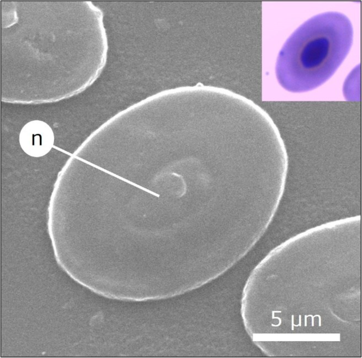
The mature erythrocytes of rheas observed by SEM (Figure 2) were presented as flat cells, with a smooth and relatively uniform surface, without evident projections or large roughnesses. The elliptical shape was well defined, with a prominent, elliptical, and centrally located internal structure corresponding to the nucleus, which showed the distinct texture of the cell membrane, providing morphological contrast between these structures. Similar results were reported by Narkkong, Aengwanich and Tanomthong (2010) in a study of the Indian crane (Grus antigone sharpii), in which erythrocytes were also characterized as flat cells with a smooth surface, although SEM images did not clearly show a nucleus, unlike in the present study.
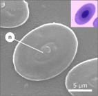
Greater rhea (Rhea americana) erythrocyte morphology observed by scanning electron microscopy (SEM), with 10,000x magnification, based on blood smears. The erythrocyte is oval in shape, with an evident nucleus (n), smooth plasma membrane surface, and regular cell contours.
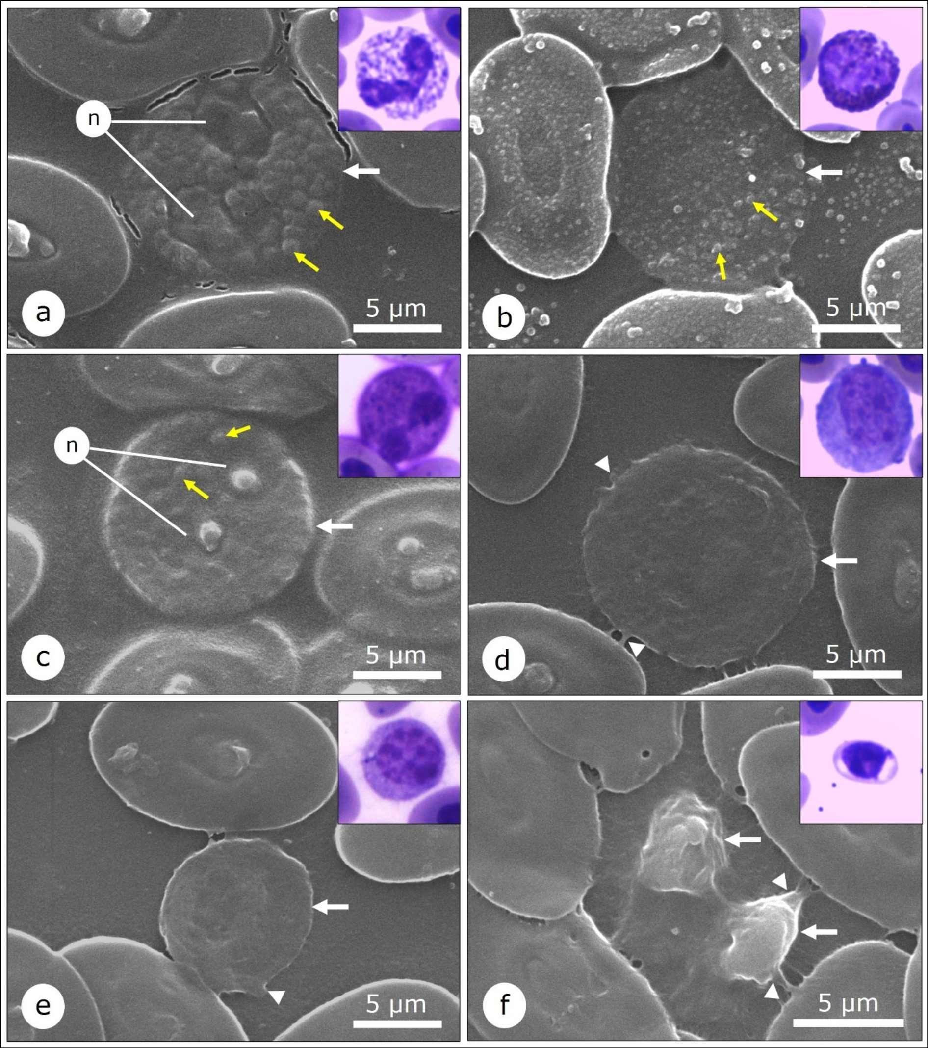
Among leukocytes, granulocytes and heterophiles (Figure 3a) had a rounded shape and an irregular membrane surface, with roughness and protuberances associated with cytoplasmic granulations and submembrane structures that outlined the conformation of the segmented nucleus. The basophils (Figure 3b) also presented a rounded shape, with an intensely textured surface, marked by roughness, elevations, and small projections compatible with microvilli, sometimes giving the cell a deformed or dentate appearance. Cytoplasmic granulations were evident as dense areas under the plasma membrane. On the other hand, eosinophils (Figure 3c) presented an irregular surface, with pronounced roughness and multiple submembrane granules or villi-like structures protruding from the cell surface. These findings are consistent with those reported by Narkkong, Aengwanich and Tanomthong (2010), who described granulocytes with rough surfaces and spicular projections, associated with cytoplasmic granulations, in G. antigone sharpii.
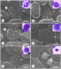
Morphology of greater rhea (Rhea americana) blood cells observed by scanning electron microscopy (SEM), with 10,000x magnification, from blood smears. (a) Heterophile (white arrow) with its nucleus (n) and granules (yellow arrows) evident. (b) Basophile (white arrow) with its granules (yellow arrows) evident. (c) Eosinophil (white arrow) with its nucleus (n) and granules (yellow arrows) evident. (d) Monocyte (white arrow) with cytoplasmic projections (arrow heads). (e) Lymphocyte (white arrow) with cytoplasmic projections (arrow heads). (f) Thrombocytes (white arrows) with cytoplasmic projections (arrow heads).
The monocytes had a larger cell size and an irregular, rough membrane surface with folds and projections (Figure 3d), characteristics consistent with their high phagocytic activity and tissue-migration capacity. These topographic modifications of the plasma membrane may favor particle adhesion, recognition, and internalization, as described for monocytes in other birds and vertebrates analyzed by SEM (NARKKONG; AENGWANICH; TANOMTHONG, 2010; CHOUDHARY et al., 2021). In contrast, lymphocytes had a smaller cell size and a relatively more homogeneous surface, although they showed a slightly rough texture and discrete fine projections (Figure 3e). This surface conformation may be related to its predominantly regulatory and antigenic recognition functions, which demand lower membrane plasticity than in monocytes (EROLES et al., 2023).
Thrombocytes, in turn, stood out for their highly complex surface, with multiple irregularities, folds, and membrane extensions resembling pseudopods (Figure 3f), characteristics that are not visible by light microscopy, underscoring the importance of SEM for identifying these morphological details. These structures are classically associated with cell adhesion, aggregation, and interaction with the extracellular matrix, reinforcing the functional role of avian thrombocytes in hemostatic and inflammatory processes (MANDEL et al., 2022).
CONCLUSION
The blood of the species R. americana presents all major cell types, including red blood cells, monocytes, lymphocytes, eosinophils, heterophils, basophils, and thrombocytes. The leukocytes of this species differ in morphology from those described for other birds, and the thrombocytes are elliptical, a characteristic previously described exclusively in rheas. Additionally, the unprecedented analysis of greater rhea blood cells using scanning electron microscopy enabled detailed characterization of the surface morphology of different cell types, revealing variations in surface texture among them.
Data Availability:
The data that support the findings of this study can be made available, upon reasonable request, from the corresponding author.
REFERENCES
- ALMEIDA, A. J. et al. Clinical aspects, morphometry, and morphology of blood cells of the emu (Dromaius novaehollandiae) reared in captivity. Comparative Clinical Pathology 24: 1553-1558, 2015.
- BHATTACHERJEE, A. Avian Erythrocytes and Agranulocytes-a Review. Science Reviews. Biology, 2: 21-29, 2023.
-
BIRDLIFE INTERNATIONAL. 2022. Rhea americana. The IUCN Red List of Threatened Species 2022: e.T22678073A219615764. Available at: <https://www.iucnredlist.org/species/22678073/219615764>. Access on: Mai. 17, 2024.
» https://www.iucnredlist.org/species/22678073/219615764 - CLARK, P. Hematology of Ratites. In: BROOKS, M. B. et al. (Eds.). Schalm's Veterinary Hematology Hoboken, NJ: John Wiley & Sons, 2022. cap. 127, p. 1159-1165.
- CAMPBELL, T. W. Hematologia das Aves. In: THRALL, M. A.; WEISER, G.; ALLISON, R. W.; CAMPBELL, T. W. (Eds.). Hematologia e Bioquímica Clínica Veterinária Rio de Janeiro, RJ: Guanabara Koogan, 2015. cap. 19, p. 507-586.
- CHANIE, M.; HAILE, Y. Comparative aspects of the clinical hematology of birds: a review. British Journal of Poultry Sciences, 3: 88-95, 2014.
- CHOUDHARY, O. P. et al. Preparation of blood samples for electron microscopy: The standard protocol. Annals of Medicine and Surgery, 70: 1-4, 2021.
- CLAVER, J. A. El trombocito aviar. InvestigaciónVeterinaria,7: 139-146, 2005.
- COSTA SARAIVA, A. L. B.; VALE, C. C.; ZANELLA, M. E. Comportamento dos Elementos Climáticos no Município de Mossoró (RN) e os Impactos na Saúde Humana. Revista GeoInterações, 1: 87-105, 2017.
- EROLES, M. et al. Coupled mechanical mapping and interference contrast microscopy reveal viscoelastic and adhesion hallmarks of monocyte differentiation into macrophages. Nanoscale, 15: 12255-12269, 2023.
- FORTES, E. A. M. et al. Morfologia das células do sangue periférico ememas (Rhea americana) Brazilian Journal of Veterinary Research and Animal Science, 46: 215-221, 2009.
- GALLO, S. S. M. et al. Hematological, morphological and morphometric characteristics of blood cells from rhea, Rhea americana (Struthioniformes: Rheidae): a standard for Brazilian birds. Brazilian Journal of Biology, 75: 953-962, 2015.
- JONES, M. P. Avian Hematology, Clinics in Laboratory Medicine, 35: 649-659, 2015.
- LÈCHE, A. et al. First assessment of persistent organic pollutants in the Greater rhea (Rhea americana), a near-threatened flightless herbivorous bird of the Pampas grasslands. Environmental Science and Pollution Research, 28: 27681-27693, 2021.
- MANDEL, J. et al. Beyond Hemostasis: Platelet Innate Immune Interactions and Thromboinflammation. International Journal of Molecular Sciences, 23: 3868, 2022.
- MITCHELL, E. B.; JOHNS, J. Avian hematology and related disorders. Veterinary Clinics of North America: Exotic Animal Practice, 11: 501-522, 2008.
- NARKKONG, N.A.; AENGWANICH, W.; TANOMTHONG, A. Morphology and morphometrics of hematological cells from eastern sarus crane, Grus antigone sharpii Comparative Clinical Pathology, 20: 299-304, 2010.
- PICASSO, M. B. J.; MOSTO, C. The new taxonomic status of Rhea anchorenensis (Ameghino and Rusconi, 1932) (Aves, Palaeognathae) from the Pleistocene of ArgentinaLe nouveau statut taxonomique de Rhea anchorenensis (Ameghino et Rusconi, 1932) (Aves, Palaeognathae) du Pléistocène d’Argentine. Annales de Paléontologie, 102: 237-241, 2016.
- PINTO, F. M. S. C.; HOLUM, S. B.; GONDIM, L. S. Q. Atlas virtual de hematologia comparada de aves mantidas sob cuidado humano. In: XVI SEMINÁRIO ESTUDANTIL DE PRODUÇÃO ACADÊMICA, 2017, Salvador. Anais… Salvador: UNIFACS, 2017. p. 381-393.
- SAMOUR, J. Avian Medicine 3. ed. Amsterdam: Elsevier, 2016. 699 p.
- SANTOS, T. F. et al. Interações entre aves e plantas: um estudo bibliométrico sobre a contribuição das aves como polinizadoras e dispersoras da flora. Contribuciones a Las Ciencias Sociales, 16: 24745-24759, 2023.
- SOUSA, R. P. et al. Morfogênese do sistema respiratório da ema (Rhea americana) em diferentes estágios embrionários e fetais. Pesquisa Veterinária Brasileira, 38: 154-166, 2018.
- TADJALLI, M. et al. Histomorphometric study on blood cells in male adult ostrich. Veterinary Research Forum, 4: 199-203, 2013.
- VALKIÜNAS, G. Avian malaria parasites and other haemosporidia 1 ed. Washington: CRC Press, 2004. 932 p.
-
VILA, L. G. Hematologia em Aves: Revisão de literatura 2013. Available at: https://files.cercomp.ufg.br/weby/up/67/o/2013_Laura_Garcia_Seminario1corrig.pdf Access on: May 17, 2024.
» https://files.cercomp.ufg.br/weby/up/67/o/2013_Laura_Garcia_Seminario1corrig.pdf
-
Editor in Chief:
Aurélio Paes Barros Júnior