 Laboratory evaluation of an optical fiber illuminator on the adhesive bond strength of resin cements to high-translucency zirconia
ABSTRACT
Objective: To evaluate in vitro the bond strength of resin cements in high-translucency zirconia restorations as a function of the light-curing system and the type of resin cement.
Methods: Forty-eight third molars received high-translucency zirconia (KatanaTM – Kota – Tokyo, Japan) and were randomly distributed into four groups (n=12) according to the light-curing system (indirect with commercial equipment or direct with the new optical fiber illuxminator) and resin cement type (light- or dual-cured). The optical fiber device conducts LED light to hard-to-reach areas. A micro shear bond strength test applied a 50 Kgf load at 0.5 mm/min until failure. Two-way ANOVA and Tukey’s test (5% significance) analyzed results.
Results: The highest bond strength occurred in the direct optical fiber system with dual-cure cement (p<0.05), also increasing bond strength for light-cured cement.
Conclusion: Within the limitations of this laboratory study, the use of the optical fiber illumination system was associated with higher bond strength values of resin cements to high-translucency zirconia compared with the conventional system, depending on the type of cement used. These findings indicate that the device may favor adhesion in situations with limited light access; however, further studies are required to establish its clinical relevance.
Indexing terms
Dental curing lights; Polymerization; Resin cements
RESUMO
Objetivo: Avaliar in vitro a resistência de união de cimentos resinosos em restaurações de zircônia de alta translucidez em função do sistema de fotopolimerização e do tipo de cimento resinoso.
Métodos: Quarenta e oito terceiros molares receberam restaurações indiretas de zircônia de alta translucidez (Katana™ – Kota – Tóquio, Japão) e foram distribuídos aleatoriamente em quatro grupos (n= 12 ), de acordo com o sistema de fotopolimerização (indireto com equipamento comercial ou direto com o novo iluminador de fibra óptica) e o tipo de cimento resinoso (fotoativado ou dual). O dispositivo de fibra óptica conduz a luz LED até áreas de difícil acesso. Um teste de resistência de união por microcisalhamento foi realizado com carga de 50 Kgf a 0,5 mm/min até a falha. Resultados: Os resultados foram analisados por ANOVA dois fatores e teste de Tukey (nível de significância de 5%).
Resultados: A maior resistência de união foi observada no sistema direto com fibra óptica associado ao cimento dual (p< 0,05), com aumento também da resistência de união para o cimento fotoativado.
Conclusão: Dentro das limitações deste estudo laboratorial, o uso do sistema de iluminação por fibra óptica esteve associado a maiores valores de resistência de união dos cimentos resinosos à zircônia de alta translucidez, quando comparado ao sistema convencional, dependendo do tipo de cimento utilizado. Esses achados indicam que o dispositivo pode favorecer a adesão em situações de acesso restrito à luz, embora estudos adicionais sejam necessários para estabelecer sua relevância clínica.
Termos de indexação
Cimento resinoso; Luz de cura dentária; Polimerização
INTRODUCTION
Dental ceramics are materials routinely used in aesthetic restorative procedures due to their excellent properties, such as high compressive strength, high translucency and fluorescence, chemical stability, low electrical conductivity, and a thermal expansion coefficient similar to dental structure [1] Adequate cementation and adhesion between the dental substrate and the ceramic are decisive factors to achieve successful prosthetic treatment with long-term restoration durability. Furthermore, the primary photoinitiator, camphorquinone, is the most commonly used in resin-based restorative materials, as it is sensitive to the visible spectrum of blue light (425–510 nm) [2]. Manufacturers use co-initiators to enhance the degree of conversion of resin cement in regions which are not directly exposed to light since it acts in the chemical conversion of cements [3]. However, even in dual-cure cements, the efficiency of photoactivation also depends on adequate exposure to ensure the correct conversion of monomers and thus improved physical properties [4-6]. The conversion of monomers into polymers is never complete, leaving considerable amounts of unreacted methacrylate groups or residual monomers within the material, and studies have reported a degree of conversion ranging from 35% to 80% [6-9].
An inadequate degree of conversion results in low wear resistance, microleakage, tissue inflammatory reactions [10], increased sorption and solubility, color instability, and a higher likelihood of failures and restoration loss [8,11-14]. This degradation can consequently lead to the restoration dislocating due to the marginal seal dissolving, resulting in fractures and/or secondary caries, along with the risk of a local inflammatory response in the pulp caused by residual monomers [15].
The distance between the light -curing equipment and the cement leads to reduced light dissipation from the restorative material thickness. The size and quantity of filler particles cause a gradual decrease in polymerization, known as the curing depth, which influences the structure of the formed polymer [16-21]. Therefore, polymerization in cervical proximal boxes is inadequate, as the light, which is already absorbed by the material, is also affected by the difficulty of reaching the region directly. Thus, the greater the restoration depth and thickness, the less light will penetrate, and consequently the monomer conversion will be lower [14,22-27].
Considering the current limitations in delivering LED light to areas of difficult access during the cementation of indirect restorations, this study aimed to develop an optical fiber–based illumination system to reach restricted regions and to evaluate its influence on the bond strength of resin cements to zirconia restorations under in vitro conditions.
METHODS
Specimen preparation
A total of 48 intact human third molars (Ethics Committee, Brazil Platform, CEP 58905222.0.0000.0077) were used and randomly assigned to 4 experimental groups (n=12) based on the type of photopolymerization system, and the type of resin cement applied (chart 1). The composition of each resin cement is described in chart 2. The sample size was determined based on a statistical power analysis applying the G*Power 3.1 software [28].
Shear bond strength sample preparation
Translucent zirconia blocks (KATANA STML, Tokyo, Japan) were fixed onto a metal base in a cutting machine (ISOMET 1000, BUEHLER, USA) and sectioned perpendicular to the long axis (perpendicular to the y-axis) using a WFR BLDE 4” X 0.12” X 1/2” diamond disc (EXTEC-ERIOS, Juiz de Fora, Minas Gerais, Brazil) under constant water cooling, obtaining 48 slices of 2 mm thickness. Both sides of each disc were manually polished using water abrasive papers with grits 800 and 1200 (3M, Saint Paul, Minnesota, USA) on an Ecomet 250 polishing machine (Buehler, Lake Bluff, Illinois, USA) under water cooling until reaching 2 mm thickness and rounded angles were achieved.
After polishing, the zirconia samples were partially embedded in a PVC cylinder with a height of 3 cm in order to obtain support. The PVC cylinders with the zirconia slice positioned at the center of the cylinder were embedded with chemically activated acrylic resin (JET, Juiz de Fora, Minas Gerais, Brazil).
The 48 teeth were fixed onto a metal base in an ISOMET 1000 cutting machine (Buehler, Lake Bluff, Illinois, USA) and sectioned perpendicular to the long axis (perpendicular to the y-axis) using a WFR BLDE 4” X 0.12” X 1/2” diamond disc (EXTEC-ERIOS) under constant water cooling. The first cut was made 1 mm from the crown, removing the enamel portion, followed by a second cut to 3 mm in thickness. Both sides of each disc were manually polished using water abrasive papers with grits 800 and 1200 (3M, Saint Paul, Minnesota, USA) on an Ecomet 250 polishing machine (Buehler, Lake Bluff, Illinois, USA) under water cooling until the 3 mm thickness and rounded angles were achieved.
Optical fiber illuminator device system
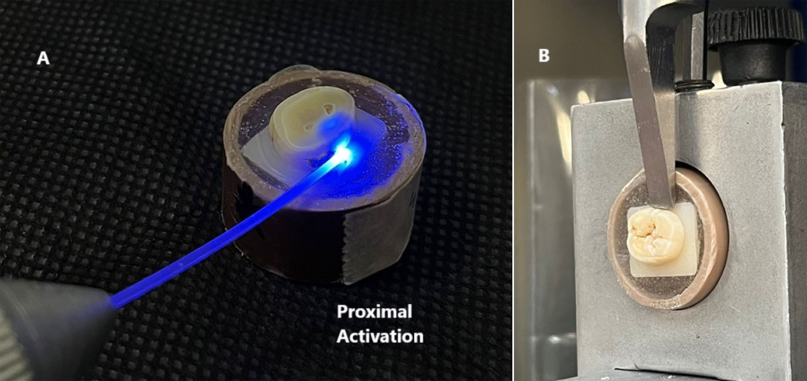
The optical illuminator device was co-developed with Photonita Metrologia Óptica Ltda company (Florianópolis, Santa Catarina, Brazil). A metal device was used, which has an opening through which a 1 mm diameter, circular-section optical fiber cable passed through. The optical fiber conducted the LED light from the Radii Cal-SDI light-curing unit (SDI Limited, Bayswater, Victoria, Australia) at the respective power levels, aiming to more effectively activate the restoration area in the proximal preparation toward the cervical area, near the cementoenamel junction (CEJ). The design of the innovative optical illuminator device system (figure 1).

A) Complete fiber optic illuminating device being activated by a curing light. B) Fiber optic conducting the advised light from the curing light.
Calculation of the photopolymerization time for the new optical lighting system
The time needed for radiant exposure of 16J was calculated to determine the photopolymerization time for the new system. A power meter (Nova Handheld Laser Power Meter, OphirOptronics, Jerusalem, Israel) was used to measure the power of the LED Radii Cal device associated with the optical fiber illuminator system, reaching a value of 1.626 mW. The optical fiber area (0.00785 cm²) was considered to calculate the irradiance, resulting in 207.13 mW/cm², or 0.207 J/cm². The photopolymerization time was calculated using the following equation:
Radiant Exposure (j) = Irradiance (J/cm²) x time (s)
Time (s) = Radiant Exposure (j) / Irradiance (J/cm²)
Time (s) = 16 (j)/ 0.207 (J/cm²)
Time (s) = 80 Seconds
Thus, complementary photopolymerization was performed on the preparation notches of the proximal box for 80 seconds in each third in the buccal-lingual direction, followed by an indirect complimentary activation for 45 seconds at a distance of 6 mm per proximal without the use of the optical fiber lighting system. The photopolymerization of the resin cement without the optical fiber lighting system was performed according to the manufacturer’s recommendations and photopolymerized for 45 seconds at a 6 mm distance per proximal. The photopolymerization was conducted in a dark room, protected from any external ambient light.
Specimen cementation protocol
The cementation step was performed according to the manufacturer’s recommendations for the resin cement (FGM – Dental Group – Santa Catarina – Brazil) using the Allcem Veneer APS (light-cured) and Allcem Dual dual-body cements. The zirconia slices were subjected to sandblasting with aluminum oxide and then washed in an ultrasonic bath for 5 minutes with deionized water to eliminate debris. The universal primer Monobond (Ivoclar, Liechtenstein) was then applied with the microbrush for 60 seconds. The dentin structure was treated by applying 37% phosphoric acid (Condac 37, FGM Dental Group) for 15 seconds, followed by thorough rinsing with water. Then, the adhesive system (Ambar, FGM Dental Group) was applied actively with the microbrush, and excess material was removed. Next, a dark barrier was placed to block light interference, and photoactivation was performed using or not using the new fiber optic photoactivation system as described in chart 1 (figure 2A).
A) Sample being photopolymerized in the proximal region with the fiber optic illuminating device. B) Sample in the microshear test in EMIC.
Micro-shear adhesive bond strength test
After cementation, the samples were stored in an incubator at 37°C, protected from light. For the mechanical test, the cylinders were mounted on a universal mechanical testing machine (EMIC DL 1000, EMIC, Brazil). The micro-shear test was performed using a knife-shaped blade and a 50 Kgf load cell at a speed of 0.5 mm/min until sample failure occurred (figure 2B). The adhesive strength (MPa) was calculated using the formula R = C/A, where C = rupture load (N), and A = bonded interfacial area (mm²). The adhesive strength values were subjected to statistical analysis.
Statistical analysis
The values obtained in the tests were subjected to the respective statistical analyses suitable for each test. A normality test (Shapiro-Wilk) was first conducted, and based on the parametric distribution, a two-way ANOVA was performed with a comparison of two independent variables: (Photopolymeralization System x Cement), followed by Tukey’s multiple comparison test, with a significance level of 5%.
RESULTS
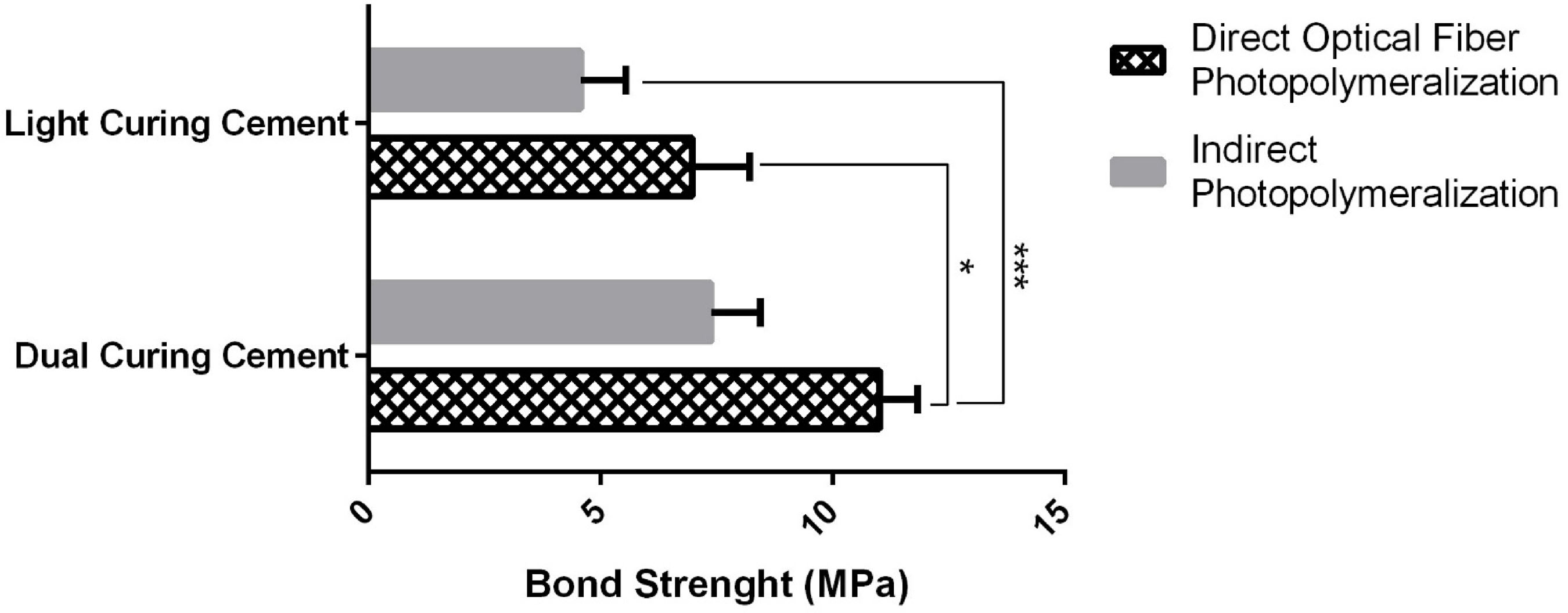
Two-way ANOVA showed that both the photoactivation system and the type of resin cement significantly influenced micro-shear bond strength values (p<0.05). The highest bond strength values related to the photoactivation system were obtained with direct photoactivation using the optical fiber device combined with the dual-cured resin cement (p<0.05).
For the light-cured resin cement, a significant increase in bond strength was observed when the optical fiber photoactivation system was used, resulting in values comparable to those obtained with the dual-cured cement under indirect photoactivation (table 1 and figure 3).
Considering the type of cement, the dual-cured resin cement showed significantly higher bond strength values than the light-cured resin cement, regardless of the photoactivation system (p< 0.05).
DISCUSSION
The success of a restoration is directly related to adequate photopolymerization. This study presented a fiber optic illuminator system, which proved to be an effective alternative to improve photopolymerization, as both types of tested cements showed a significant increase in bond strength due to better LED light conduction to areas with difficult access in the posterior region.
The literature shows that proper monomer conversion is related to the good performance of the photopolymerizer [29]. In addition, Sulaiman et al. [30] and Farah et al. [31] observed that light exposure through zirconia is affected by its opacity, so that the thicker the material, the lower the irradiance received. In this context, the increase in bond strength observed in both groups when using the new photopolymerization system suggests greater monomer-to-polymer conversion due to improved light penetration.
In order to better conduct light, the research group developed the fiber optic illuminator device, designed to deliver light to areas with difficult access. In the study by Inagati et al. [32], this system showed higher monomer-to-polymer conversion in the apical third of the root canal, a region widely reported in the literature as difficult to reach with light [33]. In the present study, the device also resulted in higher bond strength in both groups subjected to direct photopolymerization, expanding the potential clinical applications of the system. These findings corroborate the study by Ho et al. [34], which demonstrated that groups exposed to higher light intensity, even in more distant regions, showed a higher degree of conversion.
Another relevant point is that the comparison between light-cured and dual-cured cements has limitations, as dual-cured cement naturally shows higher strength due to chemical settings. However, analyzing cement that sets exclusively by light allows for better evaluation of the photopolymerization system’s effectiveness. In addition, the light-cured cement group with direct activation showed bond strength (6.98 MPa) similar to that of the dual-cured cement group with indirect activation (7.38 MPa). This finding has clinical relevance, as it allows considering the use of exclusively light-cured cements for restoration cementation, providing more treatment options for the prosthodontist in daily practice.
This study presents limitations inherent to the in vitro model. The simulation of proximal preparation does not fully reproduce the natural barrier represented by the adjacent tooth, which may influence light distribution and bond strength. Furthermore, as this is a pilot study, the primary objective was to evaluate bond strength under these conditions. The device, however, is part of an ongoing sequence of studies, including different types of restorations, other photopolymerizers, and additional analyses such as degree of conversion, fracture tests, aging, and microscopy. As this is the first study to propose the use of a new alternative photopolymerization system for indirect zirconia restorations, further mechanical and aging evaluations should be performed to better understand its behavior, in association with different ceramic restoration materials or resin cements, aiming to enable its potential clinical application in the future.
CONCLUSION
Within the limitations of this laboratory study, the preliminary findings suggest that the use of a direct optical fiber–based lighting system was associated with higher adhesive bond strength values, likely due to improved light delivery in areas of limited access, for both resin cement types. These results indicate that this approach may represent a potential alternative for light delivery in posterior or hard-to-reach regions; however, further studies are required to confirm its clinical applicability.
-
Article based on the dissertation by CG França, entitled “Influência de um novo sistema de fotopolimerização na resistência adesiva dos cimentos resinosos em uma restauração Inlay em zircônia de alta translucidez na região posterior”. Universidade Estadual Paulista Júlio de Mesquita Filho; 2024.
-
How to cite this article
França CG, Paes Junior TJA, Gomes LCL. Laboratory evaluation of an optical fiber illuminator on the adhesive bond strength of resin cements to high-translucency zirconia. RGO, Rev Gaúch Odontol. 2026;74:e20260020. doi: http://dx.doi.org/10.1590/1981-86372026002020250059
-
Support
Conselho Nacional de Desenvolvimento Científico e Tecnológico for granting the PIBITI Scholarship (announcement 03/2022; proposal 5600) and the Photonita – Metrologia Óptica company for providing the optical fiber.
Data Availability
The research data are available from the corresponding author upon reasonable request.
REFERENCES
-
1 Borges GA, Sophr AM, De Goes MF, Sobrinho LC, Chan DCN. Effect of etching and airborne particle abrasion on the microstructure of different dental ceramics. J Prosthet Dent. 2003 [cited 2024 Nov 25];89(5):479-88. Available from: https://pubmed.ncbi.nlm.nih.gov/12806326/
» https://pubmed.ncbi.nlm.nih.gov/12806326/ -
2 Caldarelli PG, Beltrani FC, Pereira SK, Cardoso SA, Hoeppner MG. Aparelhos fotopolimerizadores: evolução e aplicação clínica: uma revisão da literatura. Odontol Clín-Cient. 2011 [cited 2025 Aug 24];10(4):317-21. Available from: http://revodonto.bvsalud.org/scielo.php?script=sci_arttext&pid=S1677-38882011000400003&lng=pt&nrm=iso&tlng=pt
» http://revodonto.bvsalud.org/scielo.php?script=sci_arttext&pid=S1677-38882011000400003&lng=pt&nrm=iso&tlng=pt -
3 Vrochari AD, Eliades G, Hellwig E, Wrbas KT. Curing efficiency of four self-etching, self-adhesive resin cements. Dent Mater. 2009 [cited 2024 Nov 25];25(9):1104-8. Available from: https://pubmed.ncbi.nlm.nih.gov/19427029/
» https://pubmed.ncbi.nlm.nih.gov/19427029/ - 4 Aldhafyan M, Silikas N, Watts DC. Influence of curing modes on conversion and shrinkage of dual-cure resin-cements. Dental Mater. 2022;38(1):194-203.
- 5 Pereira JR, Lins Do Valle A, Ghizoni JS, Lorenzoni FC, Barbosa MR, Dos Reis Só MV. Push-out bond strengths of different dental cements used to cement glass fiber posts. J Prosthet Dent. 2013;110(2):134-40.
-
6 Pedreira APRV, Pegoraro LF, de Góes MF, Pegoraro TA, Carvalho RM. Microhardness of resin cements in the intraradicular environment: effects of water storage and softening treament. Dent Mater. 2009 [cited 2024 Nov 25];25(7):868-76. Available from: https://pubmed.nc bi.nlm.nih.gov/19217152/
» https://pubmed.nc bi.nlm.nih.gov/19217152/ -
7 Asmussen E. Factors affecting the quantity of remaining double bonds in restorative resin polymers. Scand J Dent Res. 1982 [cited 2024 Nov 25];90(6):490-6. Available from: https://pubmed.ncbi.nlm.nih.gov/6218604/
» https://pubmed.ncbi.nlm.nih.gov/6218604/ -
8 Ferracane JL. Correlation between hardness and degree of conversion during the setting reaction of unfilled dental restorative resins. Dent Mater. 1985 [cited 2024 Nov 25];1(1):11-4. Available from: https://pubmed.ncbi.nlm.nih.gov/3160625/
» https://pubmed.ncbi.nlm.nih.gov/3160625/ -
9 Noronha Filho JD, Brandão NL, Poskus LT, Guimarães JGA, da Silva EM. A critical analysis of the degree of conversion of resin-based luting cements. J Appl Oral Sci. 2010 [cited 2024 Nov 25];18(5):442-6. Available from: https://www.scielo.br/j/jaos/a/3xvjfT3S8vnCc3VQ8JgxCpm/?lang=en
» https://www.scielo.br/j/jaos/a/3xvjfT3S8vnCc3VQ8JgxCpm/?lang=en -
10 Kondo T, Kakinuma H, Ambo S, Otake K, Sato Y, Egusa H. Influence of dual-cure resin-cement curing modes on gingival cytotoxicity and inflammatory responses. J Dent Sci. 2024 [cited 2026 Jan 12];20(1):586. Available from: https://pmc.ncbi.nlm.nih.gov/articles/PMC11762211/
» https://pmc.ncbi.nlm.nih.gov/articles/PMC11762211/ -
11 Örtengren U, Wellendorf H, Karlsson S, Ruyter IE. Water sorption and solubility of dental composites and identification of monomers released in an aqueous environment. J Oral Rehabil. 2001 [cited 2024 Nov 25];28(12):1106-15. Available from: https://pubmed.ncbi.nlm.nih.gov/11874509/
» https://pubmed.ncbi.nlm.nih.gov/11874509/ -
12 Rosenstiel SF, Land MF, Crispin BJ. Dental luting agents: a review of the current literature. J Prosthet Dent. 1998 [cited 2024 Nov 25];80(3):280-301. Available from: https://pubmed.ncbi.nlm.nih.gov/9760360/
» https://pubmed.ncbi.nlm.nih.gov/9760360/ -
13 Magão PH, Dos Santos AF, Chiok Ocaña LR, Rizzante FAP, Furuse AY. Water sorption and color stability of dental composites light-cured with a broadband LED device and different radiant exposures. Braz Dent Sci. 2024 [cited 2025 May 27];27(3). Available from: https://ojs.ict.unesp.br/index.php/cob/article/view/4379/4911
» https://ojs.ict.unesp.br/index.php/cob/article/view/4379/4911 -
14 Meleka NM, El-Banna A, El-Korashy DI. Color stability and degree of conversion of amine-free dual cured resin cement used with two different translucencies of lithium disilicate ceramics. Braz Dent Sci. 2023 [cited 2025 May 27];26(2). Available from: https://ojs.ict.unesp.br/index.php/cob/article/view/3614/4769
» https://ojs.ict.unesp.br/index.php/cob/article/view/3614/4769 - 15 De Souza G, Braga RR, Cesar PF, Lopes GC. Correlation between clinical performance and degree of conversion of resin cements: a literature review. J Appl Oral Sci. 2015;23(4):358-68.
-
16 Pick B, Gonzaga CC, Junior WS, Kawano Y, Braga RR, Cardoso PEC. Influence of curing light attenuation caused by aesthetic indirect restorative materials on resin cement polymerization. Eur J Dent. 2010 [cited 2024 Nov 25];4(3):314. Available from: https://pmc.ncbi.nlm.nih.gov/articles/PMC2897866/
» https://pmc.ncbi.nlm.nih.gov/articles/PMC2897866/ -
17 Roberts HW, Leonard DL, Vandewalle KS, Cohen ME, Charlton DG. The effect of a translucent post on resin composite depth of cure. Dental Mater. 2004 [cited 2024 Nov 25];20(7):617-22. Available from: https://pubmed.ncbi.nlm.nih.gov/15236935/
» https://pubmed.ncbi.nlm.nih.gov/15236935/ -
18 Asmussen E, Peutzfeldt A. Polymer structure of a light-cured resin composite in relation to distance from the surface. Eur J Oral Sci. 2003 [cited 2024 Nov 25];111(3):277-9. Available from: https://onlinelibrary.wiley.com/doi/full/10.1034/j.1600-0722.2003.00044.x
» https://onlinelibrary.wiley.com/doi/full/10.1034/j.1600-0722.2003.00044.x -
19 Bilge K, İpek İ. Effects of different LED light curing units on the degree of conversion and microhardness of different composites: FT-IR and SEM-EDX analysis. Polymer Bull. 2024 [cited 2025 Aug 24];81(15):13825-38. Available from: https://link.springer.com/article/10.1007/s00289-024-05362-2
» https://link.springer.com/article/10.1007/s00289-024-05362-2 -
20 Marcos J, Silva C, Barreto De Negreiros A, Silva R, De A, Brandim S, et al. Influence of Using Different Light Curing Devices on the Microhardness of a Bulk-Fill Resin. J Health Sci. 2021 [cited 2025 Aug 24];23(3):234-7. Available from: https://journalhealthscience.pgsscogna.com.br/JHealthSci/article/view/8745
» https://journalhealthscience.pgsscogna.com.br/JHealthSci/article/view/8745 -
21 Braga SSL, Price RB, Juckes SM, Sullivan B, Ribeiro MTH, Soares CJ. Effect of thickness and surface finish on light transmission through zirconia-reinforced lithium silicate and lithium disilicate dental ceramics. Braz Dent J. 2025 [cited 2026 Jan 12];36:e25-6572. Available from: https://pmc.ncbi.nlm.nih.gov/articles/PMC12696804/
» https://pmc.ncbi.nlm.nih.gov/articles/PMC12696804/ -
22 Heffernan MJ, Aquilino SA, Diaz-Arnold AM, Haselton DR, Stanford CM, Vargas MA. Relative translucency of six all-ceramic systems. Part I: Core materials. J Prosthet Dent. 2002 [cited 2024 Nov 26];88(1):4-9. Available from: https://pubmed.ncbi.nlm.nih.gov/12239472/
» https://pubmed.ncbi.nlm.nih.gov/12239472/ -
23 Peixoto RTRC, Paulinelli VMF, Sander HH, Lanza MD, Cury LA, Poletto LTA. Light transmission through porcelain. Dent Mater. 2007 [cited 2024 Nov 26];23(11):1363-8. Available from: https://pubmed.ncbi.nlm.nih.gov/17207526/
» https://pubmed.ncbi.nlm.nih.gov/17207526/ -
24 Meng X, Yoshida K, Atsuta M. Influence of ceramic thickness on mechanical properties and polymer structure of dual-cured resin luting agents. Dent Mater. 2008 [cited 2024 Nov 26];24(5):594-9. Available from: https://pubmed.ncbi.nlm.nih.gov/17669482/
» https://pubmed.ncbi.nlm.nih.gov/17669482/ -
25 Kojic DD, El-Mowafy O, Falenchuk O, Felix CJ, Moisdelli RFL, Bombonatti JFS. Radiant-exposure attenuation through Class-2 proximal slots. Am J Dent. 2021 [cited 2024 Nov 26];34(2):116-20. Available from: https://pubmed.ncbi.nlm.nih.gov/33940671/
» https://pubmed.ncbi.nlm.nih.gov/33940671/ -
26 De J, Filho M. Dental zirconia generations: a comparative study of translucency parameters and light transmittance across varying thicknesses. Braz Dent Sci. 2024 [cited 2025 May 27];27(3):4021. Available from: https://ojs.ict.unesp.br/index.php/cob/article/view/4021/4887
» https://ojs.ict.unesp.br/index.php/cob/article/view/4021/4887 -
27 Santos MCJ, Pauna HF, Graziano KU, Voegels RL. Efficacy of disinfection of rigid endoscope by ethyl alcohol 70%. Int Arch Otorhinolaryngol. 2022 [cited 2025 May 7];26(3):E460-6. Available from: https://pubmed.ncbi.nlm.nih.gov/35846816/
» https://pubmed.ncbi.nlm.nih.gov/35846816/ -
28 Faul F, Erdfelder E, Lang AG, Buchner A. G*Power 3: a flexible statistical power analysis program for the social, behavioral, and biomedical sciences. Behav Res Methods. 2007 [cited 2025 Jan 29];39(2):175-91. Available from: https://pubmed.ncbi.nlm.nih.gov/17695343/
» https://pubmed.ncbi.nlm.nih.gov/17695343/ -
29 Lopes PC, Amorim MLA, Oliveira AR, Rocha AM, Martins VM. Influência das propriedades dos fotopolimerizadores na polimerização da resina composta. Anais COPAM. 2022 [cited 2026 Jan 12];1:63. Available from: https://anais.unipam.edu.br/index.php/copam/article/view/1804
» https://anais.unipam.edu.br/index.php/copam/article/view/1804 -
30 Sulaiman TA, Abdulmajeed AA, Donovan TE, Ritter AV, Lassila LV, Vallittu PK, et al. Degree of conversion of dual-polymerizing cements light polymerized through monolithic zirconia of different thicknesses and types. J Prosthet Dent. 2015 [cited 2025 Jan 21];114(1):103-8. Available from: http://www.thejpd.org/article/S0022391315000918/fulltext
» http://www.thejpd.org/article/S0022391315000918/fulltext -
31 Farah RI, Alblihed IA, Aljuoie AA, Alresheedi B. Light polymerization through glass ceramics: influence of light-polymerizing unit’s emitted power and restoration parameters (Shade, Translucency, and Thickness) on transmitted radiant power. Contemp Clin Dent. 2024 [cited 2024 Nov 27];15(1):35-43. Available from: https://journals.lww.com/cocd/fulltext/2024/15010/light_polymerization_through_glass_ceramics_.7.aspx
» https://journals.lww.com/cocd/fulltext/2024/15010/light_polymerization_through_glass_ceramics_.7.aspx -
32 Inagati CM, Grangeiro MTV, Rossi NR, Tribst JPM, Nakano LJN, de Carvalho PCK, et al. Effect of light emission through an optical fiber device on the bond strength of a hollow experimental intraradicular post. J Adhes Dent. 2022 [cited 2024 Apr 24];24(1):325-33. Available from: https://www.quintessence-publishing.com/deu/en/article/3240659
» https://www.quintessence-publishing.com/deu/en/article/3240659 -
33 Haralur SB, Alasmari TA, Alasmari MH, Hakami HM. Light Transmission of various aesthetic posts at different depths and its effect on push-out bond strength, microhardness of luting cement. Medicina (Kaunas). 2022 [cited 2026 Jan 12];58(1). Available from: https://pubmed.ncbi.nlm.nih.gov/35056383/
» https://pubmed.ncbi.nlm.nih.gov/35056383/ -
34 Ho YC, Lai YL, Chou IC, Yang SF, Lee SY. Effects of light attenuation by fibre posts on polymerization of a dual-cured resin cement and microleakage of post-restored teeth. J Dent. 2011 [cited 2025 Aug 12];39(4):309-15. Available from: https://www.sciencedirect.com/science/article/pii/S0300571211000236?via%3Dihub
» https://www.sciencedirect.com/science/article/pii/S0300571211000236?via%3Dihub
Edited by
-
Assistant editor
Luciana Butini Oliveira