 Correction of the gummy smile when there is vertical growth of the maxilla and resistance to orthognathics
ABSTRACT
This report aims to present a new technical approach for correcting excessive gingival exposure (gummy smile) associated with upper lip hypermobility and vertical maxillary excess, through the combination of digital planning, clinical crown lengthening surgery, and the application of the Biovolume technique. Patient DD, 40 years old, presented for evaluation and treatment, reporting dissatisfaction with her smile due to excessive gingival exposure when smiling. Based on a detailed clinical evaluation, combined with tomographic analysis, standardized intraoral and extraoral photographs, and dynamic smile recordings, it was found that the gingival smile had a multifactorial etiology, related to three main factors: involving upper lip hypermobility, altered passive eruption, and excessive vertical growth of the lower third of the face. Digital planning of the ideal dental morphology was performed using the Digital Smile Design protocol (.PSD®️), guided the performance of the clinical crown augmentation surgical procedure. Cone-beam computed tomography identified a concavity in the premaxilla region, which was corrected through the installation of a CAD/CAM-designed Biovolume prosthesis in synthetic resin, limiting the apical migration of the upper lip during smiling. Subsequently, minimally invasive ceramic veneers were installed to finalize the esthetic rehabilitation. Two months after the operation and with the installation of minimally invasive ceramic veneers, a satisfactory aesthetic result was achieved, with tooth shapes and gum exposure levels harmonized with facial aesthetics. Based on the treatment plan and proposed approaches, it was found that the Biovolume technique, made possible by digital dentistry, is not only predictable and safe, but also allows for quick and efficient correction of gummy smiles, with minimal risk of recurrence.
Indexing terms
Digital dentistry; Biovolume technique; Gummy smile
RESUMO
Este relatório tem com o finalidade apresentar uma nova abordagem técnica para a correção da exposição gengival excessiva (sorriso gengival) associada à hipermobilidade do lábio superior e ao excesso vertical maxilar, por meio da associação entre planejamento digital, cirurgia de aumento de coroa clínica e aplicação da técnica de Biovolume. A paciente DD, 40 anos, apresentou-se para avaliação e tratamento, relatando insatisfação com o seu sorriso, devido à grande exposição gengival ao sorrir. A partir da avaliação clínica detalhada, associada à análise tomográfica, às fotografias intra e extraorais padronizadas e aos registros dinâmicos do sorriso, constatou-se que o sorriso gengival apresentava etiologia multifatorial, estando relacionado a três fatores principais: hipermobilidade do lábio superior, erupção passiva alterada e crescimento excessivo do terço inferior da face. O planejamento digital prévio das novas formas dentárias usando (.PSD®) orientou a realização do procedimento cirúrgico de aumento de coroa clínica. A tomografia computadorizada permitiu identificar a presença de concavidade na região pré-maxilar, onde foi instalada uma prótese protótipo, em resina sintética, evitando dessa forma, a projeção apical excessiva do lábio superior durante o sorriso. As novas formas dentárias foram obtidas usando facetas cerâmicas minimamente invasivas. Dois meses após a operação e com a instalação das facetas cerâmicas minimamente invasivas, obteve-se um resultado estético satisfatório, com formas dentárias e níveis de exposição gengival harmonizados com a estética facial. Com base no plano de tratamento e nas abordagens propostas, verificou-se que a técnica Biovolume, viabilizada pela odontologia digital, além de ser previsível e segura, permite a correção eficiente do sorriso gengival de forma rápida, com mínimo risco de recorrência.
Termos de indexação
Odontologia digital; Técnica Biovolume; Sorriso gengival
INTRODUCTION
The smile is seen as a universal communication tool, regardless of language, belief, and race. Smiling is a dynamic process that goes beyond dental aspects related to the shapes and proportions of dental and gingival tissues, enhancing each individual’s beauty, especially when it is in harmony with the face [1,2].
Among the factors interrupting this harmony is the gummy smile, characterized by a gingival margin exposed, beyond 3 mm, when smiling moderately [3]. Even though it does not bring harm to oral health, this circumstance may cause a negative aesthetic impact [4].
The gummy smile has multifactorial causes, and the etiological factors can occur alone or in combinations. Therefore, it is essential to make the correct diagnosis in order to outline an adequate treatment plan.
Among the causes, we can mention excessive growth of the lower third of the face, thin upper lip, altered passive eruption, and hypermobility of the upper lip [5,6]. The last one is caused by lip hyperfunction during the elevator muscles’ action. The diagnosis, therefore, requires clinical reasoning and theoretical knowledge to correctly identify the causes since all those factors may even be present simultaneously in some cases [7].
Maxillary impaction or dentoalveolar intrusion is indicated for cases with excessive vertical growth of the maxilla. However, through this technique, the gummy smile tends to recur, and, in most cases, this approach is not well accepted by patients, especially due to financial reasons and higher morbidity, leading them to opt for less invasive techniques, even if they present less favorable results [8].
As a way to correct upper lip hypermobility, the Lip Stabilization Technique was proposed, which consists of a conservative surgical procedure to correct Excessive Gingival Exposure (EGD). However, recurrence has been observed in most cases treated using this technique [9].
With the same objective, the orthopedic cement technique was previously proposed to level the concavity in the premaxilla region, responsible for accommodating the upper lip in an excessively apical position in the spontaneous smile condition. Thus, functioning as a physical barrier for the upper lip [10].
Currently, this barrier is manufactured using the Computer-Aided Design/Computer-Aided Manufacturing (CAD/CAM) method. Its design is constructed based on Computed Tomography (CT) information and digitally printed from a synthetic resin, which is a rigid, radiolucent, photoactivated, and biocompatible material. This procedure, known as Biovolume, is therefore innovative and consists of installing a prototyped, personalized physical barrier positioned in the premaxilla region when there is a concavity that accommodates, apically, the upper lip when smiling spontaneously [11].
Therefore, this study aims to describe the biovolume technique as a proposal for gummy smile correction and illustrate the method based on a case report.
CASE REPORT
A female patient DD, 40 years old, attended the office, reporting her main complaint as a “smile showing excessive gummy”. During the anamnesis, the patient reported good general health and no drug allergic reaction.
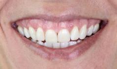
On extra-oral clinical examination, an exposure of the gingival margin of approximately 4 mm was observed when smiling moderately and 7 mm when smiling forcefully. On intraoral examination, we stated the general health of soft and hard tissues, thick gingival biotype, and a wide range of keratinized mucosa.
Intraoral and extraoral photographs were taken using a professional camera (Canon T2i), capturing images of the moderate and forced smile, in the frontal and lateral planes (figures 1 and 2), in addition to dynamic recordings of the same circumstances.

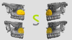
A high-resolution computed tomography (CT) was requested, performed using the i-CAT Next Generation tomograph, with an acquisition field of 8 cm, an acquisition time of 26.9 seconds, and a slice thickness of 200 microns of the entire maxilla. It was later exported in DICOM format to the CAD software for designing the biovolume, denominated Mimics, from the company Materialise, in addition to intra-oral scanning (Scanner Trios 3, 3Shape). With the help of CT, it was possible to notice the presence of a concavity in the anterior region of the premaxilla, covering the areas of premolars, canines, and incisors (figure 3).

Through the tomography images in Mimics, it is possible to read the patient’s bone anatomy, being able to identify and diagnose skeletal defects and the need for a volume increase in the premaxillary region. As there is a projection of the apex of the roots through the buccal cortex, a three-dimensional segmentation of the patient’s bone and tooth volume is created to produce projections in these regions of the bone plate and, thus, in the biovolume, causing a subtraction of the material on the inner face that will be in intimate contact with the maxilla and in the final piece, so that when in contact with the patient’s maxilla, there is a space that avoids the pressure of the thin cortical tissue and subsequent absorption of the bone plate. Therefore, the biovolume is planned to be supported in areas with at least 2 mm of bone thickness, ensuring the absence of long-term bone resorption.
From clinical, photographic, and tomographic examinations, it was possible to relate the cause of gummy smile to three factors, as follows: excessive growth of the lower third of the face, hypermobility of the upper lip with a marked depression in the anterior process of the maxilla, and altered passive eruption type 1A (figures 4 and 5).


Given the multifactorial diagnosis and the patient’s expectations, a staged treatment plan was proposed, consisting of clinical crown lengthening surgery, followed by the installation of the Biovolume prosthesis and, subsequently, minimally invasive ceramic veneers, in order to meet the patient’s esthetic expectations.
Initially, a digital planning of the new dental shapes was requested, called .PSD® (Digital Smile Project), proposed by Dr. Marcos Rogério Fonseca. This tool uses well-known proportions called the Golden Proportion, and the entire smile design was designed in complete harmony with the patient’s facial proportions.
With the approval of the entire clinical team involved in the process, in addition to the patient, the .PSD® project, including the last layer referring to all the measures foreseen in the dentogingival transformations of the smile, were transferred, via digital flow, to the dental prosthesis laboratory. Then, the model was printed in laboratory resin, and a 3D model with high replicability was obtained (figure 6). Based on this model, three silicone guides were fabricated: one incisal-palatal, one buccal, and a full-coverage guide. The latter was used for intraoral testing of the planned dental morphology and served as a reference for guiding the position of the future gingival margin during the crown lengthening surgery (figure 7).


The design of the biovolume was requested in the Meshmixer software and manufactured in a 3D printed with a synthetic resin based on methacrylic esters, which is Food and Drug Administration (FDA) approved and class 6 in relation to biocompatibility, which was based on the information from the CT scan and the intraoral scanning, respecting the concavity present in the premaxilla region, making the surgical procedure faster and more predictable. In addition, we included perforations, as recommended, in the design of the biovolume so that an exchange is possible between the installed area and the covering tissues (figure 8). Then, this prototype was sterilized with ethylene oxide (figure 9).

Preoperative medication was prescribed to the patient to control pain and edema, in addition to factors related to trans-surgical infection. A tablet of Dexamethasone 4 mg and a tablet of Amoxicillin + Potassium Clavulanate (875 mg + 125 mg), both 12 hours before the procedure, and Dipyrone 1 g an hour before the procedure, were administered.
We administered local anesthesia with 4% articaine associated with 1:100,000 epinephrine and waited for the onset of action. The procedure started with a primary incision, which consisted of an intrasulcular incision, preserving the contour of the papillae, and a full-thickness flap was made. The presence of several areas of vertical bone loss, already noticed in the CT scan, indicated no need for intervention in the alignment of the marginal bone contour.
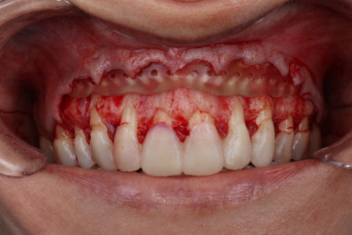
The prototype’s passive adaptation was fitted and attested on the premaxilla’s concavity area (figure 10). From there, the biovolume was fixed and stabilized through two screws for self-drilling bone graft, measuring 8 mm x 1.5 mm x 12 mm, and the screw head expanded by 2.0 mm. Then, the soft tissues were positioned within the planned proposal for the new alignment of the concave gingival arch, and sutured, using a simple suture, using 5.0 polypropylene.

Intrasulcular incision, with passive adaptation of the prototype adjusted and verified in the concavity area of the premaxilla.
After 10 postoperative days, the suture was removed, and the postoperative edema was practically imperceptible. After waiting 45 days for the soft tissues to mature, the tissue margin was already aligned, and the upper lip settled in a less apical position while the patient was smiling. Thus, we started the enamel cavity preparation. Then the case was scanned, and the models were sent to the laboratory. There were manufactured 08 ceramic laminate veneers, which completely adapted to the respective preparations. Afterward, they were cemented with the assistance of conventional photoactivatable resin cement, allowing a harmonious smile was achieved, with dental proportions and gingival exposure levels balanced in relation to the facial features (figures 11 and 12).

Cementation of laminated ceramic veneers using conventional light-cured resin cement on the front plane.

Cementation of laminated ceramic veneers using conventional light-curing resin cement on the lateral plane.
DISCUSSION
Gummy smile has numerous treatment alternatives; identifying the etiology is essential for a correct diagnosis and adequate treatment plan. Among the causes, we can mention the altered passive eruption, excessive growth of the maxilla, and hypermobility of the upper lip, which is caused by hyperfunction of the lip through the elevator muscles [1].
The spontaneous smile should, therefore, be observed in the patient’s facial evaluation to quantify the amount of exposed gingival tissue. In addition, static and dynamic records are indicated to support the treatment plan and communication with the patient and the team involved.
Some techniques were proposed when the gummy smile is related to the apical positioning of the upper lip. Surgical therapy for lip stabilization, known as “LipStaT,” is used according to the amount of gingival exposure. A horizontal incision is made along the mucogingival line, and another incision, above and parallel to the first, distant as required by the case, removes a mucosa strip. The technique is finished with a continuous suture from molar to molar, and this maneuver of joining the two incised margins will stop the mobility of the upper lip. However, this technique presents recurrence, making it unfavorable [9-12].
Botulinum toxin is used as an alternative to surgical treatment represented by upper lip hypermobility containment. It allows for smoothing lip muscle hypertonicity. However, it is not indicated in cases of concavity in the premaxilla region, as it is ineffective for correcting the gummy smile. Likewise, the use of botulinum toxin has the disadvantage of relapse episodes due to its absorption. On average, it lasts up to 4 to 8 months and may remain for less time, depending on muscle tone, requiring the procedure to be repeated frequently [13-16].
The biovolume fixation technique is a procedure whose function is to fill the concavity presented in the premaxilla region through a biocompatible material, hydroxymethyl methacrylate [7]. In addition to filling the premaxilla region, the biovolume has also been indicated for the chin region to enable the correction of its format, causing an improvement in its contour and projection, thus providing a harmonic result [9].
The synthetic resin BioMed Clear is a rigid, radiolucent, and light-curable material with low thermal conductivity. It is based on methacrylic esters and is certified by Universidade de São Paulo (USP) class VI, making it suitable for procedures that require wear resistance and low water absorption over time. This material is ISO 10993-5 and 13485 certified and supported by an FDA master file. The clear resin is a rigid and transparent material suitable for long-term contact with mucous membranes or skin [17].
The biovolume is manufactured using CAD/CAM technology through the additive prototyping technique, with layer thickness of 100 μm. After printing, the material undergoes several steps before reaching the sterilization stage. The biovolume is washed using isopropyl alcohol ≥ 99%, with this process lasting approximately 20 minutes. It is then air-dried at room temperature for 30 minutes before being placed in a post-curing device at 60°C for 60 minutes. The clear resin can be sterilized using four different methods: E-beam radiation at 35 kGy, ethylene oxide 100% at 55°C for 180 minutes, gamma radiation ranging from 29.4 to 31.2 kGy, and steam sterilization using an autoclave set at 134°C for 20 minutes or 121°C for 30 minutes [18].
The great advantage of the biovolume technique is that it promotes an immediate, predictable result with no risk of recurrence, unlike the previously mentioned techniques. It does not use absorbable medicinal material such as botulinum toxin. In addition, its fixation in the maxilla is easily done with screws, as its adaptation is entirely passive. As the biovolume technique uses digital resources, it offers accuracy in the procedures, allowing adjustments to the virtual model without needing a physical prototype. This proposed technique has additional costs related to the prosthesis’s CAD design and CAM prototyping [18].
Based on the presented case study, we can affirm that the current scenario for gummy smile correction benefits from the digital method, which enabled the personalized construction of a volume named Biovolume, fitting the premaxilla region perfectly and correcting the apical position of the upper lip definitively when smiling spontaneously. Furthermore, in the discussed case, given the multifactorial condition, we associated the enlargement of the clinical crown and ceramic veneers, which provided a harmonious, long-lasting, and youthful smile at the end of the therapies.
-
How to cite this article Silva LD, Murta A, Santos IF, Silva A, Maffille Gomes JA, Fonseca MR, et al. Correction of the gummy smile when there is vertical growth of the maxilla and resistance to orthognathics. RGO, Rev Gaúch Odontol. 2026;74:e20260008. http://dx.doi.org/10.1590/1981-86372026000820230090
Data Availability
The research data are available in the body of the document.
REFERENCES
-
1 Jreige CS, Kimura RN, Segundo ÂRTC, Coachman C, Sesma N. Esthetic treatment planning with digital animation of the smile dynamics: a technique to create a 4-dimensional virtual patient. J Prosthet Dent. 2022;128(2):130-8. doi: https://doi.org/10.1016/j.prosdent.2020.10.015
» https://doi.org/10.1016/j.prosdent.2020.10.015 -
2 Nomura S, Freitas KMS, Silva PPC, Valarelli FP, Cançado RH, Freitas MR, et al. Evaluation of the attractiveness of different gingival zeniths in smile esthetics. Dental Press J Orthod. 2018;23(5):47-57. doi: https://doi.org/10.1590/2177-6709.23.5.047-057.oar
» https://doi.org/10.1590/2177-6709.23.5.047-057.oar -
3 de Castro LF, de Andrade PF, Leite GG, de Andrade AJS, Valentim GL, de Souza ET. 3D-printed PMMA implant for gingival smile treatment through the VISTA technique: a report of a new approach. Clin Adv Periodontics. 2023;13(1):27-32. doi: https://doi.org/10.1002/cap.10200.
» https://doi.org/10.1002/cap.10200 -
4 Abdullah WA, Khalil HS, Alhind MM, Marzook H. Modifying gummy smile: a minimally invasive approach. J Contemp Dent Pract. 2014;15(6):821-6. doi: https://doi.org/10.5005/jp-journals-10024-1619
» https://doi.org/10.5005/jp-journals-10024-1619 -
5 Andrade PF, Meza-Mauricio J, Kern R, Faveri M. Labial repositioning using print manufactured polymethylmethacrylate-based cement for gummy smile: case report. Case Rep Dent. 2021;2021:7607522. doi: https://doi.org/10.1155/2021/7607522
» https://doi.org/10.1155/2021/7607522 -
6 Jananni M, Sivaramakrishnan M, Libby TJ. Surgical correction of excessive gingival display in class I vertical maxillary excess: mucosal strip technique. J Nat Sci Biol Med. 2014;5(3):494. doi: https://doi.org/10.4103/0976-9668.136171
» https://doi.org/10.4103/0976-9668.136171 -
7 Torres EM, Neto JV, Bernades KO, Naldi LF, Torres HM, Carvalho AL, et al. Facial profile changes due to bone cement graft to manage the hyperactive muscles of the gingival smile. Dental Press J Orthod. 2020;25(1):44-51.doi: https://doi.org/10.1590/2177-6709.25.1.044-051.oar
» https://doi.org/10.1590/2177-6709.25.1.044-051.oar - 8 Suma T, Shashikumar HC, Lokesh NK, Siddarth A, Shwetha GS. Orthodontic surgical treatment of gummy smile with vertical maxillary excess. IOSR J Dent Med Sci. 2014;13:68-74.
-
9 Bhola M, Fairbairn PJM, Chu SJ, Morris T. LipStaT: the lip stabilization technique - indications and guidelines for case selection and classification of excessive gingival display. Int J Periodontics Restorative Dent. 2015;35(5):549-59. doi: https://doi.org/10.11607/prd.2413
» https://doi.org/10.11607/prd.2413 -
10 Revilla-León M, Özcan M. Additive manufacturing technologies: an overview of current knowledge and applications in prosthodontics. J Prosthodont Res. 2019;63(1):26-34. doi: https://doi.org/10.1016/j.jpor.2018.09.002
» https://doi.org/10.1016/j.jpor.2018.09.002 - 11 Recchioni C, Pawlenko AE, Haidar GF, Murta A, Araujo FC, Santos JAV, et al. Procedimentos cirúrgicos em harmonização orofacial. Belo Horizonte: Nativa; 2022. p.564-6.
-
12 Freitas de Andrade P, Meza-Mauricio J, Kern R, Faveri M. Labial repositioning using print manufactured polymethylmethacrylate -(PMMA-) based cement for gummy smile. Case Rep Dent. 2021;2021:7607522. doi: https://doi.org/10.1155/2021/7607522
» https://doi.org/10.1155/2021/7607522 - 13 Kamak G, Keklik H. Use of botulinum toxin in gummy smile. e-J Dent. 2014;4(3):643-7.
-
14 Zafar MS. Prosthodontic applications of polymethyl methacrylate (PMMA): an update. Polymers (Basel). 2020;12(10):2299. doi: https://doi.org/10.3390/polym12102299
» https://doi.org/10.3390/polym12102299 - 15 Esteves LS, Rodrigues DB, Ávila C, Campos PSF, Santos JN. Preenchimentos estéticos na cirurgia ortognática: há indicações. Rev Clín Ortod Dent Press. 2016;15(1):33-59.
- 16 Torres EM, Naldi LF, Bernades KO, Carvalho AL. Four-year follow-up of a polymethyl methacrylate-based bone cement graft for optimizing esthetics in maxillary anterior implants: a case report. Gen Dent. 2017;65(1):48-53.
-
17 Formlabs. High performance 3D printers, finally within reach. São Paulo: 3DCriar; c2011 [cited 2023 Aug 31]. Available from: https://formlabs.com/
» https://formlabs.com/ - 18 Coachman C, Calamita M. Digital smile design: a tool for treatment planning and communication in esthetic dentistry. Quintessence Dent Technol. 2012;35(2):103-11.
Edited by
-
Assistant editor
Luciana Butini Oliveira