 Escitalopram induces morphometric and collagen changes in salivary glands of pregnant mice
ABSTRACT
Objective: This study aimed to evaluate the effects of exposure to Escitalopram Oxalate on morphometric parameters of salivary glands of pregnant mice.
Methods: Twenty pregnant Swiss mice (Mus musculus) were equally divided into treated and control groups, which received 20 mg/kg of Escitalopram and sterile saline, respectively, via gavage, daily, from the 5th to the 17th day of pregnancy. On the 18th day, after euthanasia, salivary glands were collected and passed through histological procedures. Under optical microscopy, acinar diameter (in µm) and area (in µm2), ductal thickness (in µm), and quantity of type I collagen (area, in µm2) from parotid, sublingual, and submandibular glands were measured. Statistical analysis was carried out with the Student’s t test for parametric data and the Mann-Whitney test for non parametric data, with 5% significance (GraphPad Prism 5).
Results: Acinar areas (C: 1207.0 ± 315.2; T: 1662.0 ± 508.0; p=0.002) and diameters (C: 34.3 [33.3-37.8]; T: 41.0 [34.9-47.4]; p=0.008) from sublingual glands and acinar areas (C: 1009.0 ± 406.3; T: 1378.0 ± 464.2; p=0.005) and diameters (C: 37.9 ± 10.1; T: 45.3 ±6.8; p=0.05) from submandibular glands were statistically bigger in the treated group than the control group. The quantity of type I collagen was statistically lower in the treated group compared to the control group (C: 4399[3346.0-7092.0]; T: 3695.0 [2948.0-4792.0]; p=0.0003).
Conclusion: Escitalopram promotes an adverse effect, causing morphological and structural alterations in the salivary glands of pregnant mice.
Indexing terms
Antidepressive agents; Sublingual gland; Submandibular gland; Mice; Pregnancy
RESUMO
Objetivo: Este estudo teve como objetivo avaliar os efeitos da exposição ao Oxalato de Escitalopram sobre os parâmetros morfométricos das glândulas salivares de camundongos prenhes.
Métodos: Vinte camundongos Swiss prenhes (Mus musculus) foram igualmente divididos em grupos controle e tratado, os quais receberam, respectivamente, 20 mg/kg de Escitalopram e solução salina estéril, por via gavagem, diariamente, do 5º ao 17º dia de gestação. No 18º dia, após a eutanásia, as glândulas salivares foram coletadas e submetidas a procedimentos histológicos. Em microscopia óptica, foram mensurados o diâmetro acinar (em µm) e a área (em µm2), a espessura ductal (em µm) e a quantidade de colágeno tipo I (área, em µm2) das glândulas parótida, sublingual e submandibular. A análise estatística foi realizada pelo teste t de Student para dados paramétricos e pelo teste de Mann-Whitney para dados não paramétricos, com nível de significância de 5% (GraphPadPrism 5).
Resultados: As áreas acinares (C: 1207,0 ± 315,2; T: 1662,0 ± 508,0; p=0,002) e os diâmetros (C: 34,3 [33,3-37,8]; T: 41,0 [34,9-47,4]; p=0,008) das glândulas sublinguais, bem como as áreas acinares (C: 1009,0 ± 406,3; T: 1378,0 ± 464,2; p=0,005) e os diâmetros (C: 37,9 ± 10,1; T: 45,3 ± 6,8; p=0,05) das glândulas submandibulares, foram estatisticamente maiores no grupo tratado em comparação ao grupo controle. A quantidade de colágeno tipo I foi estatisticamente menor no grupo tratado em comparação ao grupo controle (C: 4399 [3346,0-7092,0]; T: 3695,0 [2948,0-4792,0]; p=0,0003).
Conclusão: O Escitalopram promove um efeito adverso, causando alterações morfológicas e estruturais nas glândulas salivares de camundongos prenhes.
Termos de indexação
Antidepressivos; Glândula sublingual; Glândula submandibular; Camundongos; Gestação
INTRODUCTION
Depression is a disease that affects 4.4% of the world population, according to data from the World Health Organization (WHO) 2020 [1]. In addition to being present in the population as a whole, depression affects up to 20% of women during pregnancy [2] Depression can cause fetal development problems and hormonal alterations during pregnancy by increasing the production of cortisol, the stress hormone [3]. For the treatment of depression during pregnancy, the most commonly prescribed antidepressant is Escitalopram (ESC), since it presents greater acceptability on the part of patients when compared to other medications [4]. ESC belongs to the class of Selective Serotonin Reuptake Inhibitors (SSRI). Its action occurs in the pre-synaptic transport molecules of serotonin, increasing its concentration and residence time in the synaptic cleft [5]. Although the use of SSRI class antidepressants has a lower incidence of xerostomia when compared to other classes of antidepressants, the sensation of a dry mouth still persists and may be related to other factors such as body weight, administered dose, and pregnancy [6]. Xerostomia can be classified according to its pathogenesis, as either true xerostomia, due to alterations in the functioning of the salivary glands, or pseudo-xerostomia, where the sensation of dry mouth occurs, resulting from alterations in the salivary content [7]. During the gestational period there are several hormonal alterations that influence the feeling of dry mouth in women and the quality of saliva, since changes occur in the pH and composition [8,9]. These alterations, combined with poor hygiene, can cause problems in the oral cavity, such as caries and periodontal disease [10]. There are no studies in the literature that correlate pregnancy, the use of ESC, and alterations in salivary glands. Thus, the current study aimed to investigate the morphometric alterations in salivary glands of pregnant mice exposed to the drug during pregnancy.
METHODS
All procedures described are in accordance with the Brazilian Guideline for the Care and Use of Animals for Scientific and Didactic Purposes, proposed by the National Council for the Control of Animal Experimentation, 2016 [11], and were approved by the Committee of Ethics in the Use of Animals, of the State University of Londrina, under number: 17937.2017.35. Animals and Treatment Swiss mice, male and female, from the central vivarium of the State University of Londrina, were used as the parental generation. All animals had free access to water and feed (Nuvital™, Londrina, Brazil). During the execution of this work, the animals were maintained in a controlled environment, with a 12-hour light/dark cycle and 22 ± 2ºC. The animals were mated (two females and one male per cage) and pregnancy was determined by the presence of a vaginal plug [12].
Experimental design the sample was composed of 20 pregnant female mice, divided equally and randomly into a treated and control group. The drug Oxalate ESC (Medley 20 mg) was used, compressed, macerated, and diluted in saline. A dose of 20 mg/kg of ESC was administered to the animals in the treated group by gavage, from the 5th to the 17th day of pregnancy [13]. The control group received saline in a volume equivalent to that of the animals in the treated group. The females were euthanized by cervical dislocation on the 18th day of pregnancy and their salivary glands were collected for histological analysis. Histology of salivary glands − morphometric analysis After collection and dissection, the submandibular, sublingual, and parotid salivary glands were fixed in Bouin’s solution for 24 hours and then in 70% ethyl alcohol until the moment of processing for inclusion on slides. The cuts were made in the sagittal direction and stained with hematoxylin and eosin before being photographed by a digital camera under an optical microscope (Moticam, Motic Co, Xiamen, China). The images were analyzed using the MoticImage Plus 2.0 program (Motic Co, Xiamen, China) with a 100-fold magnification. For each gland, one field was analyzed, in which the following morphometric parameters were measured in triplicate: acinar perimeter (µm), largest acinar diameter (µm), ductal wall thickness (µm2), and acinar area (µm2).
Histology of salivary glands − analysis of the quantity of type I collagen Serial sections were performed in a microtome in the sagittal direction of the salivary glands, with each slide containing 4 sections of 7 µm. These cuts were stained using the Picrosirius red technique for the detection of type I collagen. The slides were photographed using a polarized light microscope and analyzed using the Image Pro Plus software (Media Cybernetics, Rockville, United States), with an objective of 20x.
Sample calculation to calculate the sample size, a preliminary pilot study was conducted with six female mice that made up the final selected sample. The analyzed image of each gland was considered as a sampling unit. The analyses were performed in triplicate, that is, for each salivary gland, three histological sections were considered. The mean and standard deviation values of the diameter of the acini (one of the morphometric parameters, chosen at random) of the salivary glands of these animals were used to calculate the effect magnitude. Subsequently, the sample calculation was performed in G* Power Software (Heinrich Heine University in Düsseldorf, Düsseldorf, Germany) considering: a magnitude of effect of 1.4, statistical power of 0.8, and α of 0.05.
Statistical analysis of the data the data were analyzed using the GraphPad Prism 5 program (GraphPad Software, Inc., La Jolla, CA, USA) and data normality was assessed using the D’Agostino Pearson test. Parametric data were analyzed using the unpaired Student’s t test and expressed as mean and standard deviation. Nonparametric data were analyzed using the Mann-Whitney U test and expressed as median and interquartile range. The level of significance adopted was 5% (p<0.05).
RESULTS
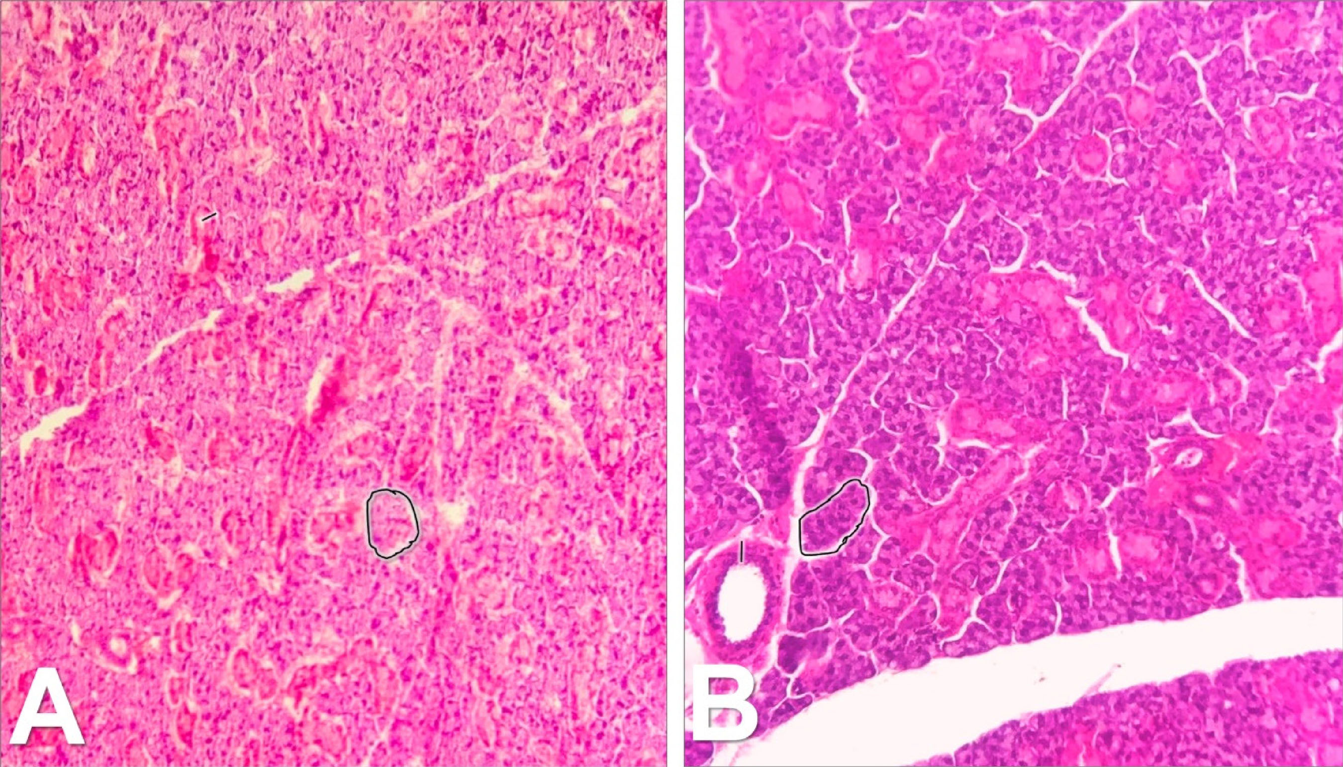
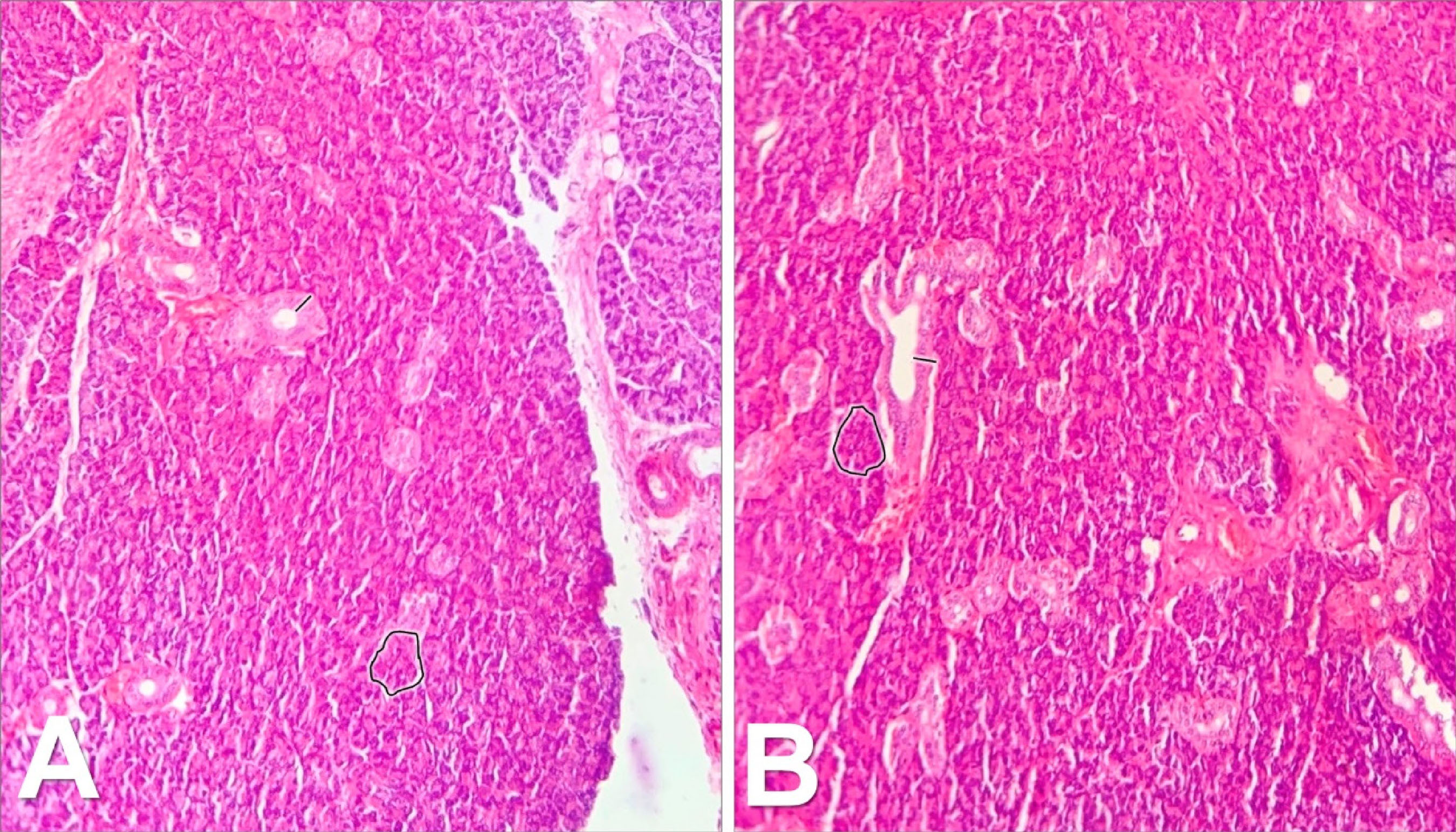
The morphometric parameters of the sublingual glands are presented in table 1. The acinar areas and diameters of the treated group were larger than those of the control group. Likewise, the acinar areas of the submandibular glands and the acinar diameters of the treated group were also larger than the control group (table 2). In turn, the thickness of the duct of the sublingual and submandibular glands of the acinar and the parameters of the parotid glands did not demonstrate significant alterations (table 3). Figure 1 represents the image of the sublingual salivary gland, separated by a control group in quadrant A and a treated group in quadrant B. Each quadrant has a contoured acinus and a measured duct. Similarly, figure 2 represented by the image of the submandibular gland and figure 3 the image of the parotid gland.
Comparison of the Sublingual Gland between the animals of the control (n=10) and treated (n=10) groups.
Comparison of the Submandibular Gland between the animals of the control (n=10) and treated (n=10) groups.
Comparison of the Parotid Gland between the animals of the control (n=10) and treated (n=10) groups.

Sublingual salivary gland in 100x magnification. A) control group, containing an outlined normal-sized acinus and a normal-thickness duct indicated with a dash. B) treated group, containing an enlarged acinus of contoured size and a duct of increased thickness indicated with a line.
Submandibular salivary gland in 100x magnification. A) control group, containing an outlined normal-sized acinus and a normal-thickness duct indicated with a dash. B) treated group, containing an enlarged acinus of contoured size and a duct of increased thickness indicated with a line.
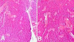
Parotid salivary gland in 100x magnification. A) control group, containing an outlined normal-sized acinus and a normal-thickness duct indicated with a dash. B) treated group, containing an outlined normal-sized acinus and a normal-thickness duct indicated with a dash.
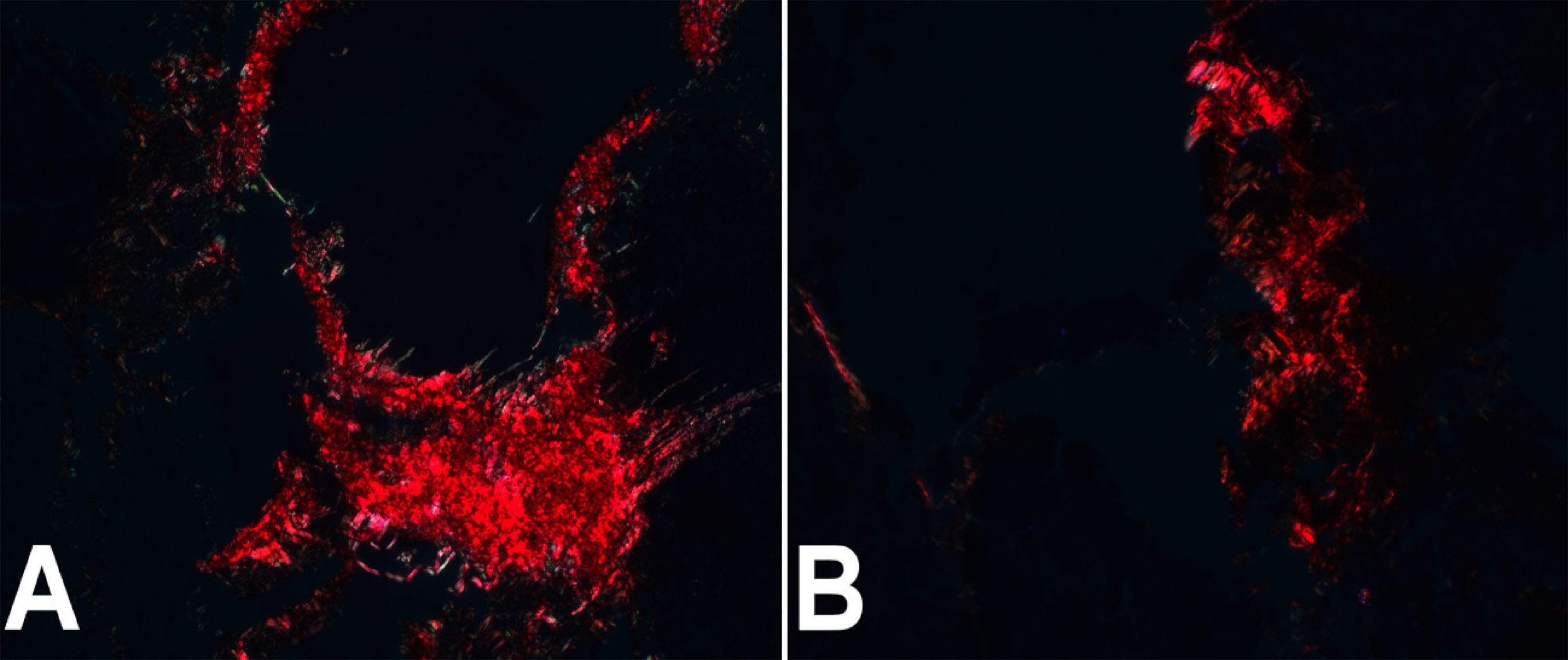
There was a decrease in type I collagen fibers per area analyzed in the treated group in relation to the control group, as observed in figure 4. Figure 5 represents the image of the collagen area stained by picro sirius, quadrant A being the control group and quadrant B being the treated group.

The amount of collagen per area in the control and treated groups. The box represents the median and quartiles from 25 to 75%, the horizontal bars represent the breadth of the data.

Amount of collagen per area in the control and treated groups. A) Imagem represents the control group with normal amounts of collagen. B) Imagem represents the group treated with reduced amounts of For Peer Review collagen.
DISCUSSION
The gestational period is marked by hormonal changes, such as the increase in estrogen in the second trimester of pregnancy, which is linked to the stimulus of salivary production and, as a consequence, dry mouth sensation [14,15]. The use of SSRI class antidepressants also demonstrated an influence on the feeling of dry mouth in humans who used the drug [16]. Xerostomia may or may not be related to insufficient saliva secretion, often caused by hyperfunction of the salivary glands. Saliva has important functions in the oral cavity, such as lubricating and preparing food for digestion and maintaining the pH between 6.8 and 7.2, as well as which, enzymes produced by the major salivary glands, such as lactoferrin and lysozyme, cause degradation of the cell wall of some bacteria [17].
Alterations in the quantity of saliva or salivary properties can have harmful consequences for the patient [18]. Saliva is produced by a system of glands which is present in both mice and humans. This system consists of smaller salivary glands, distributed throughout the oral cavity, crevicular fluid, produced in the gingival sulcus, and the major salivary glands, which are the parotid, the submandibular, and the sublingual [19]. In the current study, mice were used because they represent the most appropriate experimental model, presenting anatomy, genetics, and coexistence similar to that of humans. They also have a short and reproducible pregnancy and take up little space, which makes the experiment less costly [20].
Morphometry was used because it is an efficient method to identify changes in the shape of the organ and can measure these changes with high precision [21]. The acinar area and diameter of the sublingual and submandibular glands were statistically greater in the groups treated with ESC when compared to the control groups, while the thickness of the ducts was not altered in either gland. Regarding the parotid gland, the treated group did not demonstrate any morphometric alterations when compared to the control group. It is known that the major salivary glands are innervated by both the sympathetic and parasympathetic nervous systems. Both systems have acetylcholine as a pre-ganglion neurotransmitter. The sympathetic nervous system has the postganglionic neurotransmitter norepinephrine and the parasympathetic acetylcholine [22]. In mice it has been shown that the sympathetic nervous system has minimal action on the parotid gland, this gland being innervated by the parasympathetic nervous system [23]. According to Vissink et al. [24], the submandibular gland is more susceptible to disorders caused by antidepressant drugs. Majcherczyk et al. [25] demonstrated that the high peripheral concentration of ESC in the synaptic cleft is responsible for inhibiting the spontaneous release of noradrenaline, resulting in an antagonistic effect on secretory responses to noradrenaline in salivary glands and, as a consequence, decreases in the production of saliva dependent on this neurotransmitter. The submandibular and sublingual glands are responsible for the production of mucin, a substance that promotes lubrication of the mucosa. The change in these glands may explain the feeling of dry mouth with the continuous use of SSRIs [26].
The cellular hypertrophy present in the submandibular and sublingual glands, may be due to the fact that the morphological changes observed, such as the increase in acinar area and diameter, are related to an adaptive mechanism of the salivary glands in response to a potential reduction in salivary flow. However, this hypothesis requires confirmation through specific analyses of myoepithelial cell activity at both morphological and molecular levels, which were not addressed in this study. The results of this study corroborate the findings of Kopittke et al. [27] which demonstrate that the action of SSRI class antidepressants is related to xerostomia and hyposalivation effects after comparing the amount of induced and uninduced saliva in patients using Fluoxetine.
The salivary glands have a structural conformation composed of acini and ducts. Type I and type IV collagen compose the sustaining tissue, together with the extracellular matrix, type I being the most abundant, which provides support and basal firmness [28]. Through picrosirius staining, a decrease in type I collagen was observed when in contact with ESC. This result corroborates with the morphometric analysis, as a consequence of the increase in acini and ducts, there is a decrease in the area of the sustaining tissue.
Considering the hormonal changes during the gestational period and the onset of xerostomia, the consumption of fluids and sweets to relieve the feeling of dry mouth can worsen the picture of diseases that the pregnant woman is susceptible to, such as caries and periodontal disease [29]. This risk is increased when pregnant women use SSRI antidepressants, which are the most commonly prescribed drugs for the treatment and control of anxiety during pregnancy [30]. For this reason, it is necessary to take extra care with food, oral hygiene, and visits to the dentist for the prevention of oral diseases [31].
The data found in this work can be used as a basis for future discussions and studies, mainly because the literature is scarce in relation to the effects of ESC on salivary glands of pregnant women. It is important to highlight that clinical studies are necessary to complement the data presented in this study.
CONCLUSION
The present study demonstrates that ESC can cause important structural alterations, observed in acini, ducts, and tissue sustaining the salivary glands of mice. The data obtained in this study may lead the way for future clinical investigations in patients using this drug.
-
Article aligned with the Good Health and well-being goal of the Sustainable Development Goals (SDGs).
-
How to cite this article
Sestario CS, Mestre VF, Zeffa AC, Miqueloto CA, Neves MF, Salles MJS. Escitalopram induces morphometric and collagen changes in salivary glands of pregnant mice. RGO, Rev Gaúch Odontol. 2026;74:e20260002. http://dx.doi.org/10.1590/1981-86372026000220240003
Data Availability
The research data are available from the corresponding author upon reasonable request.
REFERENCES
-
1 Zheng W. Mental health and a novel coronavirus (2019-nCoV) in China. J Affect Disord. 2020;269:201-2. doi: https://doi.org/10.1016/j.jad.2020.03.041
» https://doi.org/10.1016/j.jad.2020.03.041 -
2 Becker M, Weinberger T, Chandy A, Schmukler S. Depression during pregnancy and postpartum. Curr Psychiatry Rep. 2016;18(3):32. doi: https://doi.org/10.1007/s11920-016-0664-7
» https://doi.org/10.1007/s11920-016-0664-7 -
3 Bae YJ, Kratzsch J. Corticosteroid-binding globulin: modulating mechanisms of bioavailability of cortisol and its clinical implications. Best Pract Res Clin Endocrinol Metab. 2015;29(5):761-72. doi: https://doi.org/10.1016/j.beem.2015.09.001
» https://doi.org/10.1016/j.beem.2015.09.001 -
4 Reis M, Källén B. Delivery outcome after maternal use of antidepressant drugs in pregnancy: an update using Swedish data. Psychol Med. 2010;40(10):1723-33. doi: https://doi.org/10.1017/s0033291709992194
» https://doi.org/10.1017/s0033291709992194 -
5 Sanchez C. The Pharmacology of citalopram enantiomers: the antagonism by R-Citalopram on the effect of S-Citalopram*. Basic Clin Pharmacol Toxicol. 2006;99(2):91-5. doi: https://doi.org/10.1111/j.1742-7843.2006.pto_295.x
» https://doi.org/10.1111/j.1742-7843.2006.pto_295.x -
6 Hunter KD, Wilson WS. The effects of antidepressant drugs on salivary flow and content of sodium and potassium ions in human parotid saliva. Arch Oral Biol. 1995;40(11):983-9. doi: https://doi.org/10.1016/0003-9969(95)00079-5
» https://doi.org/10.1016/0003-9969(95)00079-5 -
7 Millsop JW, Wang EA, Fazel N. Etiology, evaluation, and management of xerostomia. Clin Dermatol. 2017;35(5):468-76. doi: https://doi.org/10.1016/j.clindermatol.2017.06.010
» https://doi.org/10.1016/j.clindermatol.2017.06.010 - 8 Lasisi, Taye J. Pregnancy related changes in human salivary secretion and composition in a Nigerian population. Afr J Med Medical Sci. 2014;43(4):347-51.
-
9 Karnik A, Pagare S, Krishnamurthy V, Vahanwala S, Waghmare M. Determination of salivary flow rate, pH, and dental caries during pregnancy: a study. J Indian Acad Oral Med Radiol. 2015;27(3):372. doi: https://doi.org/10.4103/0972-1363.170454
» https://doi.org/10.4103/0972-1363.170454 -
10 Jain K, Kaur H. Prevalence of oral lesions and measurement of salivary pH in the different trimesters of pregnancy. Singapore Med J. 2015;56(01):53-7. doi: https://doi.org/10.11622/smedj.2015010
» https://doi.org/10.11622/smedj.2015010 -
11 Guimarães MV, Freire JE, Menezes LM. Use of animals in research: a brief review of legislation in Brazil. Rev Bioet. 2016;24(2):217-24. doi: https://doi.org/10.1590/1983-80422016242121
» https://doi.org/10.1590/1983-80422016242121 -
12 Behringer R, Gertsenstein M, Nagy KV, Nagy A. Selecting female mice in estrus and checking plugs. Cold Spring Harb Prot5. 2012;32(6):632-9. doi: https://doi.org/10.1177/0960327112462728
» https://doi.org/10.1177/0960327112462728 -
13 Anwar MJ, Pillai KK, Samad A, Vohora D. Effect of escitalopram on cardiomyopathy-induced anxiety in mice. Hum Exp Toxicol. 2012;32(6):632-9. doi: https://doi.org/10.1177/0960327112462728
» https://doi.org/10.1177/0960327112462728 -
14 Suri V, Suri V. Menopause and oral health. J Mid-life Health. 2014;5(3):115. doi: https://doi.org/10.4103/0976-7800.141187
» https://doi.org/10.4103/0976-7800.141187 -
15 Kivelä J, Laine M, Parkkila S, Rajaniemi H. Salivary carbonic anhydrase VI and its relation to salivary flow rate and buffer capacity in pregnant and non-pregnant women. Arch Oral Biol. 2003;48(8):547-51. doi: https://doi.org/10.1016/s0003-9969(03)00096-7
» https://doi.org/10.1016/s0003-9969(03)00096-7 - 16 Kumar NN, Panchaksharappa MG, Annigeri RG. Modified schirmer test: a screening tool for xerostomia among subjects on antidepressants. Arch Oral Biol. 2014;59(8):829-34.
-
17 Wiener RC, Wu B, Crout R, Wiener M, Plassman B, Kao E, et al. Hyposalivation and Xerostomia in Dentate Older Adults. J Am Dental Assoc. 2010;141(3):279-84. doi: https://doi.org/10.14219/jada.archive.2010.0161
» https://doi.org/10.14219/jada.archive.2010.0161 -
18 Tanasiewicz M, Hildebrandt T, Obersztyn I. Xerostomia of various etiologies: a review of the literature. Adv Clin Exp Med. 2016;25(1):199-206. doi: https://doi.org/10.17219/acem/29375
» https://doi.org/10.17219/acem/29375 -
19 Knosp WM, Knox SM, Hoffman MP. Salivary gland organogenesis. Wiley Develop Biol. 2011;1(1):69-82. doi: https://doi.org/10.1002/wdev.4
» https://doi.org/10.1002/wdev.4 -
20 Choi JS, Park IS, Kim SK, Lim JY, Kim YM. Analysis of age-related changes in the functional morphologies of salivary glands in mice. Arch Oral Biol. 2013;58(11):1635-42. doi: https://doi.org/10.1016/j.archoralbio.2013.07.008
» https://doi.org/10.1016/j.archoralbio.2013.07.008 -
21 Lilliu MA, Loy F, Cossu M, Solinas P, Isola R, Isola M. morphometric study of diabetes related alterations in human parotid gland and comparison with submandibular gland. Anat Rec. 2015;298(11):1911-8. doi: https://doi.org/10.1002/ar.23255
» https://doi.org/10.1002/ar.23255 -
22 Moos F, Richard P. Effects of dopaminergic antagonist and agonist on oxytocin release induced by various stimuli. Neuroendocrinology. 1979;28(2):138-44. doi: https://doi.org/10.1159/000122854
» https://doi.org/10.1159/000122854 -
23 Sayardoust S, Ekström J. Nitric oxide-dependent protein synthesis in parotid and submandibular glands of anaesthetized rats upon sympathetic stimulation or isoprenaline administration. Exp Physiol. 2004;89(2):219-27. doi: https://doi.org/10.1113/expphysiol.2003.002648
» https://doi.org/10.1113/expphysiol.2003.002648 -
24 Vissink A, Waterman HA, s-Gravenmade EJ, Panders AK, Vermey A. Rheological properties of saliva substitutes containing mucin, carboxymethylcellulose or polyethylenoxide. J Oral Pathol Med. 1984;13(1):22-8. doi: https://doi.org/10.1111/j.1600-0714.1984.tb01397.x
» https://doi.org/10.1111/j.1600-0714.1984.tb01397.x - 25 Majcherczyk J, Kulza M, Senczuk-Przybylowska M, Florek E, Jawien W, Piekoszewski W. Influence of tobacco smoke on the pharmacokinetics of citalopram and its enantiomers. J Physiol Pharmacol. 2012;63(1):95-100. PMID: 22460466.
- 26 Delgado PL. Serotonin function and the mechanism of antidepressant action. Arch Gen Psychiatr. 1990;47(5):411.
- 27 Kopittke L, Gomez R, Barros HMT. Opposite effects of antidepressants on unstimulated and stimulated salivary flow. Arch Oral Biol. 2005;50(1):17-21.
-
28 Porcheri C, Mitsiadis TA. Physiology, Pathology and Regeneration of Salivary Glands. Cells. 2019; Aug 26;8(9):976. doi: https://doi.org/10.3390/cells8090976. PMID: 31455013; PMCID: PMC6769486.
» https://doi.org/10.3390/cells8090976 - 29 El-Ashiry GM, El-Kafrawy AH, Nasr MF, Younis N. Comparative study of the influence of pregnancy and oral contraceptives on the gingivae. Oral Surg, Oral Med, Oral Pathol. 1970;30(4):472-5.
- 30 Gentile S. Untreated depression during pregnancy: short- and long-term effects in offspring. A systematic review. Neuroscience. 2017;342:154-66.
-
31 Xiao J, Alkhers N, Kopycka-Kedzierawski DT, Billings RJ, Wu TT, Castillo DA, et al. Prenatal Oral Health care and early childhood caries prevention: a systematic review and meta-analysis. Caries Res. 2019;53(4):411-21. doi: https://doi.org/10.1159/000495187
» https://doi.org/10.1159/000495187
Edited by
-
Assistant editor
Luciana Butini Oliveira