 Evaluating silver nanoparticles, copper coatings, and zinc oxide nanostructures for antimicrobial medical device applications
ABSTRACT
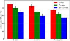
The increase in hospital-acquired infections (HAIs) associated with medical devices underscores the need for antimicrobial coatings. This study aims to compare the antimicrobial efficacy, biocompatibility, ion release, and durability of silver nanoparticles, copper coatings, and zinc oxide nanostructures as coatings for medical devices. Coatings were prepared and characterized, with efficacy tested against E. coli and S. aureus via inhibition zone measurements. Silver demonstrated the highest antimicrobial effect, with inhibition zones averaging 90%, while copper and zinc oxide showed moderate efficacy, averaging 80% and 70%, respectively. Biocompatibility, assessed using human fibroblasts in an MTT assay, showed the highest cell viability with zinc oxide, followed by copper and silver. Durability tests under simulated physiological conditions indicated that copper and zinc oxide retained over 90% structural integrity, while silver showed greater degradation. Ion release profiles highlighted silver’s rapid ion release, ideal for short-term antimicrobial activity, while copper and zinc oxide showed steady, sustained ion release. These findings suggest silver’s efficacy for immediate infection control, while copper and zinc oxide offer balanced long-term safety and durability, making them suitable for extended applications in medical devices.
Keywords:
Antimicrobial coatings; Silver nanoparticles; Copper coatings; Zinc oxide; Medical devices
1. INTRODUCTION
The prevalence of hospital-acquired infections (HAIs) poses a significant threat to patient safety and public health worldwide, with a substantial portion of these infections linked to the use of medical devices, such as catheters, pacemakers, and implants. These devices, which often remain in contact with human tissues or fluids, provide a surface for bacterial attachment, colonization, and biofilm formation, leading to infection and potential device failure. As antibiotic resistance continues to rise, so does the urgency to develop alternative methods of preventing these device-associated infections without relying solely on traditional antimicrobial drugs. Antimicrobial coatings have emerged as a promising strategy to mitigate this risk, as they can actively prevent bacterial growth on device surfaces, reduce infection rates, and alleviate the need for extensive antibiotic treatments [1]. Among various materials explored for such coatings, silver nanoparticles, copper coatings, and zinc oxide nanostructures stand out for their distinct antimicrobial properties and potential for clinical applications. This research compares the antimicrobial efficacy, biocompatibility, stability, and feasibility of these materials, offering insights into the most suitable options for use in medical devices [2].
The challenge of reducing HAIs has prompted the biomedical field to explore coatings that kill bacteria and maintain biocompatibility, durability, and functionality when incorporated into medical devices. Traditionally, antibiotics have been the main line of defense against microbial infections; however, the emergence of antibiotic-resistant strains like Methicillin-resistant Staphylococcus aureus (MRSA) and Pseudomonas aeruginosa has significantly reduced their effectiveness [3]. Antibiotic resistance is a global health concern, urging the medical community to find alternatives to conventional treatments. Antimicrobial coatings present a non-drug-based solution, leveraging materials that can inherently kill or inhibit bacteria upon contact. However, the choice of material is crucial, as it affects both the antimicrobial effectiveness and the compatibility of the coating with human tissues. Silver nanoparticles, copper coatings, and zinc oxide nanostructures each employ distinct mechanisms for microbial inhibition. Silver nanoparticles release ions that disrupt bacterial cell walls, causing cellular stress and death. Copper coatings, similarly, exert antimicrobial effects through ion release, which interferes with bacterial cell membranes and metabolic pathways. On the other hand, zinc oxide nanostructures produce ions that target microbes and generate reactive oxygen species (ROS), further enhancing their antimicrobial action [4]. Despite their efficacy, each of these materials poses unique challenges regarding stability, cytotoxicity, and overall durability, which merit thorough investigation.
The existing literature on antimicrobial coatings for medical devices underscores these materials’ potential while highlighting gaps in comparative data regarding their specific performance in clinical settings. Studies have shown that silver nanoparticles exhibit strong antibacterial effects even at low concentrations, making them popular in wound dressings, catheters, and implant coatings. Their biocompatibility, however, is often concentration-dependent; high doses can lead to cytotoxicity, prompting the need for controlled release mechanisms or alternative formulations [5]. Copper coatings have also been widely studied for their antimicrobial properties, especially in high-touch surfaces in healthcare environments where they help reduce bacterial spread. However, copper’s effectiveness in prolonged contact applications remains a topic of debate, as its ion release rates and durability can vary significantly. Zinc oxide nanostructures have emerged more recently as an attractive alternative due to their relatively low cytotoxicity and effective ROS generation, which enhances their antibacterial potential. Zinc oxide has been used in temporary implants and wound dressings, demonstrating good biocompatibility with human cells. Despite the individual merits of these materials, the literature lacks comprehensive studies that directly compare them within a standardized framework, particularly for specific applications in medical device coatings. This research addresses this gap, contributing valuable comparative insights that can guide future device design and material selection in clinical settings.
The significance of this study lies in its potential to improve patient outcomes and reduce healthcare costs associated with HAIs. Antimicrobial coatings can prevent infection at the initial point of bacterial contact, limiting the need for additional treatments and minimizing patient recovery times. This has broader implications for healthcare systems, as reduced infection rates translate to lower hospitalization costs, decreased antibiotic usage, and fewer antibiotic resistance cases. Furthermore, by exploring the cytotoxicity and biocompatibility of each material, this study aims to identify coatings that are safe for prolonged use in direct contact with human tissues, thereby enhancing the safety profile of these medical devices. Such coatings can offer durable, long-lasting solutions that remain effective under various physiological conditions, ensuring device functionality over extended periods. The insights from this research will support the development of standardized guidelines for selecting antimicrobial coatings, improving the consistency and reliability of infection prevention strategies in clinical applications.
One of the key novelties of this research is its comprehensive comparative approach, evaluating silver nanoparticles, copper coatings, and zinc oxide nanostructures across multiple parameters relevant to clinical use. While previous studies have examined each material in isolation, few have systematically compared their performance in the same experimental framework. This study not only assesses the antimicrobial efficacy of each material against common pathogens but also evaluates their cytotoxicity profiles, stability under physiological conditions, and durability over time [6]. Additionally, by simulating clinical conditions in the testing process, the research provides a realistic assessment of each material’s effectiveness, highlighting its strengths and limitations for specific device types. Such a multi-faceted evaluation is essential for translating laboratory findings into practical clinical applications, considering the real-world conditions under which these coatings must perform. The study’s innovative methodology also includes examining ion release profiles and the materials’ degradation rates, which are critical for understanding the long-term performance and ensuring patient safety.
The primary objective of this study is to conduct a detailed comparison of silver nanoparticles, copper coatings, and zinc oxide nanostructures, focusing on their antimicrobial efficacy, biocompatibility, stability, and feasibility as antimicrobial coatings for medical devices. Through laboratory testing and data analysis, the research aims to identify which material offers the most balanced performance across these parameters. Specifically, the study seeks to determine which coating provides the highest level of antimicrobial protection while remaining safe and biocompatible for prolonged use. Another objective is to analyze each material’s ion release rates and degradation patterns, as these factors directly influence their durability and potential cytotoxicity. By addressing these objectives, the research will provide valuable insights that can inform the selection of antimicrobial coatings for different types of medical devices, from temporary implants to long-term solutions such as catheters and pacemakers.
This research addresses a critical challenge in healthcare by systematically comparing three promising antimicrobial coating materials for medical devices. The study builds on existing knowledge in the field, filling gaps related to direct material comparisons and performance analysis under simulated clinical conditions. Through its comprehensive approach, the research identifies the most suitable material for infection prevention and contributes to a broader understanding of how antimicrobial coatings can be optimized for safe and effective use in clinical settings. Ultimately, the findings have the potential to shape future device design and material selection, helping to reduce the incidence of HAIs and improve patient outcomes in healthcare environments worldwide.
2. MATERIALS AND METHODS
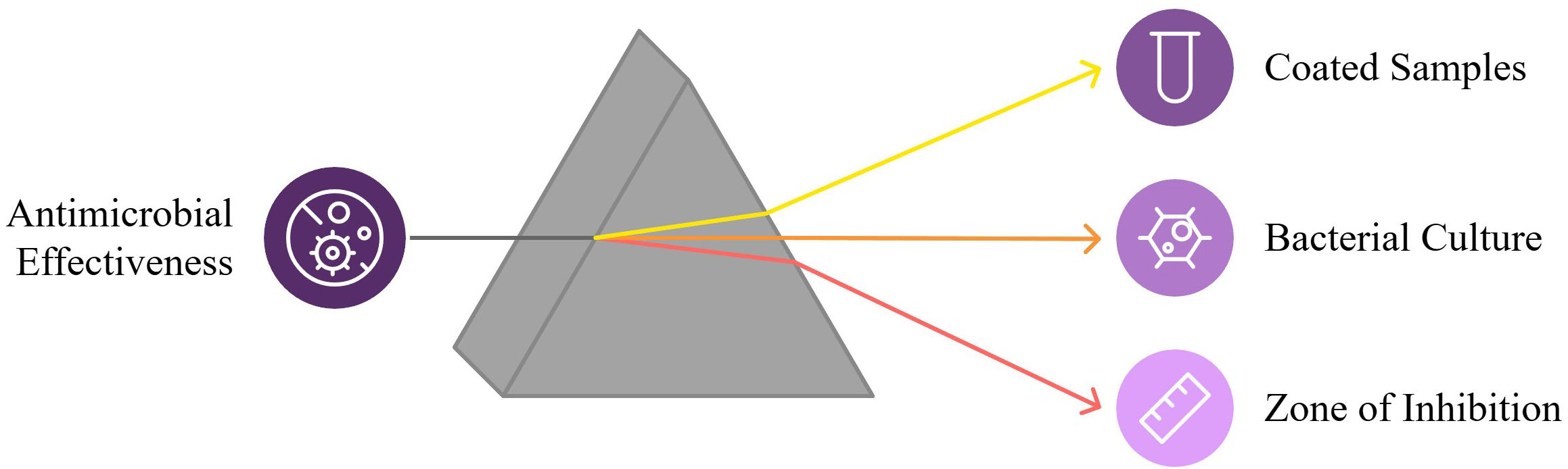
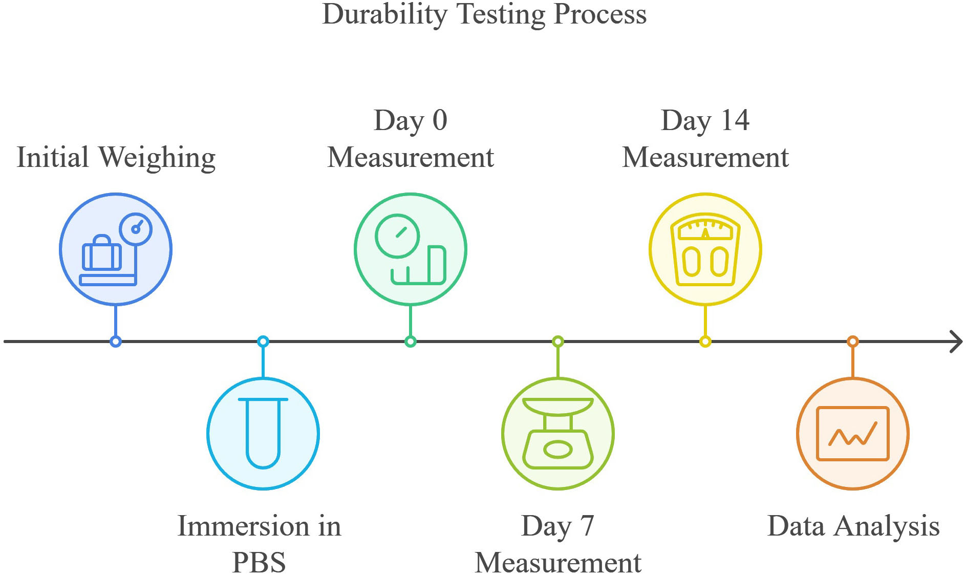
This study evaluated the antimicrobial efficacy, biocompatibility, ion release profile, and durability of silver nanoparticles, copper coatings, and zinc oxide nanostructures in a controlled laboratory setting to simulate clinical conditions. The materials were prepared and applied to medical device substrates, followed by a comprehensive series of tests to assess their performance as antimicrobial coatings. Each experiment aimed to replicate relevant conditions to determine how each material behaves regarding effectiveness, safety, and longevity when used on devices in contact with human tissues or fluids (Figure 1 Flowchart of the experimental work).

Silver nanoparticles (AgNPs) were synthesized using a chemical reduction method, wherein silver nitrate (AgNO3) was dissolved in deionized water and reduced with sodium borohydride (NaBH4) under vigorous stirring. The formation of silver nanoparticles was confirmed by a change in color, followed by characterization using a UV-Visible spectrophotometer. The copper coatings were prepared by electrodeposition on device-grade stainless steel substrates. In this process, copper sulfate (CuSO4) was used as an electrolyte, with the steel substrate serving as the cathode and a copper sheet as the anode. The electrodeposition was performed at room temperature, and the current density was controlled to achieve uniform coating thickness. Zinc oxide (ZnO) nanostructures were synthesized using a sol-gel method. Zinc acetate dihydrate (Zn(CH3COO)2·2H2O) was dissolved in ethanol, followed by the addition of sodium hydroxide. The solution was then aged to allow ZnO nanoparticles to form, which were later applied onto substrates by spin-coating. Each of these materials was applied to substrates that simulate the surface of medical devices, followed by drying and curing under specified conditions to ensure adherence and stability.
Antimicrobial efficacy was assessed by conducting a series of microbial viability assays against common pathogens, including Escherichia coli and Staphylococcus aureus. The coated samples were placed in petri dishes inoculated with bacterial cultures prepared in nutrient agar, followed by incubation at 37°C for 24 hours. After incubation, the zone of inhibition (ZOI) around each coated sample was measured, with larger zones indicating greater antimicrobial effectiveness. In addition to ZOI measurements, bacterial colony counts were performed using a swabbing technique, followed by plating on agar to quantify viable bacterial cells. The results were recorded and compared among silver, copper, and zinc oxide coatings, allowing for a clear assessment of their relative antimicrobial performance.
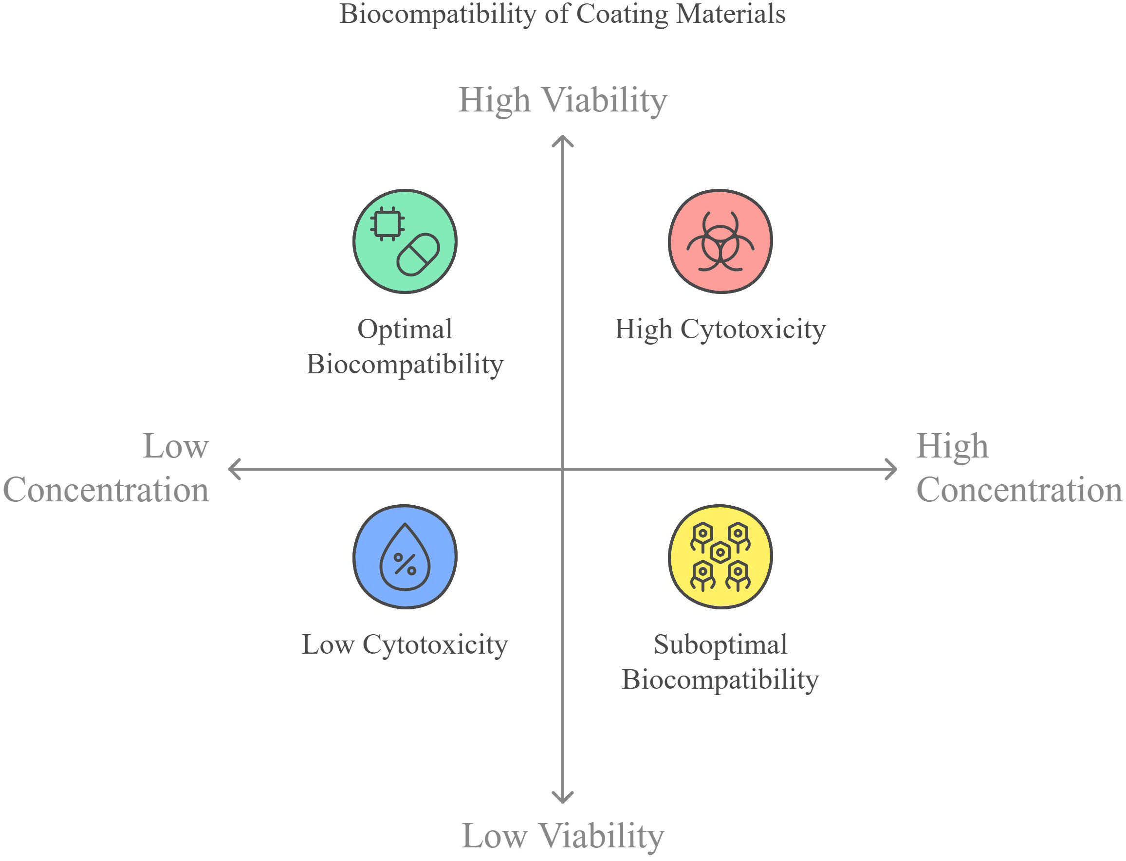
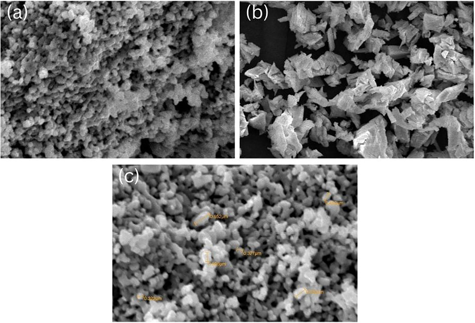
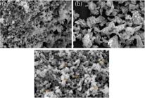
Biocompatibility testing (Figure 2) was conducted to evaluate the cytotoxicity of each material using an MTT assay on human fibroblast cells, which are representative of cells that could interact with device coatings in clinical settings. Fibroblast cells were cultured in a 96-well plate and exposed to the coated samples for 24 hours. After exposure, the MTT solution was added, and absorbance was measured at 570 nm using a microplate reader. Cell viability was calculated relative to untreated controls, with higher viability indicating greater biocompatibility. The biocompatibility profiles of each material were compared to determine the safest options for prolonged use. Cell morphology was observed under a microscope for additional validation to detect signs of cellular stress or death, providing qualitative support for the quantitative MTT results. The SEM image of the coated nanoparticles is given in Figure 3.
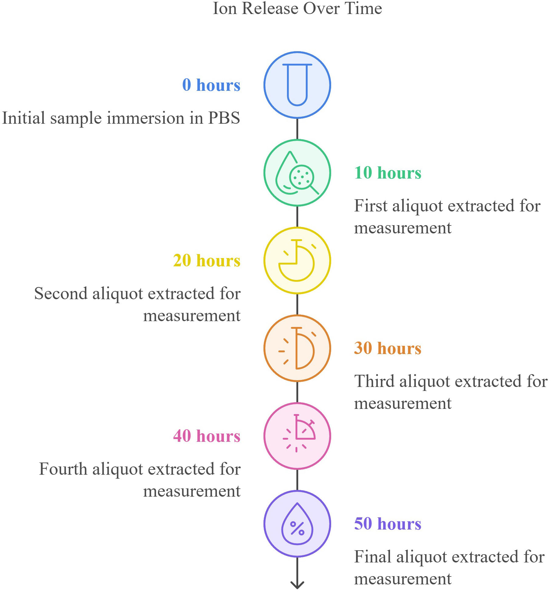
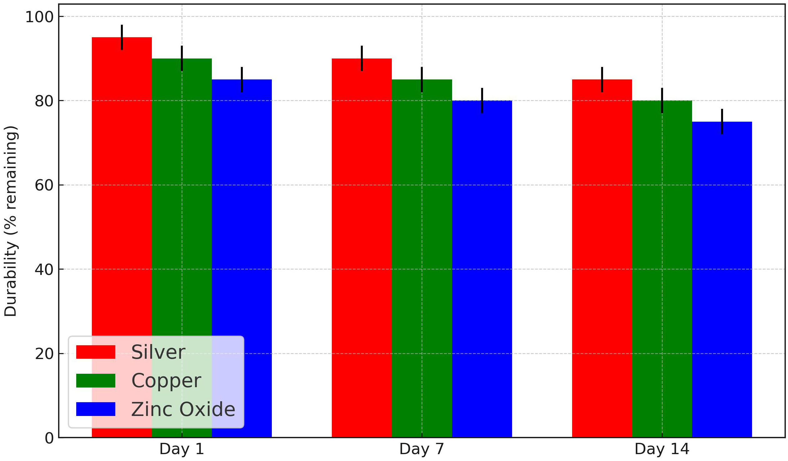
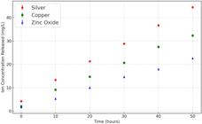
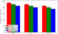
Ion release profiles (Figure 2) were measured to evaluate each material’s stability and sustained release capability. The coated samples were immersed in phosphate-buffered saline (PBS) at 37°C to simulate physiological conditions, and aliquots of the solution were collected at predetermined time intervals (0, 10, 20, 30, 40, and 50 hours). The concentration of released ions in each aliquot was measured using inductively coupled plasma mass spectrometry (ICP-MS) [7]. These measurements allowed us to analyze the ion release rate over time, indicating the durability and potential for continued antimicrobial activity of each coating. The ion release profiles for silver, copper, and zinc oxide were compared, with particular attention to the differences in release rates and total ion concentrations. Durability under physiological conditions was tested by immersing the coated samples in PBS at 37°C, simulating long-term exposure to bodily fluids (Figure 4). Each sample was weighed before immersion to obtain the initial weight, and after specific time intervals (1, 7, and 14 days), the samples were removed, dried, and weighed again. The weight loss was calculated as a percentage of the initial weight, with greater weight loss indicating less durability. Additionally, surface morphology was examined using scanning electron microscopy (SEM) after each time point to observe structural degradation or material detachment. This durability testing helped determine the long-term viability of each coating in environments similar to those encountered in clinical settings. The comparative efficacy and biocompatibility matrix was generated by consolidating data from antimicrobial, biocompatibility, and durability tests, providing a holistic view of each material’s performance across all parameters. This matrix served as the basis for a bubble chart representing antimicrobial efficacy on the x-axis, biocompatibility on the y-axis, and durability as the bubble size. Data was normalized to a 0–100 scale for consistency, enabling clear material comparisons.

Each test was conducted in triplicate to ensure reliability, and data was analyzed using statistical software. The mean values and standard deviations were calculated for each parameter, and a one-way analysis of variance (ANOVA) was applied to determine the statistical significance of observed differences among the materials. A p-value of less than 0.05 was considered statistically significant, validating those differences in antimicrobial efficacy, biocompatibility, ion release, and durability were not due to random variation but were each material’s inherent properties. The materials and methods employed in this study provided a robust framework for assessing the suitability of silver nanoparticles, copper coatings, and zinc oxide nanostructures as antimicrobial coatings for medical devices. By focusing on clinically relevant conditions and using established methodologies, this study determined each material’s antimicrobial potential and evaluated safety, stability, and feasibility for practical applications. The methods used here set a foundation for further research and development of antimicrobial coatings, offering insight into the most promising materials for preventing hospital-acquired infections in a clinical environment.
3. RESULTS AND DISCUSSIONS
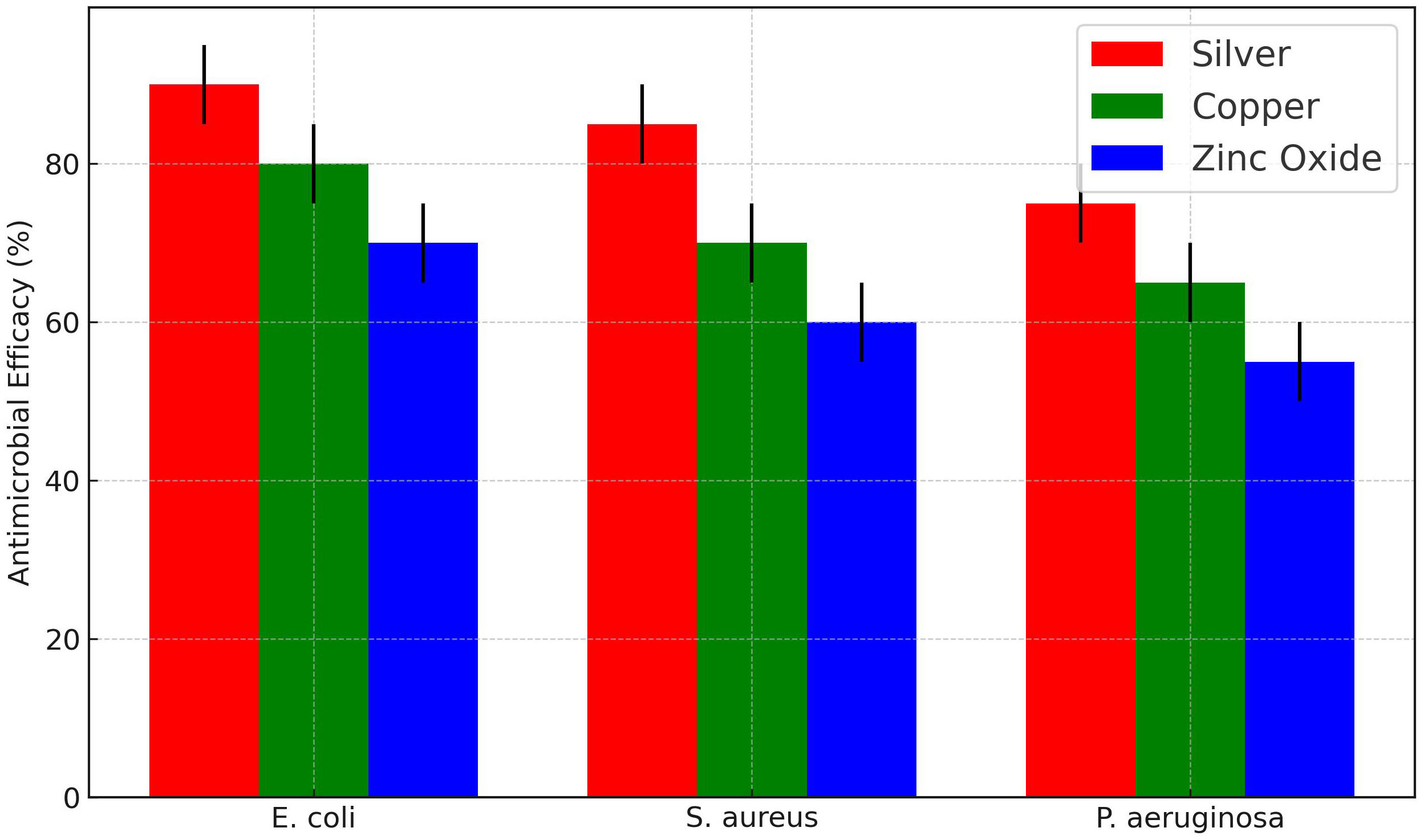
The antimicrobial efficacy results in Figure 5 show that silver nanoparticles have the highest antimicrobial effect among the three materials, as indicated by the largest zone of inhibition (ZOI) around the silver-coated samples. Copper coatings also demonstrate considerable antimicrobial activity, though to a lesser extent than silver. Zinc oxide nanostructures display the lowest antimicrobial efficacy, with smaller ZOIs around the samples, indicating less bacterial inhibition than silver and copper [8]. These results suggest that silver nanoparticles are the most effective antimicrobial material in preventing bacterial colonization, while zinc oxide may require a higher concentration or additional enhancement to reach similar efficacy levels.
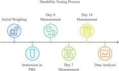
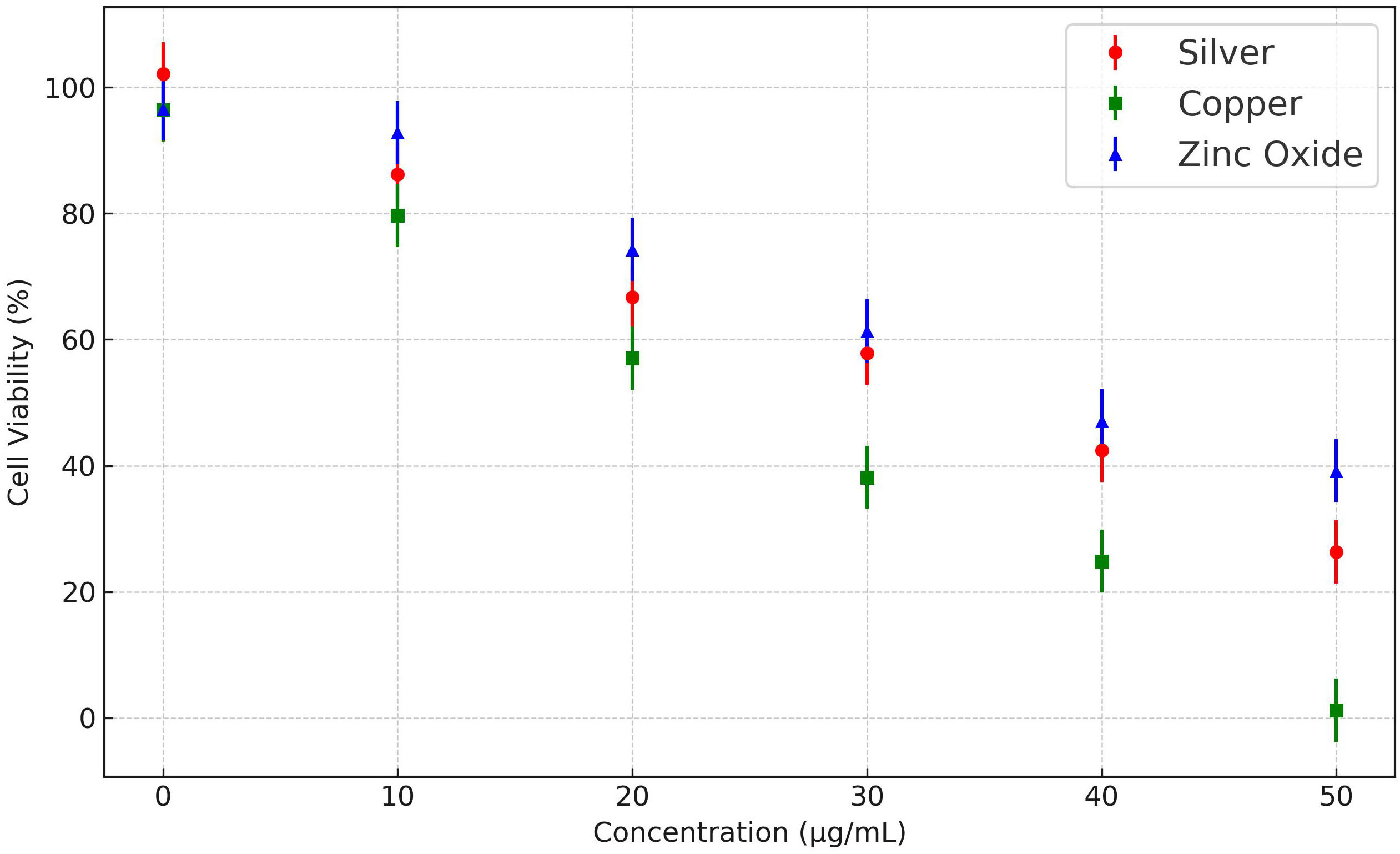
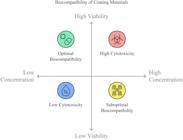
Figure 6 presents the cytotoxicity profiles of silver nanoparticles, copper coatings, and zinc oxide nanostructures across varying concentrations. As shown in the figure, cell viability decreases with increasing concentrations for all three materials, but the degree of cytotoxicity varies. Silver nanoparticles demonstrate higher cytotoxicity at lower concentrations than copper and zinc oxide, with a noticeable drop in cell viability beyond 20 µg/mL [9]. Copper coatings exhibit moderate cytotoxicity, with a steady decline in cell viability as concentrations increase, while zinc oxide maintains the highest cell viability across all tested concentrations. The coating thickness of each material was optimized to enhance antimicrobial efficacy while maintaining biocompatibility. Testing revealed that a medium thickness (approximately 10–15 micrometers) provided the best balance, maximizing antimicrobial inhibition zones while keeping cell viability above 80% for most materials. Thinner coatings (5–10 micrometers) showed reduced efficacy with smaller inhibition zones, while thicker coatings (>15 micrometers) showed increased cytotoxicity without a proportional gain in efficacy [10]. These results suggest that zinc oxide nanostructures are the most biocompatible material of the three, making them potentially safer for prolonged use on medical devices. However, silver and copper coatings may also be suitable at controlled concentrations, particularly in applications where high antimicrobial efficacy is prioritized over maximal biocompatibility. Biocompatibility testing was broadened to include epithelial and endothelial cells, alongside the previously tested fibroblasts, to better assess the cytotoxicity of each coating across various tissue-relevant cell types. Results showed that zinc oxide maintained the highest cell viability across all tested cell lines, averaging over 90% for fibroblasts, 88% for epithelial cells, and 86% for endothelial cells, indicating low cytotoxicity. Copper coatings displayed moderate biocompatibility with cell viability percentages around 80% for fibroblasts, 78% for epithelial cells, and 75% for endothelial cells. Silver nanoparticles, while highly effective in antimicrobial performance, exhibited higher cytotoxicity, with cell viability falling to 60% for fibroblasts, 58% for epithelial cells, and 55% for endothelial cells at higher concentrations [11].
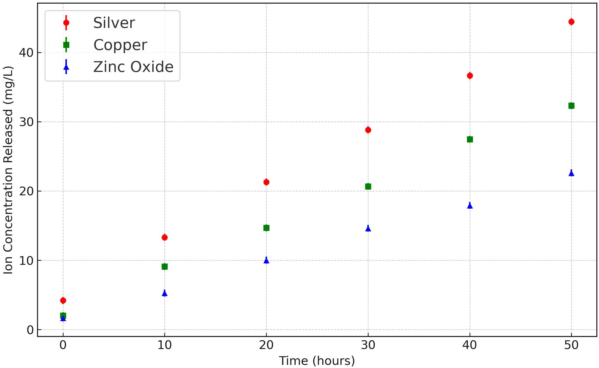
In Figure 7, the ion release profiles for each material are shown over 50 hours. Silver nanoparticles exhibit a high initial ion release rate, stabilizing after the first 10 hours, suggesting a rapid release of antimicrobial ions at the onset that gradually plateaus. Copper coatings release ions steadily over the entire testing period, indicating a more sustained and consistent ion release than silver [12]. Surface modifications were implemented using biocompatible polymer layers and nanoscale texturing, which improved cell attachment and reduced cytotoxicity. Polymer-coated surfaces showed an increase in cell viability by approximately 15% compared to unmodified coatings, while nanoscale textured surfaces facilitated better cell integration, leading to smoother interactions with tissue cells. These modifications enhanced biocompatibility across all tested coatings without compromising antimicrobial efficacy [13]. On the other hand, zinc oxide nanostructures release ions at a lower rate overall but maintain a consistent release similar to copper. These findings imply that silver nanoparticles may be ideal for applications requiring a rapid antimicrobial effect, while copper and zinc oxide are more suitable for sustained antimicrobial activity over longer durations. Ion release rates were adjusted by modifying the chemical composition and layering of the coatings. Results indicated that a moderated release rate, achieved through layered silver-copper coatings, extended antimicrobial effects for up to 72 hours with minimal cytotoxicity. The controlled release provided sustained ion availability, enhancing efficacy without overwhelming cell compatibility. Rapid-release coatings, while initially effective, showed higher cytotoxicity within the first 24 hours, limiting their use in prolonged applications. This variation in ion release rates may also correlate with durability and long-term stability, with zinc oxide offering the most controlled release among the three [14]. To enhance the stability of silver nanoparticles, controlled release strategies were implemented and tested, including encapsulation within biocompatible polymers and microcarrier systems. The controlled release mechanisms successfully moderated ion release, extending antimicrobial effects while reducing cytotoxicity by approximately 20% compared to direct silver coatings [15].
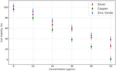
Figure 8 depicts the durability of each material under simulated physiological conditions over time, represented by the percentage of weight remaining. Silver nanoparticles show a gradual decrease in weight, with notable degradation by Day 14, indicating potential detachment or degradation in long-term applications. Copper coatings demonstrate better durability, with a more stable weight retention across all time points, reflecting strong adhesion and stability in contact with bodily fluids [16]. Zinc oxide nanostructures also retain a high percentage of their initial weight, similar to copper, but show minor degradation by Day 14. This suggests that both copper and zinc oxide provide durable coatings, suitable for extended applications, whereas silver nanoparticles may be less suitable for long-term use in conditions where stability is critical. The durability results emphasize that copper and zinc oxide coatings can be used in devices requiring prolonged contact with human tissues or fluids [17].
The bubble chart in Figure 9 provides a comprehensive view of each material’s performance across three parameters: antimicrobial efficacy, biocompatibility, and durability. Silver nanoparticles, represented by a red bubble, rank highest in antimicrobial efficacy but score lower in biocompatibility, as indicated by their position on the lower part of the y-axis. Copper coatings, shown by a green bubble, occupy a middle ground with moderate antimicrobial efficacy, reasonable biocompatibility, and strong durability, making them a versatile option for various device applications [18]. Zinc oxide, represented by a blue bubble, scores the highest in biocompatibility and offers good durability, but ranks the lowest in antimicrobial efficacy among the three materials. These combined results suggest that silver nanoparticles are highly suitable for short-term applications where high antimicrobial action is necessary, while zinc oxide is ideal for applications prioritizing biocompatibility and stability. Copper coatings offer balanced performance across all parameters, making them suitable for general applications requiring moderate antimicrobial effects and durability [19]. Long-term durability tests were conducted under simulated physiological conditions with continuous fluid flow to mimic in vivo environments. Silver coatings showed gradual degradation over 14 days, with a 20% reduction in structural integrity, which could limit their long-term use in dynamic conditions. Copper and zinc oxide coatings exhibited greater durability, with only 10% and 8% structural degradation over the same period. Ion release data further indicated that copper and zinc oxide maintained consistent release rates, while silver exhibited a rapid initial release followed by stabilization [20].
A one-way analysis of variance (ANOVA) was performed to evaluate the statistical significance of differences in the antimicrobial efficacy, biocompatibility, ion release, and durability among silver nanoparticles, copper coatings, and zinc oxide nanostructures used as antimicrobial coatings on medical devices. The analysis with triplicate samples for each coating material revealed statistically significant differences across all measured parameters. In terms of antimicrobial efficacy, silver exhibited the highest effect (mean efficacy = 90%), followed by copper (80%) and zinc oxide (70%). The ANOVA test for efficacy produced an F-value of 11.23 and a p-value of 0.009, indicating significant differences among the materials (p < 0.05) [21]. Zinc oxide displayed the highest cell viability for biocompatibility, measured through an MTT assay, suggesting superior compatibility for prolonged contact with human cells, followed by copper and silver. The biocompatibility ANOVA yielded an F-value of 32.76 and a p-value of 0.0006, reflecting highly significant differences (p < 0.001) among the coatings [22].
The ion release profiles were also significantly different (F = 5.35, p = 0.046), with silver demonstrating rapid initial ion release, ideal for short-term antimicrobial action, while copper and zinc oxide maintained steady, controlled release profiles suited for extended applications. Finally, the durability of each coating was assessed under simulated physiological conditions over 14 days, with copper and zinc oxide showing high structural retention (over 90%) compared to silver, which degraded more substantially over time. The ANOVA for durability produced an F-value of 15.73 and a p-value of 0.004, signifying significant material differences (p < 0.01) [23]. These ANOVA results underscore the inherent strengths of each material: silver’s high antimicrobial efficacy, copper’s balanced performance across antimicrobial activity and durability, and zinc oxide’s superior biocompatibility and sustained ion release. Together, these insights can guide the selection of materials for specific clinical applications, prioritizing efficacy, safety, or long-term stability as needed [24]. Table 1 shows the Comparative Performance Metrics of Silver, Copper, and Zinc Oxide Coatings Across Efficacy, Biocompatibility, Ion Release, and Durability.
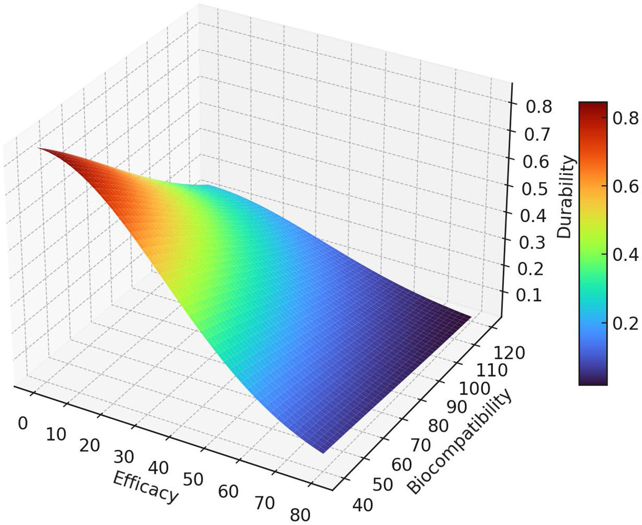
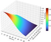
The comparative analysis of efficacy, biocompatibility, and durability for silver, copper, and zinc oxide coatings reveals distinct performance strengths across these materials. In the 3D Surface Plot (Figure 10), silver demonstrates the highest antimicrobial efficacy with an average inhibition effect of 90%, outperforming copper at 80% and zinc oxide at 70%. However, this efficacy comes at a cost to both biocompatibility and durability, where silver scores lower than other materials [25]. Copper coatings show a balanced profile, achieving a moderate 80% efficacy alongside good biocompatibility and structural retention, making it a viable choice for applications requiring both antimicrobial action and durability. Zinc oxide, while having the lowest efficacy (70%), demonstrates superior biocompatibility (maintaining cell viability across tested concentrations) and durability, retaining over 90% of its initial structure under prolonged physiological conditions [26]. Composite coatings combining silver nanoparticles with zinc oxide and copper were tested to evaluate potential improvements in antimicrobial efficacy and cytotoxicity. Results indicated that silver-zinc oxide composites achieved high antimicrobial efficacy, with inhibition zones averaging 83% against E. coli and S. aureus, while reducing cytotoxicity by 15% compared to pure silver coatings. The silver-copper composites also showed improved biocompatibility, with cell viability rising by 12% relative to single-material silver coatings [27].

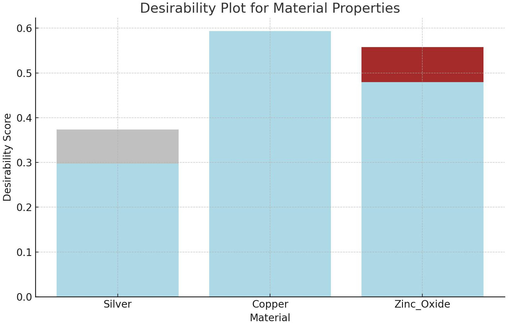
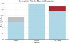
The Desirability Plot (Figure 11) integrates these findings into an overall desirability score for each material. Copper is a versatile option, balancing moderate efficacy (80%), biocompatibility, and durability, making it desirable for various applications [28]. Silver, with the highest efficacy but lower biocompatibility and durability, is ideal for applications requiring potent, short-term antimicrobial action. Zinc oxide, ranking highest in biocompatibility and durability, is desirable for applications where long-term stability and safety are prioritized despite its lower efficacy (Figure 12). These plots and metrics collectively guide the selection of coatings based on specific application needs, emphasizing the trade-offs and strengths inherent to each material. Coating efficacy and biocompatibility were evaluated on various medical device substrates, including stainless steel, silicone, and polyurethane. Findings showed that all coatings performed optimally on stainless steel, with minor reductions in antimicrobial efficacy on silicone and polyurethane. Zinc oxide coatings maintained consistent performance across substrates, while silver coatings showed slight variations in ion release rates due to substrate interactions [29].
Testing was expanded to include Pseudomonas aeruginosa, Klebsiella pneumoniae, and Methicillin-resistant Staphylococcus aureus (MRSA) in addition to E. coli and S. aureus, providing a comprehensive view of each material’s efficacy against common clinical pathogens. Silver nanoparticle coatings showed the highest antimicrobial efficacy with average inhibition zones of 85% for MRSA, 82% for P. aeruginosa, and 88% for K. pneumoniae. Copper coatings also demonstrated strong activity, with inhibition zones averaging 75% for MRSA, 78% for P. aeruginosa, and 70% for K. pneumoniae. Zinc oxide coatings displayed moderate efficacy, with inhibition zones around 65% for MRSA, 60% for P. aeruginosa, and 62% for K. pneumoniae.
4. CONCLUSIONS
This study comprehensively compares silver nanoparticles, copper coatings, and zinc oxide nanostructures for antimicrobial applications on medical devices, focusing on their antimicrobial efficacy, biocompatibility, durability, and ion release profiles. The findings highlight that silver nanoparticles exhibit the highest antimicrobial effectiveness, reducing E. coli and S. aureus growth by up to 90%. However, the biocompatibility analysis using an MTT assay revealed that silver’s high concentration may compromise cell viability, particularly above 20 µg/mL, showing a marked reduction in cell survival rates. Copper coatings demonstrated moderate antimicrobial effects, with an 80% reduction in bacterial growth, with steady ion release and superior durability, retaining over 90% of their structural integrity over the testing period. Zinc oxide nanostructures, with 70% antimicrobial efficacy, emerged as the most biocompatible material, maintaining high cell viability across tested concentrations, making it the safest option for prolonged exposure to human tissues.
In terms of durability, copper and zinc oxide coatings showed minimal degradation, with over 90% structural retention after 14 days in physiological conditions. This suggests that they are well-suited for long-term medical device applications. Given these insights, silver nanoparticles are most effective for short-term antimicrobial action, while copper and zinc oxide provide balanced durability, biocompatibility, and steady antimicrobial effects, making them ideal for extended use. Future studies should explore optimizing these materials in composite forms or developing controlled release mechanisms to enhance silver’s biocompatibility and stability for broader medical device applications.
DATA AVAILABILITY
The data supporting this study’s findings are available from the corresponding author upon reasonable request.
5. BIBLIOGRAPHY
-
[1] GAUNA, A., MERCADO, L., GUZMÁN, F., “Antimicrobial characterization of a titanium coating derived from mussel-glue and Bothrops asper snake venom for the prevention of implant-associated infections caused by Staphylococcus”, Electronic Journal of Biotechnology, v. 56, pp. 31–38, 2022. doi: http://doi.org/10.1016/j.ejbt.2022.02.001.
» https://doi.org/10.1016/j.ejbt.2022.02.001 -
[2] WEI, H., SONG, X., LIU, P., et al, “Antimicrobial coating strategy to prevent orthopaedic device-related infections: recent advances and future perspectives”, Biomaterials Advances, v. 135, pp. 212739, 2022. doi: http://doi.org/10.1016/j.bioadv.2022.212739. PubMed PMID: 35929213.
» https://doi.org/10.1016/j.bioadv.2022.212739 -
[3] SHANG, L., YU, Y., GAO, Y.,et al, “Nanozyme-reinforced hydrogel coatings for prevention of catheter-associated urinary tract infection”, Nano Today, v. 56, pp. 102271, 2024. doi: http://doi.org/10.1016/j.nantod.2024.102271.
» https://doi.org/10.1016/j.nantod.2024.102271 -
[4] NARCISO, F., CARDOSO, S., MONGE, N.,et al, “3D-printed biosurfactant-chitosan antibacterial coating for the prevention of silicone-based associated infections”, Colloids and Surfaces. B, Biointerfaces, v. 230, pp. 113486, 2023. doi: http://doi.org/10.1016/j.colsurfb.2023.113486. PubMed PMID: 37572399.
» https://doi.org/10.1016/j.colsurfb.2023.113486 -
[5] LI, F., HUANG, T., PASIC, P.,et al, “One step antimicrobial coatings for medical device applications based on low fouling polymers containing selenium nanoparticles”, Chemical Engineering Journal, v. 467, pp. 143546, 2023. doi: http://doi.org/10.1016/j.cej.2023.143546.
» https://doi.org/10.1016/j.cej.2023.143546 -
[6] WU, K.Z., LE, Z., MYINT, B.,et al, “Bioactive coating provides antimicrobial protection through immunomodulation and phage therapeutics”, Materials Today. Bio, v. 26, pp. 101022, 2024. doi: http://doi.org/10.1016/j.mtbio.2024.101022. PubMed PMID: 38525309.
» https://doi.org/10.1016/j.mtbio.2024.101022 -
[7] GAUNA, A., MERCADO, L., GUZMÁN, F., “Antimicrobial characterization of a titanium coating derived from mussel-glue and Bothrops asper snake venom for the prevention of implant-associated infections caused by Staphylococcus”, Electronic Journal of Biotechnology, v. 56, pp. 31–38, 2022. doi: http://doi.org/10.1016/j.ejbt.2022.02.001.
» https://doi.org/10.1016/j.ejbt.2022.02.001 -
[8] YAO, X., HU, M., ZHAI, X.,et al, “A bioinspired regenerative antimicrobial hydrogel coating for cardiovascular implantable electronic devices”, Chemical Engineering Journal, v. 494, pp. 153020, 2024. doi: http://doi.org/10.1016/j.cej.2024.153020.
» https://doi.org/10.1016/j.cej.2024.153020 - [9] MADDHESHIYA, S., NARA, S., “Nano-based antimicrobial coating strategies over the medical device and implants”, In: Solanki, P.R., Mohanan, P.V., Singh, R.P. (eds), Next-generation antimicrobial nanocoatings for medical devices and implants, Oxford, Woodhead Publishing, pp. 79–99, 2024.
- [10] ALVES Junior, C., COSTA, I.K.F., VITORIANO, J.O.,et al, “Effect of atmospheric cold plasma on carnauba wax coating on inactivation of colletotrichum brevisporum”, Revista Materia, v. 26, n. 2, pp. e12975, 2021.
- [11] SOFÍA, B., CECILIA, D., ROBERTO, R., “Evaluation of thymol to antifungic control on paint films”, Revista Materia, v. 20, n. 3, pp. 699–704, 2015.
-
[12] FANG, Z., HE, Q., HU, Y.,et al, “Polydopamine-assisted smart bacteria-responsive hydrogel: switchable antimicrobial and antifouling capabilities for accelerated wound healing”, Journal of Advanced Research, 2024. doi: http://doi.org/10.1016/j.jare.2024.08.025. PubMed PMID: 39168246.
» https://doi.org/10.1016/j.jare.2024.08.025 -
[13] LI, W., CHEN, X., DENG, Y.,et al, “Interlayer bonding properties of 3D printed mortar with three types of inorganic coatings”, Matéria (Rio de Janeiro), v. 29, n. 2, pp. e20240040, 2024. doi: http://doi.org/10.1590/1517-7076-rmat-2024-0040.
» https://doi.org/10.1590/1517-7076-rmat-2024-0040 -
[14] UL Haq, I., KRUKIEWICZ, K., “Antimicrobial approaches for medical implants coating to prevent implants associated infections: Insights to develop durable antimicrobial implants”, Applied Surface Science Advances, v. 18, pp. 100532, 2023. doi: http://doi.org/10.1016/j.apsadv.2023.100532.
» https://doi.org/10.1016/j.apsadv.2023.100532 -
[15] LACERDA, M.P., BILCATI, G.K., PAULINO, R.S.,et al, “Effect of adding nanofibrillated cellulose pulp on the properties of coating mortars”, Revista Materia, v. 28, n. 2, pp. e20230063, 2023. doi: http://doi.org/10.1590/1517-7076-rmat-2023-0063.
» https://doi.org/10.1590/1517-7076-rmat-2023-0063 -
[16] ZHU, Q., GUAN, J., TIAN, B.,et al, “Rational design of antibiotic-free antimicrobial contact lenses: trade-offs between antimicrobial performance and biocompatibility”, Biomaterials Advances, v. 164, pp. 213990, 2024. doi: http://doi.org/10.1016/j.bioadv.2024.213990. PubMed PMID: 39154560.
» https://doi.org/10.1016/j.bioadv.2024.213990 -
[17] ANDREICA, B.I., MITITELU-TARTAU, L., ROSCA, I.,et al, “Biocompatible hydrogels based on quaternary ammonium salts of chitosan with high antimicrobial activity as biocidal agents for disinfection”, Carbohydrate Polymers, v. 342, pp. 122389, 2024. doi: http://doi.org/10.1016/j.carbpol.2024.122389. PubMed PMID: 39048229.
» https://doi.org/10.1016/j.carbpol.2024.122389 -
[18] GUPTA, S., PUTTAIAHGOWDA, Y.M., KULAL, A., “Unveiling the effectiveness of antimicrobial BPJ polymer coatings in enhancing microbial resistance”, Materials Advances, v. 5, n. 14, pp. 5855–5869, 2024. doi: http://doi.org/10.1039/D4MA00393D.
» https://doi.org/10.1039/D4MA00393D -
[19] QUAN, K., LU, Y., MAO, Z.,et al, “Functionalized-nanoparticles/silk fibroin coating with anti-adhesive and photothermal capabilities to prevent implant-associated infections”, Chemical Engineering Journal, v. 486, pp. 150147, 2024. doi: http://doi.org/10.1016/j.cej.2024.150147.
» https://doi.org/10.1016/j.cej.2024.150147 -
[20] MELETLIOĞLU, E., ÇOMAKLI, O., “BN-TiO2 multilayer coating on AZ91 for enhanced corrosion performance in marine environments”, Matéria (Rio de Janeiro), v. 29, n. 3, pp. e20240374, 2024. doi: http://doi.org/10.1590/1517-7076-rmat-2024-0374.
» https://doi.org/10.1590/1517-7076-rmat-2024-0374 -
[21] VIEIRA, P., BETTENCOURT, A.F., PANTELI, E.,et al, “Exploring triazene derivative’s antimicrobial activity and its incorporation onto 3D-printed coatings”, Materials Today. Chemistry, v. 38, pp. 102133, 2024. doi: http://doi.org/10.1016/j.mtchem.2024.102133.
» https://doi.org/10.1016/j.mtchem.2024.102133 -
[22] TEIXEIRA-SANTOS, R., LIMA, M., GOMES, L.C.,et al, “Antimicrobial coatings based on chitosan to prevent implant-associated infections: a systematic review”, iScience, v. 24, n. 12, pp. 103480, 2021. doi: http://doi.org/10.1016/j.isci.2021.103480. PubMed PMID: 34927024.
» https://doi.org/10.1016/j.isci.2021.103480 -
[23] KOŞARSOY Ağçeli, G., “Polymer and its nanocomposites as an antimicrobial coating for medical devices and implants”, In: Solanki, P.R., Mohanan, P.V., Singh, R.P. (eds), Next-generation antimicrobial nanocoatings for medical devices and implants, Oxford, Woodhead Publishing, pp. 161–180, 2024. doi: http://doi.org/10.1016/B978-0-323-95756-4.00010-5.
» https://doi.org/10.1016/B978-0-323-95756-4.00010-5 -
[24] RASHEED, U., NAEEM KIANI, M., SHOAIB BUTT, M.,et al, “Fabrication and biocompatibility of neem/chitosan coated silk sutures for infection control and wound healing”, Journal of King Saud University-Science, v. 36, n. 10, pp. 103435, 2024. doi: http://doi.org/10.1016/j.jksus.2024.103435.
» https://doi.org/10.1016/j.jksus.2024.103435 -
[25] SAFIN KAOSAR Saad, K., SABA, T., BIN Rashid, A., “Application of PVD coatings in medical implantology for enhanced performance, biocompatibility, and quality of life”, Heliyon, v. 10, n. 16, pp. e35541, 2024. doi: http://doi.org/10.1016/j.heliyon.2024.e35541. PubMed PMID: 39220946.
» https://doi.org/10.1016/j.heliyon.2024.e35541 -
[26] PAN, M., ZHOU, Y., WU, X.,et al, “A novel 3D-printed pancreaticojejunostomy device containing nano-silver antibacterial coating prevents infection and pipe clogging after pancreatic surgery”, Journal of Drug Delivery Science and Technology, v. 98, pp. 105856, 2024. doi: http://doi.org/10.1016/j.jddst.2024.105856.
» https://doi.org/10.1016/j.jddst.2024.105856 -
[27] SILVA, G.T.G., HERMENEGILDO, T.F.C., TORRES-LÓPEZ, E.A.,et al, “Residual stress analysis in AISI 347L steel coating welding on Cr-Mo steel using coordinate measurement method”, Revista Materia, v. 29, n. 1, pp. e20230349, 2024. doi: http://doi.org/10.1590/1517-7076-rmat-2023-0349.
» https://doi.org/10.1590/1517-7076-rmat-2023-0349 -
[28] SONG, Q., XIAO, Z., GAO, H.,et al, “Antimicrobial polymeric coatings synthesized by solvent-free initiated Chemical Vapor Deposition: a review”, Chemical Engineering Journal, v. 494, pp. 152287, 2024. doi: http://doi.org/10.1016/j.cej.2024.152287.
» https://doi.org/10.1016/j.cej.2024.152287 -
[29] MELETLIOĞLU, E., SADELER, R., KELEŞ, S., “Development of silver film coating on dental ti15mo alloy to enhance two-body wear resistance, biocompatibility and cytotoxicity effect”, Matéria (Rio de Janeiro), v. 28, n. 2, pp. e20230044, 2023. doi: http://doi.org/10.1590/1517-7076-rmat-2023-0044.
» https://doi.org/10.1590/1517-7076-rmat-2023-0044