
 Antiglycative effect of olive leaf (olea europaea) against dietary advanced glycation end products in rats
ABSTRACT
Objective The aim was to examine the antiglycation effect of (Olive Leaf Extract) OLE in rats fed a diet rich in Advanced Glycation End Products (AGEs).
Methods Rats were divided into six groups: a group standard diet, a group standard diet + 500 mg/kg/wk OLE, a group standard diet + 1000 mg/kg/wk OLE, a group H-AGEs diet, a group H-AGEs diet + 500 mg/kg/wk OLE and a group H-AGEs diet + 1000 mg/kg/wk OLE.
Results Following an 8-week intervention, serum AGE and receptor of AGE (RAGE) amounts of rats in the High Advanced Glycation End Products (HAGE) group were higher than the control group (p<0.001). The serum AGE amount of rats in the HAGE group was significantly higher than those receiving 1000 mg/kg/wk OLE supplementation (p<0.001). 1000 mg/kg/wk OLE supplementation was effective on inflammatory markers in serum. Histopathological changes in the liver were reduced by OLE supplementation. However, it did not significantly affect liver Receptor Advanced Glycation End Products (RAGE) and nuclear factor kappa B (NF-ĸB) gene expressions.
Conclusion This research has revealed that OLE can be an essential phytotherapeutic agent in sustainable nutrition, as it is a plant waste and a potential antiglycation agent.
Keywords
Advanced glycation end products; Gene expression; Glycation; Inflammation; Olive leaf
RESUMO
Objetivo O objetivo era examinar o efeito antiglicação do Extrato de Folha de Oliveira (EFO) em ratos alimentados com uma dieta rica em Advanced Glycation End Products (AGEs, Produtos Finais de Glicação Avançada).
Métodos Os ratos foram divididos em seis grupos: um grupo dieta padrão, um grupo dieta padrão + 500 mg/kg/semana de EFO, um grupo dieta padrão + 1000 mg/kg/semana de EFO, um grupo dieta H-AGEs, um grupo dieta H-AGEs + 500 mg/kg/semana de EFO e um grupo dieta H-AGEs + 1000 mg/kg/semana de EFO.
Resultados Após uma intervenção de 8 semanas, as quantidades séricas de AGE e receptor de AGE (RAGE) de ratos no grupo High Advanced Glycation End Products (HAGE, Níveis elevados de produtos finais de glicação avançada) foram maiores do que no grupo controle (p<0,001). A quantidade sérica de AGE de ratos no grupo HAGE foi significativamente maior do que aqueles que receberam suplementação de EFO de 1000 mg/kg/semana (p<0,001). A suplementação de EFO de 1000 mg/kg/semana foi eficaz em marcadores inflamatórios no soro. As alterações histopatológicas no fígado foram reduzidas pela suplementação de EFO. No entanto, não afetou significativamente as expressões gênicas de Receptor Advanced Glycation End Products (RAGE, receptores de produtos finais de glicação avançada) e fator nuclear kappa B (NF-ĸB) do fígado.
Conclusão Esta pesquisa revelou que o EFO pode ser um agente fitoterápico essencial na nutrição sustentável, pois é um resíduo vegetal e um potencial agente antiglicação.
Palavras-chave
Produtos finais de glicação avançada; Expressão gênica; Glicação; İnflamação; Folha de oliveira
INTRODUCTION
Advanced glycation end products (AGEs) are a group of heterogeneous compounds formed irreversibly by a non-enzymatic reaction between free amino groups and carboxyl groups [1]. In addition to being synthesized in the body, it is also found in foods through many cooking and processing methods, including frying, heating, and storage, and contributes to the body pool [2]. AGEs, which accumulate excessively in body tissues along with a diet rich in AGEs, form the basis for many inflammation-based chronic diseases [3-6]. In addition to inflammation, several health problems, such as oxidative stress [7], aging [8], cognitive disorders [9], and allergy development [10], have been directly or indirectly associated with AGEs.
Approximately 40 AGE compounds have been identified [11]. The most common of these in foods are carboxymethyl lysine, carboxyethyl lysine, and methylglyoxal (MG) [12]. However, more AGE compounds are anticipated to be detected in future studies. The AGE content in foods varies significantly depending on the type of food, heat treatment, and method applied [12,13]. Foods of animal origin and foods exposed to high heat or alkaline conditions during processing have exceptionally high AGEs [13].
After understanding the adverse effects of high AGEs in foods on health, methods that would have an antiglycation effect began to be investigated [14-16]. Some nutritional components, especially those with antioxidant and anti-inflammatory effects, could reduce non-enzymatic glycation. With many studies, evidence for the inhibition effect of phenolic compounds and some natural nutrients has increased [2,17-20]. It is emphasized that natural nutritional ingredients are especially preferable for several reasons, such as having a lower risk of complications compared to synthetic inhibitors and having additional antioxidant and antiinflammatory benefits [21]. The process of AGE formation is quite complex. Therefore, these effects of antiglycative natural food components may be challenging to understand due to this complex structure of AGE formation [22].
Olive leaf (OL) has been used as a therapeutic natural ingredient in the regions where it grows for centuries. The most important reason for its use as a pharmacological agent is its many phenolic components [23]. The most researched phenolic compounds identified in OL Extract (OLE) are oleuropein, hydroxytyrosol, verbascoside, apigenin 7-glucoside, and luteolin 7-glucoside [23,24]. Oleuropein is the most dominant phenolic compound in olives and is found in more than 140 mg/g in the dry matter of unripe olives and reaches a concentration of 60-90 mg/g in the dry matter of leaves [25]. There is much evidence in the literature for the antiatherogenic, antiviral, anti-inflammatory, and antimicrobial effects of both oleuropein and OL directly [26-28]. A few recent in vitro studies have indicated that it may also have antiglycation properties in addition to these effects [16,17].
When the relevant literature is examined, while there is plenty of evidence that OLE is anti-inflammatory, only a few in vitro studies on its antiglycation effects were found. This study was planned to evaluate the in vivo effects of the results obtained from in vitro studies. The study aimed to investigate the amounts of some inflammatory markers, AGE, and RAGE, in the serum of rats fed a diet rich in OLE and AGEs, the RAGE and NF-ĸB gene expressions in the livers of rats, and the histopathological images of the liver.
METHODS
Animals
A 7 week old healthy male Wistar rat was obtained from Karabuk University Experimental Medicine Application and Research Center. A power analysis was performed to determine the number of samples, considering previous studies [29,30]. Accordingly, it was calculated that a total of 36 rats, a minimum of six from each group, would be sufficient for α=0.05, 80% power rate, and 0.80 effect size. However, considering possible losses during the study, seven were from each group. The study was started with a total of 42 rats. While the study was in progress, one rat died during oral gavage, and two rats died due to failure to adapt to environmental conditions. The study was completed with 39 rats. Rats were housed in polycarbonate cages with an ambient temperature of 20±2 °C, a 12-hour light and 12-hour dark cycle, 50-55% relative humidity, and appropriate ventilation. Standard rat chow (AIN-93 G) and feed containing high AGE (HAGE) were provided ad libitum. This study was approved by the Karabuk University Animal Experiments Local Ethics Committee (protocol number 2023/5/16) and was carried out by the ethical rules of the Declaration of Helsinki.
Experimental Design
After a one-week adaptation period, the rats were randomized into six groups: Group 1 (n=7) (CON), standard diet; Group 2 (n=7) (CON500), standard diet + 500 mg/kg/wk OLE; Group 3 (n=6) (CON1000), standard diet + 1000 mg/kg/wk OLE; Group 4 (n=6) (HAGE), HAGE diet; Group 5 (n=7) (HAGE500), HAGE diet + 500 mg/kg/wk OLE; Group 6 (n=6) (HAGE1000) received HAGE diet + 1000 mg/kg/wk OLE.
The HAGE diet was obtained by heating standard rat chow at 130°C for 1 hour, considering similar studies after reviewing the relevant literature [4,31,32]. The AGE content of the feeds was evaluated using the Enzyme-Linked ImmunoSorbent Assay (ELISA) method before the start of the study. Accordingly, the AGE contents of the standard and HAGE diets were 693.70 ng/mL and 1897.98 ng/mL, respectively. Commercially, OLE containing 20% (100 mg/500 mg) oleuropein (ZadeVital, Türkiye) was used as a potential antiglycation agent. The content of commercial extracts is presented in Table 1. When determining the dose, the amount of oleuropein contained in the extract was considered, and two different doses were adjusted: 500 mg/kg/wk and 1000 mg/kg/wk. The equivalent of these doses for humans has been calculated to be 3083 mg/kg/wk and 6166 mg/kg/wk [33]. The extracts were in powder capsule form, dissolved with distilled water within an hour before being given to the rats, and administered via oral gavage at the same time on the same day of the week. In order to mimic the treatment, distilled water was applied to groups 1 and 4 by oral gavage.
At the end of the 8th week, after 18 hours of fasting, the animals were first weighed, then placed under general anesthesia with 50 mg/kg ketamine (ip) and 5 mg/kg xylazine (ip), and euthanasia was performed by cardiac puncture method. Serum samples were collected for ELISA tests and liver samples were taken for gene expression analysis by Reverse-Transcriptase Polymerase Chain Reaction (RT-PCR) and histopathological imaging. The samples were transferred to tubes for appropriate analysis and stored at – 80 °C until analysis.
Biochemical Analysis
Total plasma levels of inflammatory markers (Tumor necrosis factor-alpha (TNF-α), Interleukin-6 (IL-6), Interleukin-10 (IL-10)), AGE, and RAGE from serum samples were determined by ELISA. All antibodies and standards were purchased according to the commercial ELISA kit (BT-LAB, China), and analyses were performed according to the manufacturer’s procedures. The absorbance of the samples was measured using a multimode microplate reader (Thermo Scientific Multiskan GO, Finland).
Gene Expression Analysis
Similar studies showed that the organ where dietary AGE caused the most damage was the liver [6,34,35]. For this reason, in the study, the expression of RAGE and NF-ĸB genes in the livers of rats was analyzed by the semi-quantitative multiplex RT-PCR method. Since the RAGE/NF-ĸB interaction has been well evaluated in the literature and the mechanism in this regard is relatively better understood [36-38], NF-ĸB, in addition to RAGE gene expression, was also examined. Total RNA was obtained from rat tissues using the DiaRex Total RNA Extraction kit (Catalog No: TR-0877, Diagen, Türkiye). Briefly, after 5-30 mg of tissue was homogenized in a 1.5 mL homogenization tube, extraction was performed according to the kit manufacturer’s instructions, and finally, 30-50 μl of total RNA was obtained. Total RNAs were stored at -80°C until the study was performed. cDNA processing was performed on the RNA samples, the amounts of which were calculated using the SensiFAST cDNA Synthesis Kit (Bioline, England) following the manufacturer’s instructions. Briefly: 4 μl 5X TransAmp Buffer, 1 μl reverse transcription enzyme, 7 μl RNA (1μg), and 8 μl ddH2O in a total volume of 20 μl. The PCR protocol consisted of primer binding at 25 °C for 10 min, reverse transcription at 42 °C for 15 min, and enzyme inactivation at 85 °C for 5 min. SensiFAST™ SYBR NoROX Kit (Bioline, UK) was used to determine gene expression levels. For the 1X PCR reaction, the total volume consisted of 10 μl master mix, 5 μl mixB (0.5mM, Table-1) and 5 μl cDNA, making the total volume 20 μl. The reaction was performed on a real-time PCR device (BioRAD CFX-96, Germany) with initial denaturation at 95 °C for 5 min, 40 repetitions at 95 °C for 5 sec, 55°C (RAGE and NF-ĸB)/63°C (actin beta) for 30 sec (reading). In the study, actin beta (ACTB), a house-keeping gene, was used as a normalizer. Relative mRNA expression levels obtained for specific genes via the device were determined using the 2- ΔΔ Ct method.
The following primers were used:
-
• Rat RAGE
Forward (5’-3’): GAGTCTACCAGATTCCTG
Reverse (5’-3’):CCTTATTAGGGACATTGG
-
• Rat NF-ĸB
Forward (5’-3’): GAACTGGGCAAATGTTTCA
Reverse (5’-3’): TGCTGTTGACAGTGGTAT
Histopathological Analyses
Liver samples were kept in 10% buffered formalin solution (pH=7.2-7.4) for 24 hours. After the fixed tissues were cassetted, they were taken to a routine tissue tracking device (Epredia/Thermo Scientific STP 120-2) and blocked in paraffin (Epredia/Thermo Scientific Histostar A81000001). Sections prepared with a microtome (Epredia/Thermo Scientific HM355S) in 5 µm thickness from each block were deparaffinized and dehydrated in an automatic staining machine (Leica ST5020), stained with Harris’ Hematoxylin-Eosin method, and covered with a coverslip in an automatic coverslipping machine (Leica CV5030). All stainings were evaluated under a light microscope, and those deemed necessary were photographed (Olympus).
Data were analyzed with SPSS 27.0 and GraphPad Prism programs. Results are expressed as mean±SEM. One Way ANOVA test was used to compare the means of the research groups. Post-Hoc (Bonferroni) analysis was performed to examine the groups’ differences. All statistical analyses evaluated data at a 95 % confidence interval and p<0.05 significance level.
RESULTS
When the body weights of the rats were examined, there was no significant difference between the groups (p=0.919). When the Lee index calculated with the weight reached at the end of the study was evaluated, a statistically significant difference was detected between the groups (p=0.002). The Lee index of the control group was significantly higher than that of the HAGE1000 group. However, all groups were at normal weight levels. When the weight change amounts at the end of 8 weeks were examined, the amount of weight change in the HAGE1000 group was significantly lower than the control group (p<0.001) (Table 2).
Figure 1 shows the amounts of AGE and RAGE in the serum of rats. While the average amount of AGE in the serum of rats in the HAGE group was 386.59±32.19 ng/mL, the average amount of AGE in the serum of rats in the CON group was 209.24±18.07 ng/mL (p<0.001). At the same time, the average AGE amounts in the HAGE500 and HAGE1000 groups are 221.48±9.96 ng/mL and 205.40±40.82 ng/mL, respectively. This difference is statistically significant (p<0.001) (Figure 1a). The average amount of RAGE in the serum of rats in the HAGE group was 2272.98±195.72 ng/L, and that in the CON group was 1611.31±107.01 ng/L. This difference is statistically significant (p=0.002). However, no significant difference was detected between the average serum RAGE amounts of the supplemented HAGE500 and HAGE1000 groups and the HAGE group (Figure 1b).
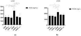
Evaluation of the amounts of AGE (a), RAGE (b) in the serum of the study groups. Data are given as mean±SEM. Expressed as *p<0.01.
Evaluation of some inflammatory markers in the serum of rats is shown in Figure 2. While the average serum TNF-α amount of rats in the HAGE group was 331.48±17.20 ng/L, the average serum TNF-α amount of rats in the HAGE1000 group was 250.95±20.81 ng/L. This difference is statistically significant (p<0.001) (Figure 2a). Similarly, in Figure 2b, the average amount of IL-6 in the serum of rats in the HAGE group is 13.04±0.40 ng/L, and that of rats in the HAGE1000 group is 10.00±0.60 ng/L (p<0.001). Figure 2c shows the average values of IL-10, an antiinflammatory marker. Accordingly, the serum mean IL-10 values of those in the HAGE group (203.46±8.02 ng/L) were significantly lower than those in the HAGE1000 group (252.22±9.2 ng/L) (p=0.013).
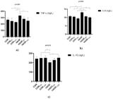
Evaluation of the amounts of TNF-α (a), IL-6 (b) and IL-10 (c) in the serum of the study groups. Data are given as mean±SEM. Expressed as *p<0.01.
Figure 3 shows the RAGE and NF-ĸB gene expression levels in the livers of rats. The RAGE expression level in the livers of those in the HAGE group was higher than that of those in the CON, HAGE500, and HAGE1000 groups, but this difference was not statistically significant (p=0.596) (Figure 3a). Similarly, the NF-ĸB gene expression level in the HAGE group is higher than that in the CON, HAGE500, and HAGE1000 groups, but this difference is not significant (p=0.109) (Figure 3b).
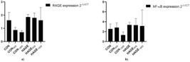
Evaluation of RAGE (a) and NF-ĸB (b) gene expressions in liver samples of the study groups by RT-PCR. Results were normalized using the 2-ΔΔCt method. Data are given as mean±SEM.
Histopathological changes such as acute cell swelling, hyperemia, and sinusoid swelling were more in the HAGE group than in the control group. OLE supplementation reduced these histopathological changes (Figure 4, Table 3).
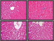
Histopathological images of liver tissue. (a) Hyperemia and widening of sinusoids detected in the HAGE group (b) Mononuclear cell infiltrations detected in the HAGE group (c) Acute cell swelling in hepatocytes seen in the HAGE group (d) Reduced appearance of infiltrations and hyperemia in the liver tissue of animals in the HAGE1000 group.
DISCUSSION
This study investigated the antiglycation effect of OLE, rich in phenolic compounds, whose antiinflammatory and antioxidant effects are frequently emphasized, in rats in the context of biochemical, gene expression levels and histopathological images. This study is the first known study to examine the antiglycation effect of OLE in vivo.
In the study, serum AGE levels of rats receiving the HAGE diet increased approximately two times compared to the control group. The results were seen to be parallel to the literature. In the study by Chen et al., serum AGE levels of rodents receiving the HAGE diet increased at a level similar to our study compared to the control diet [39]. In a study conducted with mice, serum AGE values of mice receiving the HAGE diet for 24 weeks increased 1.5-2 times compared to the control group [40].
The antiinflammatory effect of OLE is generally well known. One study examined the antiinflammatory effect of oleacin and oleuropein-aglycone extracted from olive leaves. As a result, the antiinflammatory effect of oleacin and oleuropein has been proven in both acute and chronic inflammation and the pathogenesis of viral infections [41]. Another study found that OLE reduced inflammation caused by carrageenan-induced paw edema in rats in a dose-dependent manner [42]. The cardioprotective properties of OLE were examined in diabetic rats. It was determined that OLE applied at doses of 100, 200, and 400 mg/kg for six weeks positively affected inflammation in the heart tissue [43]. In this study, the positive effects of OLE on serum inflammatory parameters were proven, which aligns with the literature.
A few in vitro studies have previously been published showing that OLE may have an antiglycation effect in addition to its antiinflammatory properties. Kontogianni et al. extracted aqueous and methanolic extracts from OLs. They found that the methanolic extract inhibited the formation of fluorescent AGEs in the bovine serum albumin (BSA)-ribose system. When the phytochemical composition of OLE was examined, they found that luteolin and luteolin-4’- O - β - d -glucopyranoside were potent AGE inhibitors [17]. In a study evaluating plant-based commercial nutritional supplements, it was found that the antiglycation effect of OLE, which contains the active ingredient oleuropein at different doses, was higher than other supplements, and the antiglycation effect increased as the oleuropein dose increased [16]. Another recent study determined that the antiglycation effect of oleuropein-enriched OLE was higher than pure OLE [44]. These in vitro studies reported that the potential antiglycation effect of OLE is due to the phenolic compounds it contains. Especially in structure-activity studies, it has been shown that the antiglycation activity strengthens as the number of hydroxyl groups in the 3’-, 4’-, 5-, and 7-positions of phenolic compounds increases. It has also been reported that the activities of flavones are more potent than the corresponding flavonols, flavanones, and isoflavones [45,46]. This information indicates that the phenolic compounds found in OLE may be potent antiglycation agents [17]. The primary mechanism at this point is that phenolic compounds block the free amino groups on proteins or the carbonyl groups on reducing sugars, so they cannot be bound [16]. Another mechanism is that the phenolic compounds in OLE may reduce AGE formation by indirectly reducing inflammation and oxidative stress. The results obtained from these in vitro studies in the literature were confirmed, and it was determined that OLE reduced the levels of AGE and RAGE in serum.
Exogenous AGEs in tissues and organs cause an increased expression and activation of RAGE. Physiologically, increased RAGE activation induces transcription factors such as NF-κB, an activator of transcription signal transducer (STAT3) and hypoxia-inducible factor 1-α (HIF1α) by accelerating the cytokine secretion, increasing the production of Reactive Oxygen Species (ROS) and the inflammatory response. Moreover, ROS can further increase the formation of AGEs, and a cyclical inflammatory response occurs [39,47,48]. For this reason, the RAGE value in the liver was examined together with NF-κB in the study. At the end of the study, OLE reduced the expressions of RAGE and NF-ĸB in liver samples of rats, but this difference was not found to be statistically significant. The fact that the intervention period is relatively short for diet studies may be effective in obtaining these results at the gene level.
In addition to these results, when the literature is examined, studies report that biocomponents such as triterpenes and phenolic acids, which are also found in OLs but extracted from different plants, have antiglycation effects alone [49-51]. The main limitation of such in vitro studies is that the bioavailability of the biocomponent or nutrient and its interaction with other nutrients are unknown. For this reason, it is essential to test the results obtained from in vitro studies in vivo animal models.
CONCLUSION
As a result, the study found that the OLE supplement of rats receiving a HAGE diet for eight weeks positively affected serum inflammatory markers and AGE and RAGE levels; thus, it could be a potential antiglycation and antidiabetic agent. This is an important result for understanding the potential therapeutic activities of OL for humans. Evaluating the antiglycative effect of different and long-term OLE doses on different chronic disease models in future studies will contribute to obtaining more accurate results.
ACKNOWLEDGMENT
Thanks to the staff of the Karabuk University Experimental Medicine Application and Research Center for their help in breeding experimental animals and their care during the experiment.
-
Article based on the doctoral thesis by B DEMİRER, entitled “Effect of olive leaf extract added to the diet on glycation, ınflammatıon and gene expression ın rats feeding a diet rich in advanced glycation end products”. Hacettepe University, Türkiye.
-
How to cite this article:
Demirer B, Özdemir M, Samur G. Antiglycative effect of olive leaf (olea europaea) against dietary advanced glycation end products in rats. Rev Nutr. 2026;39:e250025. https://doi.org/10.1590/1678-9865202639e250025
-
Support
This study was funded by the Karabuk University Scientific Research Projects Support Program with the number KBÜBAP-23-KP-125 and by TÜBİTAK with the number 124S203 within the scope of module 1002.
Data Availability
The research data are available on request from the corresponding author.
REFERENCES
- 1 Semba RD, Bandinelli S, Sun K, Guralnik JM, Ferrucci L. Relationship of an advanced glycation end product, plasma carboxymethyl-lysine, with slow walking speed in older adults: the InCHIANTI study. Eur J Appl Physiol. 2010;108(1):191-5.
- 2 Liu H, Zhang R, Wang W, Xia X, Xu Z, Xiang X. Inhibitory effects and mechanisms of phenolic compounds in rapeseed oil on advanced glycation end product formation in chemical and cellular models in vitro. Food Chem. 2024;447:139056.
- 3 Demirer B, Yardımcı H, Erem Basmaz S. Inflammation level in type 2 diabetes is associated with dietary advanced glycation end products, Mediterranean diet adherence and oxidative balance score: A pathway analysis. J Diabetes Complications. 2023;37(1):108354.
- 4 Guo WA, Davidson BA, Ottosen J, Ohtake PJ, Raghavendran K, Mullan BA, Dayton MT, Knight PR. Effect of high advanced glycation end-product diet on pulmonary inflammatory response and pulmonary function following gastric aspiration. Shock. 2012;38(6):677-84.
- 5 Hegab Z, Gibbons S, Neyses L, Mamas MA. Role of advanced glycation end products in cardiovascular disease. World J Cardiol. 2012;4(4):90-102.
- 6 Litwinowicz K, Waszczuk E, Gamian A. Advanced Glycation End-Products in Common Non-Infectious Liver Diseases: Systematic Review and Meta-Analysis. Nutrients. 2021;13(10).
- 7 Nowotny K, Jung T, Höhn A, Weber D, Grune T. Advanced glycation end products and oxidative stress in type 2 diabetes mellitus. Biomolecules. 2015;5(1):194-222.
- 8 Rungratanawanich W, Qu Y, Wang X, Essa MM, Song BJ. Advanced glycation end products (AGEs) and other adducts in aging-related diseases and alcohol-mediated tissue injury. Exp Mol Med. 2021;53(2):168-88.
- 9 Kobori A, Miyashita M, Miyano Y, Suzuki K, Toriumi K, Niizato K, et al. Advanced glycation end products and cognitive impairment in schizophrenia. Plos One. 2021;16(5):e0251283.
- 10 Venter C, Pickett K, Starling A, Maslin K, Smith PK, Palumbo MP, et al. Advanced glycation end product intake during pregnancy and offspring allergy outcomes: A Prospective cohort study. Clin Exp Allergy. 2021;51(11):1459-70.
- 11 Tian Z, Chen S, Shi Y, Wang P, Wu Y, Li G. Dietary advanced glycation end products (dAGEs): An insight between modern diet and health. Food Chemistry. 2023;415:135735.
- 12 Chen G. Dietary N-epsilon-carboxymethyllysine as for a major glycotoxin in foods: A review. Compr Rev Food Sci Food Saf. 2021;20(5):4931-49.
- 13 Uribarri J, Woodruff S, Goodman S, Cai W, Chen X, Pyzik R, et al. Advanced glycation end products in foods and a practical guide to their reduction in the diet. J Am Diet Assoc. 2010;110(6):911-16.e12.
- 14 Asif A, Zeeshan N, Mehmood S. Antioxidant and antiglycation activities of traditional plants and identification of bioactive compounds from extracts of Hordeum vulgare by LC-MS and GC-MS. J Food Biochem. 2020;44(9):e13381.
- 15 Khan M, Liu H, Wang J, Sun B.Inhibitory effect of phenolic compounds and plant extracts on the formation of advance glycation end products: A comprehensive review. Food Research International. 2020;130:108933.
- 16 Koch ER, Deo P. Nutritional supplements modulate fluorescent protein-bound advanced glycation endproducts and digestive enzymes related to type 2 diabetes mellitus. BMC Complement Altern Med. 2016;16(1):338.
- 17 Kontogianni VG, Charisiadis P, Margianni E, Lamari FN, Gerothanassis IP, Tzakos AG. Olive leaf extracts are a natural source of advanced glycation end product inhibitors. J Med Food. 2013;16(9):817-22.
- 18 Vijaykrishnaraj M, Wang K. Dietary natural products as a potential inhibitor towards advanced glycation end products and hyperglycemic complications: A phytotherapy approaches. Biomed Pharmacother. 2021;144:112336.
- 19 Song Q, Liu J, Dong L, Wang X, Zhang X. Novel advances in inhibiting advanced glycation end product formation using natural compounds. iomed Pharmacother. 2021;140:111750.
- 20 Spagnuolo L, Della Posta S, Fanali C, Dugo L, De Gara L. Antioxidant and Antiglycation Effects of Polyphenol Compounds Extracted from Hazelnut Skin on Advanced Glycation End-Products (AGEs) Formation. Antioxidants (Basel). 2021;10(3):424.
- 21 Jia W, Ma R, Zhang R, Fan Z, Shi L. Synthetic-free compounds as the potential glycation inhibitors performed in in vitro chemical models: Molecular mechanisms and structure requirements. Trends Food Sci. Technol. 2022;128:147-59.
- 22 Asgharpour Dil F, Ranjkesh Z, Goodarzi MT. A systematic review of antiglycation medicinal plants. Diabetes Metab Syndr. 2019;13(2):1225-29.
- 23 Gürbüz M, Öğüt S. Potential Health Benefits of Olive Leaf. Turkiye Klinikleri J Health Sci. 2018;3(3):242-53.
- 24 Omar SH. Oleuropein in Olive and its Pharmacological Effects. Sci Pharm. 2010;78(2):133-54.
- 25 Morelló JR, Vuorela S, Romero MP, Motilva MJ, Heinonen M. Antioxidant activity of olive pulp and olive oil phenolic compounds of the arbequina cultivar. J Agric Food Chem. 2005;53(6):2002-8.
- 26 Alirezaei M, Dezfoulian O, Neamati S, Rashidipour M, Tanideh N, Kheradmand A. Oleuropein prevents ethanol-induced gastric ulcers via elevation of antioxidant enzyme activities in rats. J Physiol Biochem. 2012;68(4):583-92.
- 27 Barbaro B, Toietta G, Maggio R, Arciello M, Tarocchi M, Galli A, et al. Effects of the Olive-Derived Polyphenol Oleuropein on Human Health. Int J Mol Sci. 2014;15(10):18508-24.
- 28 Janahmadi Z, Nekooeian AA, Moaref AR. Oleuropein Offers Cardioprotection in Rats with Acute Myocardial Infarction. Cardiovasc Toxicol. 2015;15(1):61-8.
- 29 Muellenbach EA, Diehl CJ, Teachey MK, Lindborg KA, Archuleta TL, Harrell NB, et al. Interactions of the advanced glycation end product inhibitor pyridoxamine and the antioxidant alpha-lipoic acid on insulin resistance in the obese Zucker rat. Metabolism. 2008;57(10):1465-72.
- 30 Zhang X, Xu L, Chen W, Yu X, Shen L, Huang Y. Pyridoxamine alleviates mechanical allodynia by suppressing the spinal receptor for advanced glycation end product-nuclear factor-κB/extracellular signal-regulated kinase signaling pathway in diabetic rats. Mol Pain.2020;16:1744806920917251.
- 31 Csongová M, Renczés E, Šarayová V, Mihalovičová L, Janko J, Gurecká R, et al. Maternal Consumption of a Diet Rich in Maillard Reaction Products Accelerates Neurodevelopment in F1 and Sex-Dependently Affects Behavioral Phenotype in F2 Rat Offspring. Foods. 2019;8(5):168
- 32 Yang S, Zhou H, Wang G, Zhong XH, Shen QL, Zhang XJ, et al. Quercetin is protective against short-term dietary advanced glycation end products intake induced cognitive dysfunction in aged ICR mice. J Food Biochem. 2020;44(4):e13164.
- 33 Reagan-Shaw S, Nihal M, Ahmad N. Dose translation from animal to human studies revisited. Faseb J. 2008;22(3):659-61.
- 34 Takata T, Sakasai-Sakai A, Takino JI, Takeuchi M. Evidence for Toxic Advanced Glycation End-Products Generated in the Normal Rat Liver. Nutrients. 2019;11(7):1612.
- 35 Yamagishi S, Matsui T. Role of receptor for advanced glycation end products (RAGE) in liver disease. Eur J Med Res. 2015;20(1):15.
- 36 Alves M, Calegari VC, Cunha DA, Saad MJ, Velloso LA, Rocha EM. Increased expression of advanced glycation end-products and their receptor, and activation of nuclear factor kappa-B in lacrimal glands of diabetic rats. Diabetologia. 2005;48:2675-81.
- 37 Tobon-Velasco JC, Cuevas E, Torres-Ramos MA. Receptor for AGEs (RAGE) as mediator of NF-kB pathway activation in neuroinflammation and oxidative stress. CNS Neurol Disord Drug Targets. 2014;13(9):1615-26.
- 38 Prasad K, Mishra M. AGE-RAGE Stress, Stressors, and Antistressors in Health and Disease. Int J Angiol. 2018;27(1):1-12.
- 39 Chen MC, Lin JA, Lin HT, Chen SY, Yen GC. Potential effect of advanced glycation end products (AGEs) on spermatogenesis and sperm quality in rodents. Food Funct. 2019;10(6):3324-33.
- 40 Wang J, Cai W, Yu J, Liu H, He S, Zhu L, et al. Dietary Advanced Glycation End Products Shift the Gut Microbiota Composition and Induce Insulin Resistance in Mice. Diabetes Metab Syndr Obes. 2022;15:427-37.
- 41 Silvestrini A, Giordani C, Bonacci S, Giuliani A, Ramini D, Matacchione G+ Anti-inflammatory effects of olive leaf extract and its bioactive compounds oleacin and oleuropein-aglycone on senescent endothelial and small airway epithelial cells. Antioxidants (Basel). 2023;12(8):1509.
- 42 Fayez N, Khalil W, Abdel-Sattar E, Abdel-Fattah AM. In vitro and in vivo assessment of the anti-inflammatory activity of olive leaf extract in rats. Inflammopharmacology. 2023;31(3):1529-38.
- 43 Asghari AA, Mahmoudabady M, Shabab S, Niazmand S. Anti-inflammatory, anti-oxidant and anti-apoptotic effects of olive leaf extract in cardiac tissue of diabetic rats. J Pharm Pharmacol. 2022;74(7):961-72.
- 44 Márquez K, Márquez N, Ávila F, Cruz N, Burgos-Edwards A, Pardo X, et al. Oleuropein-Enriched Extract From Olive Mill Leaves by Homogenizer-Assisted Extraction and Its Antioxidant and Antiglycating Activities. Front Nutr. 2022;9:895070.
- 45 Matsuda H, Wang T, Managi H, Yoshikawa M. Structural requirements of flavonoids for inhibition of protein glycation and radical scavenging activities. Bioorg Med Chem. 2003;11(24):5317-23.
- 46 Xie Y, Chen X. Structures required of polyphenols for inhibiting advanced glycation end products formation. Curr Drug Metab. 2013;14(4):414-31.
- 47 Palioura E, Palimeri S, Piperi C, Sakellariou S, Kandaraki E, Sergentanis T, et al. Impact of Androgen and Dietary Advanced Glycation End Products on Female Rat Liver. Cell Physiol Biochem. 2015;37(3):1134-46.
- 48 Poulsen MW, Andersen JM, Hedegaard RV, Madsen AN, Krath BN, Monošík R, et al. Short-term effects of dietary advanced glycation end products in rats. Br J Nutr. 2016;115(4):629-36.
- 49 Ashraf JM, Shahab U, Tabrez S, Lee EJ, Choi I, Ahmad S. Quercetin as a finer substitute to aminoguanidine in the inhibition of glycation products. International Int J Biol Macromol. 2015;77:188-92.
- 50 Li X, Zheng T, Sang S, Lishuang, Lv. Quercetin Inhibits Advanced Glycation End Product Formation by Trapping Methylglyoxal and Glyoxal. J. Agric. Food Chem. 2014;62(50):12152-58.
- 51 Vlassopoulos A, Lean MEJ, Combet E. Protein–phenolic interactions and inhibition of glycation – combining a systematic review and experimental models for enhanced physiological relevance. Food & Function. 2014;5(10):2646-55.
Edited by
-
Editors
Maria Teresa Clerici, Alex Crisp

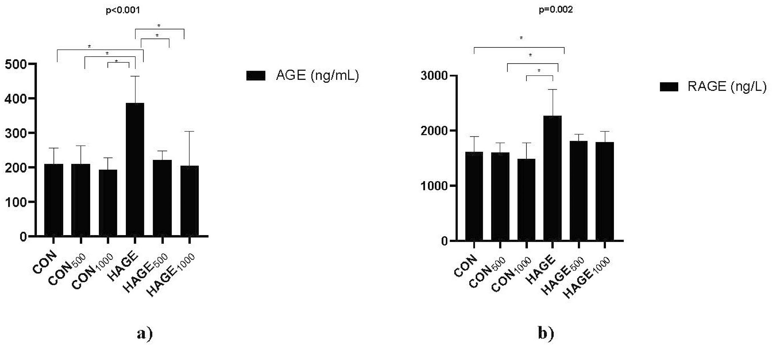 Note: AGE: Advanced glycation end product, CON: Control, HAGE: High advanced glycation end product, RAGE: Receptor advanced glycation end product.
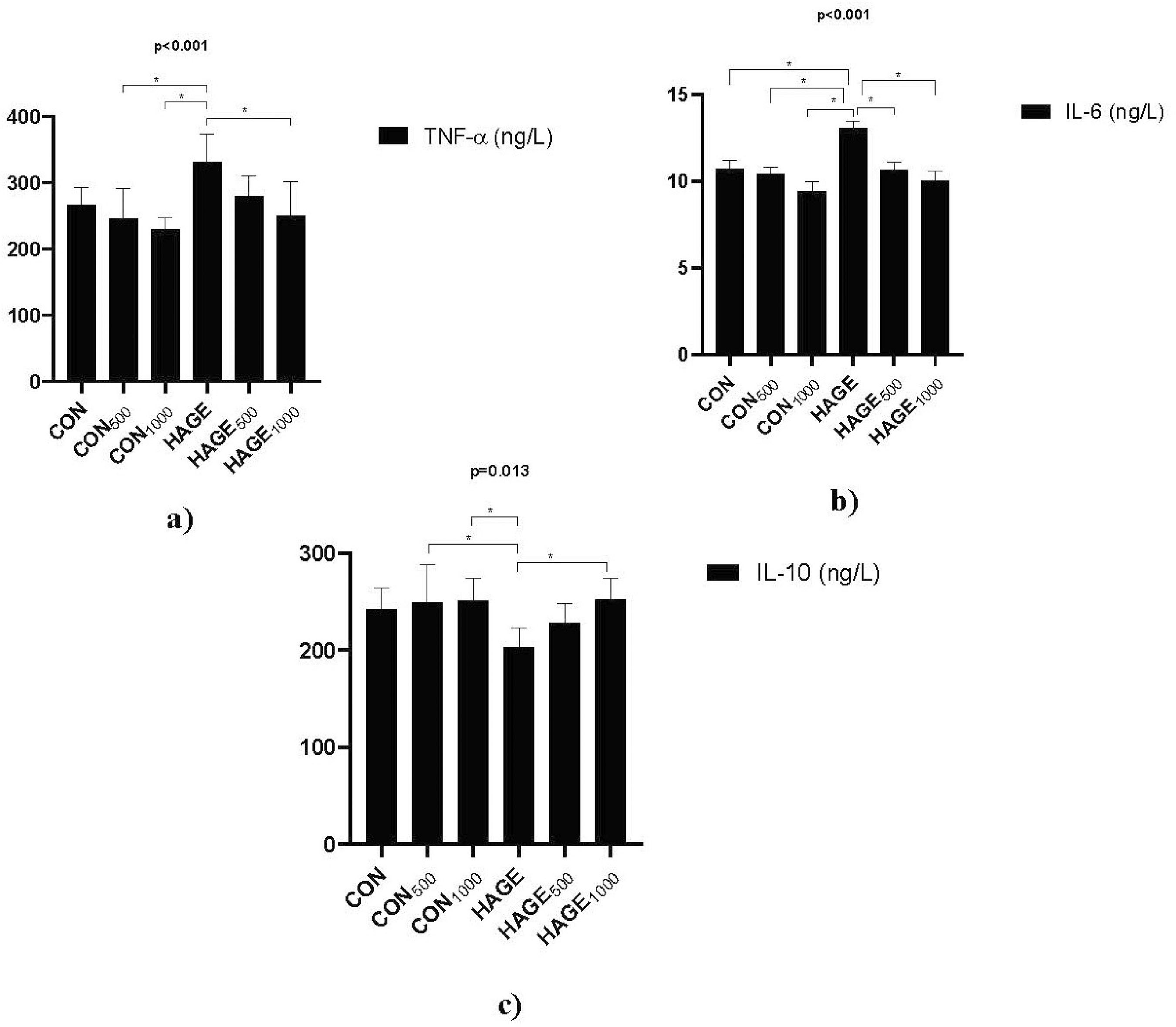 Note: CON: Control, HAGE: High advanced glycation end product, IL-10: Interleukin-10, IL-6: Interleukin-6, TNF-α: Tumor necrosis factor-alpha.
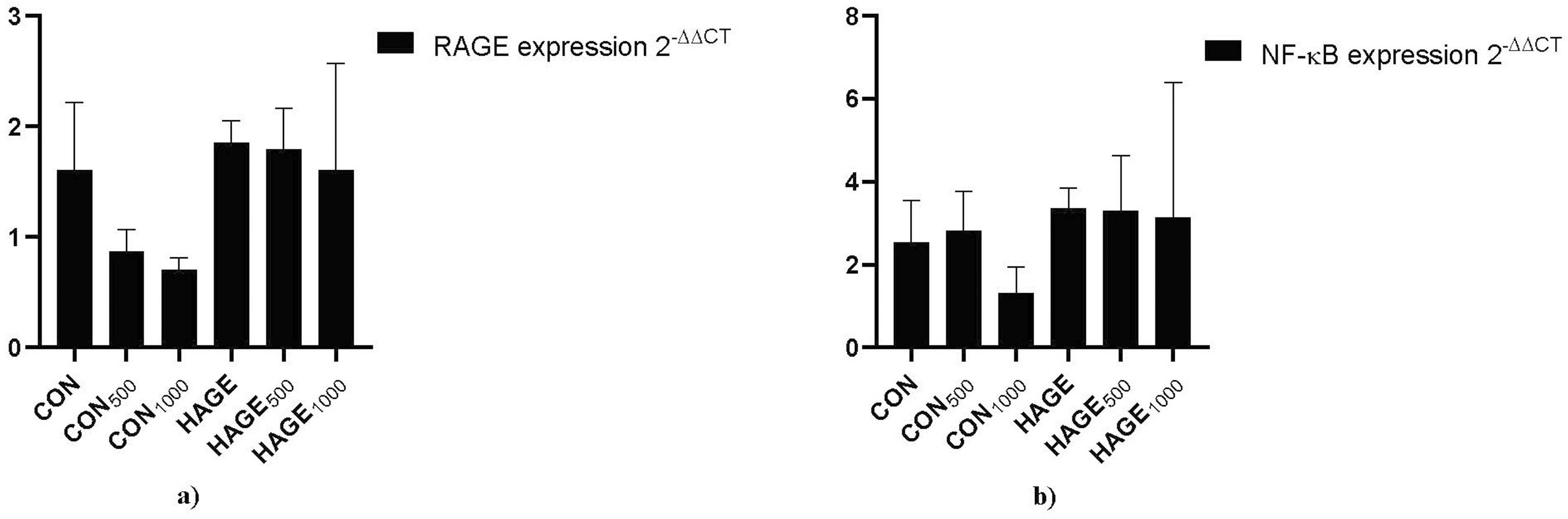 Note: CON: Control, HAGE: High advanced glycation end product, NF-ĸB: Nuclear factor kappa B, RAGE: Receptor advanced glycation end product.
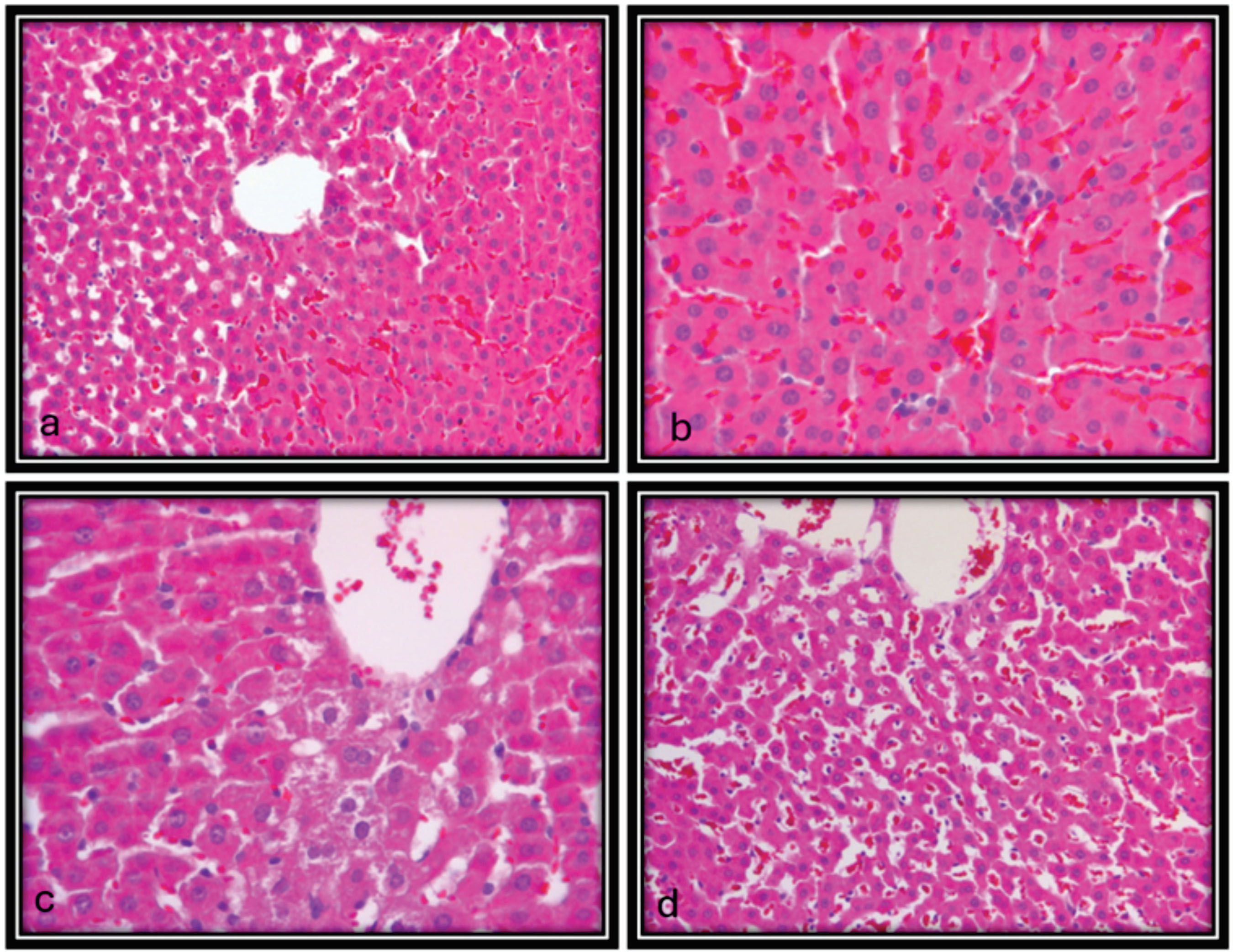 Note: HAGE: High advanced glycation end product.