 Effects of Myrcia splendens extract on innate inflammatory response
Abstract
This study investigated the anti-inflammatory activity of the crude hydroalcoholic extract of Myrcia splendens leaves. In vitro, neutrophil chemotaxis and LPS-induced inflammatory mediators were assessed in neutrophils or macrophages treated with M. splendens, salicylic acid, or gallic acid. Inflammation resolution was evaluated via efferocytosis. In vivo effects were tested using the air pouch model. M. splendens impaired macrophage secretion of TNF, IL-1β, IL-6, and NO following LPS stimulation. Neutrophils treated with LPS and incubated with the extract showed reduced migration and NO secretion. Likewise, salicylic and gallic acids decreased NO production in LPS-stimulated neutrophils. The extract enhanced efferocytosis, increased IL-10, and decreased TNF levels. In vivo, similar effects were observed, including reduced neutrophil migration and NO production. These findings suggest that M. splendens modulates neutrophil and macrophage functions, likely due to the presence of salicylic and gallic acids. This highlights its potential as a treatment for inflammatory conditions.
Key words:
inflammation; macrophage; Myrtaceae; neutrophil; resolution of inflammation
Resumo
Este estudo investigou a atividade anti-inflamatória do extrato hidroalcoólico bruto das folhas de Myrcia splendens. In vitro, a quimiotaxia de neutrófilos e os mediadores inflamatórios induzidos por LPS foram avaliados em neutrófilos e macrófagos tratados com M. splendens, ácido salicílico ou ácido gálico. A resolução da inflamação foi avaliada por eferocitose. Os efeitos in vivo foram testados usando o modelo de bolsa de ar. M. splendens reduziu a secreção de TNF, IL-1β, IL-6 e NO pelos macrófagos após a estimulação com LPS. Neutrófilos com LPS e incubados com o extrato apresentaram redução na migração e na secreção de NO. Da mesma forma, os ácidos salicílico e gálico diminuíram a produção de NO em neutrófilos estimulados com LPS. O extrato aumentou a eferocitose, IL-10 e diminuiu os níveis de TNF. In vivo, efeitos semelhantes foram observados, incluindo redução na migração de neutrófilos e na produção de NO. Esses achados sugerem que M. splendens modula as funções de neutrófilos e macrófagos, provavelmente devido à presença de ácidos salicílico e gálico. Isso destaca seu potencial como tratamento para condições inflamatórias.
Palavras-chave:
inflamação; macrófago; Myrtaceae; neutrófilo; resolução da inflamação
Introduction
Inflammation represents one of the fundamental processes in response to injuries, with the main intention of restoring homeostasis and guiding tissue healing. The inflammatory response is triggered in two ways: (a) acute, represented by a physiological process against injuries or infections, and (b) chronic, progression from the previous phase, without proper solution, making a non-physiological process. The mechanism involved in the inflammatory process is complex and dependent on organized cellular and molecular interaction (Abdulkhaleq et al. 2018).
The innate immune response is a rapid and nonspecific defense mechanism against various pathogens and harmful stimuli. It is the first line of defense and provides immediate protection, even before the adaptive immune response is triggered. The main events of innate immune response during acute phase of inflammation are: migration of granulocytic cells (basophil, neutrophil, and eosinophil) and monocyte from the blood vessels to the injured tissue; vasodilation; increased vascular permeability; platelet plugging and chemical mediators production. These events are mediated by chemical mediators as histamines, prostaglandins, free radicals, cytokines, and serotonin, with the aim of regulating the inflammatory response by removing the inflammatory stimulus and promoting the resolution of inflammation (Varela et al. 2018).
The acute phase is enough to resolve the caused initial damage, but if the inflammation persists, the chronic phase of the inflammatory process starts, and it can lead to the loss of function. This phase of inflammatory processes is labeled by infiltration of monocytes, lymphocytes, and plasma cells in the affected tissue. These cells release various substances like inflammatory cytokines, growth factors, antibodies and enzymes, contributing to the tissue damage and subsequent repair processes including fibrosis and granuloma formation. The chronic inflammatory process contributes to numerous diseases, such as arthritis, asthma, atherosclerosis, autoimmune diseases, diabetes, and cancer (Germolec et al. 2018; Pahwa et al. 2023).
Therefore, the new therapeutic strategies need to be developed with less adverse effects than conventionally available synthetic steroids or non-steroidal drugs. Thus, increasing the search for new anti-inflammatory agents of herbal source, due to the presence of their secondary metabolites, it is possible to obtain therapeutic tools with better safety, efficacy, and economy in the treatment of inflammation (Abdulkhaled et al. 2018).
In the literature, the Myrcia species presents important biological activity, including anti-inflammatory properties (Cascaes et al. 2015; Marena et al. 2022). However, just some species of the genus have been studied regarding the anti-inflammatory activity, such as Myrcia pubiflora, Myrcia bela, Myrcia rubra, Myrcia ovata, and Myrcia pubipetala. The species Myrcia splendens (Sw.) DC. is a tree found in the Brazilian cerrado and it is popularly known as “guamirim-de-folha-miúda”, ocurring from Mexico to southern Brazil [syn. M. acutata DC., M. rostrata DC., M. communis Berg., and M. fallax (Rich.) DC.] (Paganelli et al. 2020). Regarding the chemical composition of Myrcia splendens, a high amount of ellagic acid was detected, following other majority compounds such as protocatecuic acid, syringic acid, p-coumaric acid, salicylic acid, isoquercetin, gallic acid, myricetin and kaempferol (Cascaes et al. 2015; Paganelli et al. 2020) were identified in the crude hydroalcoholic extract obtained from the leaves. Some of phenolic compounds present in the Myrcia splendens are widely studied and with the anti-inflammatory activity well established, as salicylic acid (Rainsford et al. 1980; Munir et al. 2020), gallic acid (BenSaad et al. 2017; Bai et al. 2021), p-coumaric acid (Kheiry et al. 2019), and isoquercetin (Rogerio et al. 2007; Li et al. 2016).
Based on the previous phytochemical study of the crude hydroalcoholic extract of Myrcia splendens leaves and the knowledge of the isolated compounds and their biological activity, the present study aimed to evaluate the anti-inflammatory activity Myrcia splendens through in vitro and in vivo assays. The results herein exposed, demonstrated for the first time that M. splendens presents anti-inflammatory activity with driving the resolution of inflammation.
Material and Methods
Myrcia splendens
The aerial parts of Myrcia splendens were collected on Campus I of the Regional University of Blumenau (26°54’18.2”S, 49°04’44.5”W) in December 2017, under the supervision of Dr. André Luis de Gasper (FURB). The specimen was cataloged at the Roberto Miguel Klein Herbarium (FURB 000607). Leaves and branches were separated, and the crude hydroalcoholic extract was prepared from the leaves according to a previously described procedure (SisGen no. A7519AA). The phenolic profile of this extract has already been characterized in a previous publication by our group (Paganelli et al. 2020), as the same extract was used in the present study. For in vitro assays, the extract was solubilized with the culture medium used in the tests. For in vivo assays, the extract was solubilized with phosphate-buffered saline (PBS).
Drugs and reagents
Indomethacin, LPS (serotype 026:B6), carrageenan-λ, oyster glycogen type II, paraformaldehyde, MTT, gallic acid, salicylic acid and Griess reagent were obtained from Sigma-Aldrich (St. Louis, MO, USA). DMEM and FBS were purchased from Vitrocell (Campinas, SP, Brazil), and ketamine and xylazine from Vetbrands (Paulinia, SP, Brazil). All other solvents were of analytical grade. IL-1 and TNF were quantified using DuoSet kits from R&D Systems (Minneapolis, MN, USA).
Animals
Male Swiss mice (20-30 g) from the Central Animal Vivarium of FURB were housed in an enriched environment with controlled temperature (22 ± 1 °C), humidity (40-60%), and a 12 h light/dark cycle, with food and water ad libitum. A 5-day acclimatization period was observed. For experiments, mice were anesthetized with ketamine (100 mg/kg, i.p.) and xylazine (10 mg/kg, i.p.) to prevent stress. All procedures followed the Brazilian Society of Laboratory Animal Science guidelines and were approved by the Ethics Committee on Animal Use of FURB (CEUA/FURB 110/19).
Neutrophils and macrophage cell culture
Neutrophils were isolated as described by Santin et al. (2022). Male Swiss mice received an intraperitoneal injection of 1% sterile oyster glycogen (3 mL, PBS). After 4 h, mice were euthanized with ketamine (100 mg/kg, s.c.) and xylazine (15 mg/kg, s.c.), and the peritoneal cavity was washed with 3 mL PBS. Viable cells were counted using a Neubauer chamber and trypan blue exclusion assay.
RAW 264.7 macrophages (BCRJ, Rio de Janeiro, Brazil) were cultured in DMEM with 10% FBS, streptomycin (100 μg/L), and penicillin (100 UI/mL) at 37 °C, 5% CO₂. Cell viability was assessed by MTT assay. Cells (1×10⁵/well) were incubated in 96-well plates for 24 h, then exposed to M. splendens extract (1, 10, or 100 μg/mL) or dimethyl sulfoxide 10 % (DMSO, as positive control) for another 24 h. MTT (0.5 mg/mL) was added, and after 4 h, the medium was removed. The precipitate was dissolved in DMSO, and absorbance was measured at 570 nm. Results were expressed as percentages of viable cells.
LPS-stimulated macrophages
RAW 264.7 cells were plated in 96-well plates (1×10⁵ cells/well) and incubated for 24 hours at 37 °C in a 5% CO₂ environment. The medium was then replaced with fresh FBS-free DMEM. The cells were treated with M. splendens extract (1, 10, or 100 μg/mL) in FBS-free DMEM. After 1 hour of incubation, the cells were exposed to LPS (5 μg/mL) for 24 hours. The supernatant was subsequently collected for analysis of inflammatory mediators.
LPS-stimulated neutrophils
Murine neutrophils were seeded into 96-well plates at a density of 1×10⁵ cells per well and incubated for 24 hours at 37 °C in a 5% CO₂ atmosphere. The culture medium was then replaced with fresh serum-free DMEM (FBS-free DMEM). The cells were treated with M. splendens extract (1, 10, or 100 μg/mL), or the isolated compounds salicylic acid (1, 10, or 100 μM) and gallic acid (1, 10, or 100 μM), all prepared in FBS-free DMEM.
Analysis of cytokines and nitrite levels
Nitrite levels were measured in the culture supernatant to indirectly assess NO production by macrophages and neutrophils. Samples (100 μL) were incubated with 100 μL of Griess reagent (1% sulfanilamide, 0.1% naphthylethylenediamine dihydrochloride, 5% phosphoric acid) for 10 min. Absorbance was read at 540 nm (SPECTROstar Nano, BMG Labtech), and nitrite concentration was calculated using a sodium nitrite standard curve (Grisham et al. 1996).
Cytokine levels (IL-6, IL-1β, TNF) in macrophage supernatants were quantified by ELISA per the manufacturer’s instructions (R&D Systems).
Neutrophil chemotaxis assay
The study evaluated the effect of M. splendens extract at 1, 10, or 100 μg/mL on neutrophil chemotaxis. The chemotaxis assay followed the method of Nelson et al. (1975) with minor modifications. Neutrophils (1×10⁶ cells/mL) were treated with the extract or left untreated and placed in peripheral wells (10 μL) within an agarose gel in a Petri dish. The central well contained the chemotactic factor fMLP at 0.1 μM (10 μL). After incubating for 4 hours at 37 °C in a 5% CO₂ environment, the cells migrating toward the central well were counted to quantify chemotaxis.
Efferocytosis assay
Macrophages were obtained from the medullary cavity of mouse bones using sterile PBS, yielding a suspension of 1×10⁶ cells/well. Cells were incubated on circular glass coverslips in 24-well plates at 37 °C, 5% CO₂ for 2 h for adherence. Non-adherent cells were removed by washing three times with PBS.
Adherent macrophages were exposed to M. splendens extract (1, 10, or 100 μg/mL) for 1 h, then incubated with senescent neutrophils (1×10⁶ cells/well) for 20 min. Supernatants were collected for cytokine analysis, and coverslips were examined under 100x magnification. Phagocytosis was quantified as the percentage of macrophages containing neutrophils (n = 100).
TNF and IL-10 levels were measured in supernatants by ELISA, following the manufacturer’s instructions.
In vivo leukocyte migration: air pouch model
Air pouches were generated dorsally near the cervical region as described (Sin et al. 1986; Jain et al. 2011). Anesthetized animals received 3 mL of sterile air subcutaneously via a 0.22 μm filter. After 72 h, a 3 mL booster was injected. Six days later, animals received oral M. splendens (3, 30, or 300 mg/kg, in PBS), indomethacin (30 mg/kg, positive control), or vehicle (PBS, negative control). One-hour post-treatment, 2 mL of 1%-carrageenan was injected into the pouch. Naïve animals received only PBS and anesthesia.
After 4 h, exudate was collected via a small incision, washed with 2 mL PBS, and centrifuged (600 g, 10 min, 4 °C). Total and differential cell counts were performed using a Neubauer chamber and Giemsa-stained smears.
Air pouch tissue was preserved in 10% formaldehyde (24 h), transferred to 70% ethanol, dehydrated in graded alcohols, treated with xylene and paraffin, sectioned (3-4 μm), stained (H&E), and analyzed microscopically (10x, 40x).
Statistical analysis
Data are expressed as mean ± SEM and analyzed using Student’s t-test or one-way ANOVA. Dunnett’s test determined significance. GraphPad Prism® (GraphPad Software, USA) was used. p < 0.05 was considered significant.
Results and Discussion
The phytochemical analysis of Myrcia splendens, recently published by our research group, revealed similarities with other Myrcia species (Paganelli et al. 2020). Several compounds previously identified within the genus were detected, including syringic acid, p-coumaric acid, ferulic acid, syringaldehyde, salicylic acid, umbelliferone, coniferaldehyde, synapaldehyde, and carnosol, the latter of which were reported for the first time in the genus. This phytochemical characterization lays the groundwork for further investigation into the biological potential of M. splendens, particularly given the anti-inflammatory activity demonstrated by other Myrcia species, such as Myrcia pubiflora, Myrcia bela, Myrcia rubra, Myrcia ovata, and Myrcia pubipetala.
Considering this evidence, we evaluated the effects of M. splendens on neutrophil and macrophage activity. Neutrophils and macrophages are highly versatile immune cells with crucial roles in modulating inflammation, transitioning from its onset to tissue repair. These cells interact with other immune components through direct contact or the release of inflammatory mediators, significantly influencing both innate and adaptive immune responses (Filep et al. 2022; Lendeckel et al. 2022).
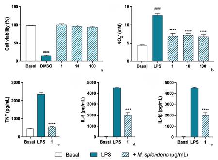
Here, we demonstrated for the first time that M. splendens leaf extract modulates key neutrophil and macrophage functions, including the reduction of neutrophil chemotaxis, cytokine release, and nitric oxide (NO) production, as well as the facilitation of inflammation resolution (Petri & Sanz 2018; Metzemaekers et al. 2020; Gierlikowska et al. 2021). Cell viability assays confirmed that M. splendens extract did not induce significant cytotoxicity at any tested concentration (Fig. 1a), supporting its safe use in pharmacological evaluations.
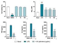
a-e. Effects of M. splendens on macrophage inflammatory mediator production. The cell viability of murine macrophage strain (RAW.264.7) incubated with M. splendens extract (1, 10 or 100 μg/mL) was accessed by MTT method (a). Cells were stimulated with LPS (5 μg/mL) and then treated with the extract for NO2- quantification performed by the Griess reaction (b). Quantification of TNF (c), IL-6 (d) and IL-1beta (e) was performed by ELISA according to the manufacturer’s standards in the 1 μg/mL treated cells. Data is expressed as the mean ± S.E.M. of tests performed with cells obtained from 4 independent experiments. Statistical analysis was performed using one-way ANOVA followed by Dunnett’s post hoc test. **** = p < 0.0001 vs LPS. #### = p < 0.0001 vs basal.
To further investigate its pharmacological effects, we analyzed the impact of M. splendens on the production of inflammatory mediators (TNF, IL-6, IL-1β, and NO2-). As illustrated in Figure 1b, lipopolysaccharide (LPS) stimulation triggered the production of inflammatory mediators by macrophages. However, treatment with M. splendens extract at 1, 10, or 100 μg/mL significantly inhibited NO2- production (48.30 ± 10.6%, 59.45 ± 6.1%, and 66.40 ± 8.9%, respectively). Additionally, at 1 μg/mL, the extract significantly reduced TNF (86.62 ± 6.32%), IL-1β (36.14 ± 4.33%), and IL-6 (54.86 ± 6.32%) levels compared to LPS-treated macrophages (Fig. 1c-e).
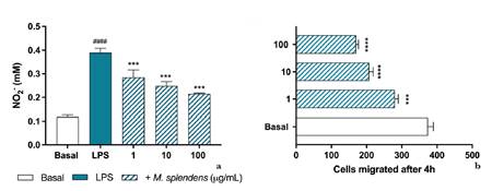
Similarly, the extract significantly inhibited NO2- secretion in neutrophils stimulated by LPS (Fig. 2a). The anti-inflammatory effect of M. splendens may be attributed to its gallic and salicylic acid content, as both compounds have been reported to suppress NO secretion in LPS-stimulated neutrophils. Notably, salicylic acid inhibits cyclooxygenase-2-dependent prostaglandin E (2) synthesis (Hinz et al. 2000). As well Gallic acid, which is the major phenolic compound in the crude hydroalcoholic extract of M. splendens leaves, exerts anti-inflammatory effects through multiple molecular mechanisms, including antioxidant activity, inhibition of pro-inflammatory cytokine releases such as TNF-α and IL-6, downregulation of the NF-κB and p38 MAPK signaling pathways, and suppression of cyclooxygenase-2 expression and nitric oxide production. These mechanisms highlight the potential of M. splendens in the treatment of inflammatory conditions (Ahn et al. 2016; Tanaka et al. 2018; Bai et al. 2021; Behl et al. 2021; Alhyari et al. 2025).
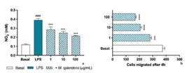
a-b. Effects of M. splendens on in vitro neutrophils chemotaxis and nitric oxide production. Neutrophils were stimulated with LPS (5 μg/mL) and then treated with the extract for NO2- quantification, which was performed by the Griess reaction (a). For the chemotaxis assay (b), neutrophil cell suspension was incubated with extract (1, 10 or 100 μg/mL) and placed in front of the fMLP (1 μM) into wells performed in an agarose plate. Cells were counted after 4 h. Quantification was made from the margin of the peripheral perforations towards the chemotactic agent (central perforation). Data is expressed as the mean ± S.E.M. of tests performed with cells obtained from 4 animals. Statistical analysis was performed using one-way ANOVA followed by Dunnett’s post hoc test. *** = p < 0.001 and **** = p < 0.0001 vs LPS or basal. #### - p < 0.0001 vs basal.
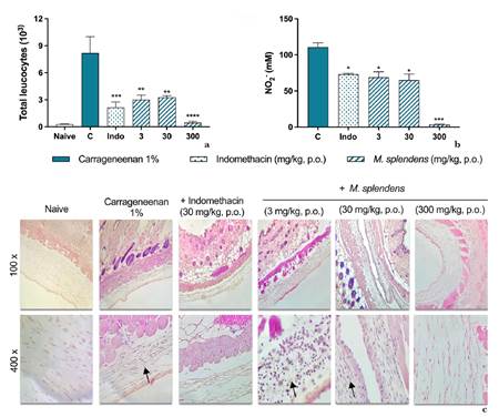
Additionally, we found that M. splendens extract significantly reduced N-formylmethionyl-leucyl-phenylalanine (fMLP)-induced neutrophil chemotaxis in vitro using the agar-overlay method. Treatment with 1, 10, or 100 μg/mL of the extract led to a marked reduction in neutrophil chemotaxis compared to untreated control cells (Fig. 2b). This inhibitory effect was further confirmed in vivo using the carrageenan-induced air pouch inflammation model in mice. Treatment with M. splendens extract (3, 30, or 300 mg/kg) significantly reduced leukocyte recruitment and NO secretion at all doses evaluated (Fig. 3a-b).
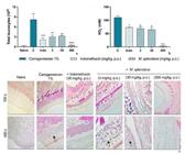
a-c. Effects of M. splendens extract on in vivo inflammatory cell migration. Air pouches were induced in the dorsal subcutaneous tissue of Swiss mice. The animals were treated orally one hour before the injection of 2 mL of carrageenan. Representative H.E. histological sections of skin biopsies obtained from mice with air pouches (100 and 400x). Black arrows indicate PMN leukocytes. The lavage of the inflammatory infiltrate was collected 4 hours after the injection of carrageenan into the air pouch. The determination of the (a) total number of exudate cells was performed on a Neubauer chamber. (b) The NO level was determined by the Griess reaction method. Values express the mean ± S.E.M. of tests performed with the inflammatory exudate obtained from 6 animals per group. * = p < 0.05 vs control group significantly different from naive group. # = p < 0.001 (One way ANOVA followed by Tukey’s post hoc test).
Histological analysis of the air pouch membranes supported these findings. In the naive group, the pouch wall consisted of a cell layer surrounded by connective tissue and extracellular matrix, primarily composed of fibroblasts (Fig. 3c). In contrast, the carrageenan group exhibited typical acute inflammatory responses, including membrane condensation, edema, polymorphonuclear cell (PMN) infiltration, and tissue damage. Indomethacin-treated animals showed a reduced inflammatory response, with less membrane condensation, limited edema, and decreased PMN infiltration. Similarly, M. splendens-treated mice exhibited significantly reduced edema and PMN infiltration compared to the carrageenan group, confirming the anti-inflammatory effects of the extract.
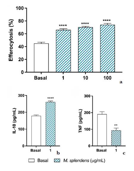
Neutrophils play an indirect yet crucial role in tissue repair by modulating macrophage activity. Through efferocytosis, macrophages engulf apoptotic neutrophils, leading to their transformation into M2 macrophages, which are essential for tissue repair and inflammation resolution (Thieblemont et al. 2018; Ge et al. 2022; Mehrotra et al. 2022). These responses are associated with increased transforming growth factor-beta (TGF-β) and interleukin-10 (IL-10) production, alongside a reduction in pro-inflammatory mediators (Loh et al. 2022; Saas et al. 2022; Purnama et al. 2023). Our data indicate that macrophages treated with M. splendens extract exhibited enhanced efferocytosis and reduced TNF levels, along with increased IL-10 production (Fig. 4a-c). These findings suggest that M. splendens contributes to inflammation resolution and tissue homeostasis.
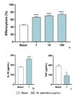
a-b. Effects of M. splendens on in vitro efferocytosis. The extract was able to promote the efferocytosis in vitro (a) and reduce the levels of TNF (c) with concomitant increase in IL-10 (b). Data is expressed as a mean ± S.E.M. of cells obtained from four animals. Statistical analysis was performed using One way ANOVA followed by Dunnett’s post hoc test. ** = p < 0.01; *** = p < 0.001 and **** = p < 0.0001 vs basal.
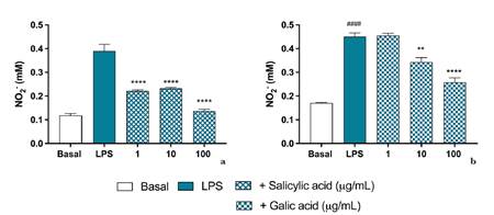
To assess the pharmacological effects of salicylic and gallic acid, cell viability assays confirmed that none of the tested concentrations (1, 10, or 100 μg/mL) induced cytotoxicity, as determined by the trypan blue exclusion method (data not shown). Further analysis of NO2- levels in neutrophil cultures revealed that salicylic acid significantly reduced NO2- levels at all tested concentrations, whereas gallic acid exerted inhibitory effects only at concentrations exceeding 10 μg/mL (Fig. 5a-b).
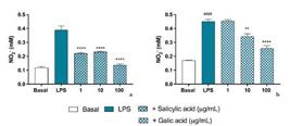
a-b. Effects of salicylic and gallic acid on in vitro nitric oxide production by neutrophils. Neutrophils were stimulated with LPS (5 μg/mL) and then treated with salicylic acid (a) or gallic acid (b) for NO2- quantification, which was performed by the Griess reaction. Data is expressed as the mean ± S.E.M. of tests performed with cells obtained from four animals. Statistical analysis was performed using one-way ANOVA followed by Dunnett’s post hoc test. ** = p < 0.01 and **** = p < 0.0001 vs LPS or basal. #### = p < 0.0001 vs basal.
Overall, these findings demonstrate the anti-inflammatory potential of M. splendens, highlighting its capacity to modulate neutrophil and macrophage activity. The observed inhibition of cytokine release, NO production, and neutrophil chemotaxis, coupled with enhanced efferocytosis and IL-10 production, underscores the therapeutic potential of M. splendens in inflammation resolution. Further studies are warranted to elucidate the molecular mechanisms underlying these effects and to explore its potential clinical applications.
The data obtained highlight the strong anti-inflammatory effects of the M. splendens extract, primarily through its ability to modulate neutrophil and macrophage functions. These effects include suppressing the production and/or release of inflammatory mediators, reducing cell migration, and promoting efferocytosis, all of which contribute to inflammation resolution. The presence of compounds such as salicylic acid and gallic acid may partially explain these actions, as they effectively inhibit the secretion of inflammatory mediators. Overall, these findings underscore the potential of M. splendens as a promising therapeutic for managing inflammatory responses.
Acknowledgements
This work was supported by grants and financial support from the Conselho Nacional de Desenvolvimento Científico e Tecnológico (CNPq); and the Fundação de Apoio à Pesquisa Científica e Tecnológica do Estado de Santa Catarina (FAPESC). The co-authors were supported by grants, as follows: L.B., Coordenação de Aperfeiçoamento de Pessoal de Nível Superior - CAPES, finance code 001); C.J.P. (FAPESC - edital 03/2017); M.D.A. (CNPq - 308155/2022-0); N.L.M.Q. (CNPq - 305550/2018-7); J.R.S. (CNPq - grant number 429505/2018-3; 310326/2020-6); I.D.M. (FAPESC - 001464/2023).
References
-
Alhyari D, Qinna NA, Sheldrake HM, Kantamneni S, Ghanem BY & Paluch KJ (2025) Antioxidant, anti-inflammatory, and oral bioavailability of novel sulfonamide derivatives of gallic acid. Antioxidants 14: 374. DOI: 10.3390/antiox14040374
» https://doi.org/10.3390/antiox14040374 -
Ahn CB, Jung WK, Park SJ, Kim YT, Kim WS & Je JY (2016) Gallic Acid-g-Chitosan modulates inflammatory responses in LPS-Stimulated RAW264.7 Cells Via NF-κB, AP-1, and MAPK Pathways. Inflammation 39: 366-374. DOI: 10.1007/s10753-015-0258-2
» https://doi.org/10.1007/s10753-015-0258-2 -
Abdulkhaleq LA, Assi MA, Abdullah R, Zamri-Saad M, Taufiq-Yap YH & Hezmee MNM (2018) The crucial roles of inflammatory mediators in inflammation: a review. Veterinary World 11: 627-635. DOI: 10.14202/vetworld.2018.627-635
» https://doi.org/10.14202/vetworld.2018.627-635 -
Bai J, Zhang Y, Tang C, Hou Y, Ai X, Chen X, Zhang Y, Wang X & Meng X (2021) Gallic acid: pharmacological activities and molecular mechanisms involved in inflammation-related diseases. Biomedicine & Pharmacotherapy 133: 110985. DOI: 10.1016/j.biopha.2020.110985
» https://doi.org/10.1016/j.biopha.2020.110985 -
Behl T, Upadhyay T, Singh S, Chigurupati S, Alsubayiel AM, Mani V, Vargas-De-La-Cruz C, Uivarosan D, Bustea C, Sava C, Stoicescu M, Radu AF & Bungau SG (2021) Polyphenols targeting MAPK mediated oxidative stress and inflammation in rheumatoid arthritis. Molecules 26: 6570. DOI: 10.3390/molecules26216570
» https://doi.org/10.3390/molecules26216570 -
BenSaad LA, Kim KH, Quah CC, Kim WR & Shahimi M (2017) Anti-inflammatory potential of ellagic acid, gallic acid and punicalagin A&B isolated from Punica granatum BMC Complementary and Alternative Medicine 17: 47. DOI: 10.1186/s12906-017-1555-0
» https://doi.org/10.1186/s12906-017-1555-0 -
Cascaes MM, Guilhon GM, Andrade EHA, Zoghbi MGB & Silva Santos L (2015) Constituents and Pharmacological Activities of Myrcia (Myrtaceae): a review of an aromatic and medicinal group of plants. International Journal of Molecular Sciences 16: 23881-23904. DOI: 10.3390/ijms161023881
» https://doi.org/10.3390/ijms161023881 -
Filep JG (2022) Targeting neutrophils for promoting the resolution of inflammation. Frontiers in immunology 13: 866747. DOI: 10.3389/fimmu.2022.866747
» https://doi.org/10.3389/fimmu.2022.866747 -
Ge Y, Huang M & Yao YM (2022) Efferocytosis and Its Role in inflammatory disorders. Frontiers in cell and developmental biology 10: 839248. DOI: 10.3389/fcell.2022.839248
» https://doi.org/10.3389/fcell.2022.839248 -
Germolec DR, Shipkowski KA, Frawley RP & Evans E (2018) Markers of inflammation. Methods in Molecular Biology 1803: 57-79. DOI: 10.1007/978-1-4939-8549-4_5
» https://doi.org/10.1007/978-1-4939-8549-4_5 -
Gierlikowska B, Stachura A, Gierlikowski W & Demkow U (2021) Phagocytosis, degranulation and extracellular traps release by neutrophils - the current knowledge, pharmacological modulation and future prospects. Frontiers in Pharmacology 12: 666732. DOI: 10.3389/fphar.2021.666732
» https://doi.org/10.3389/fphar.2021.666732 -
Grisham MB, Johnson GG & Lancaster JR (1996) Quantitation of nitrate and nitrite in extracellular fluids. Methods in Enzymology 268: 237-246. DOI: 10.1016/s0076-6879(96)68026-4
» https://doi.org/10.1016/s0076-6879(96)68026-4 -
Hinz B, Kraus V, Pahl A & Brune K (2000) Salicylate metabolites inhibit cyclooxygenase-2-dependent prostaglandin E(2) synthesis in murine macrophages. Biochemical and Biophysical Research Communications 274: 197-202. DOI: 10.1006/bbrc.2000.3123
» https://doi.org/10.1006/bbrc.2000.3123 -
Jain M & Parmar HS (2011) Evaluation of antioxidative and anti-inflammatory potential of hesperidin and naringin on the rat air pouch model of inflammation. Inflammation Research: Official Journal of the European Histamine Research Society 60: 483-491. DOI: 10.1007/s00011-010-0295-0
» https://doi.org/10.1007/s00011-010-0295-0 -
Kheiry M, Dianat M, Badavi M, Mard SA & Bayati V (2019) p-Coumaric acid protects cardiac function against lipopolysaccharide-induced acute lung injury by attenuation of oxidative stress. Iranian Journal of Basic Medical Sciences 22: 949-955. DOI: 10.22038/ijbms.2019.36316.8650
» https://doi.org/10.22038/ijbms.2019.36316.8650 -
Lendeckel U, Venz S & Wolke C (2022) Macrophages: shapes and functions. Chemtexts 8: 12. DOI: 10.1007/s40828-022-00163-4
» https://doi.org/10.1007/s40828-022-00163-4 -
Li J, Zhang J & Wang M (2016) Extraction of flavonoids from the Flowers of Abelmoschus manihot (L.) Medic by Modified Supercritical CO₂ Extraction and Determination of Antioxidant and Anti-Adipogenic Activity. Molecules (Basel, Switzerland) 21: 810. DOI: 10.3390/molecules21070810
» https://doi.org/10.3390/molecules21070810 -
Loh W & Vermeren S (2022) Anti-Inflammatory neutrophil functions in the resolution of inflammation and tissue repair. Cells 11: 4076. DOI: 10.3390/cells11244076
» https://doi.org/10.3390/cells11244076 -
Marena GD, Girotto L, Saldanha LL, Ramos MADS, De Grandis RA, Silva PB, Dokkedal AL, Chorilli M, Bauab TM, Pavan FR, Trovatti E, Lustri WR & Resende FA (2022) Hydroalcoholic extract of Myrcia bella loaded into a Microemulsion System: a study of antifungal and mutagenic potential. Planta Medica 88: 405-415. DOI: 10.1055/a-1323-3622
» https://doi.org/10.1055/a-1323-3622 -
Mehrotra P & Ravichandran KS (2022) Drugging the efferocytosis process: concepts and opportunities. Nature reviews. Drug Discovery 21: 601-620. DOI: 10.1038/s41573-022-00470-y
» https://doi.org/10.1038/s41573-022-00470-y -
Metzemaekers M, Gouwy M & Proost P (2020) Neutrophil chemoattractant receptors in health and disease: double-edged swords. Cellular & Molecular Immunology 17: 433-450. DOI: 10.1038/s41423-020-0412-0
» https://doi.org/10.1038/s41423-020-0412-0 -
Munir A, Khushal A, Saeed K, Sadiq A, Ullah R, Ali G, Ashraf Z, Ullah Mughal E, Saeed Jan M, Rashid U, Hussain I & Mumtaz A (2020) Synthesis, in-vitro, in-vivo anti-inflammatory activities and molecular docking studies of acyl and salicylic acid hydrazide derivatives. Bioorganic Chemistry 104: 104168. DOI: 10.1016/j.bioorg.2020.104168
» https://doi.org/10.1016/j.bioorg.2020.104168 - Nelson RD, Quie PG & Simmons RL (1975) Chemotaxis under agarose: a new and simple method for measuring chemotaxis and spontaneous migration of human polymorphonuclear leukocytes and monocytes. Journal of Immunology 115: 1650-1656.
-
Paganelli CJ, Siebert DA, Vitali L, Micke GA & Alberton MD (2020) Quantitative analysis of phenolic compounds in crude extracts of Myrcia splendens leaves by HPLC-ESI-MS/MS. Rodriguésia 71: e00552019. DOI: 10.1590/2175-7860202071045
» https://doi.org/10.1590/2175-7860202071045 -
Pahwa R, Goyal A & Jialal I (2023) Chronic inflammation. In: StatPearls. Treasure Island: StatPearls Publishing. Available at <https://www.ncbi.nlm.nih.gov/books/NBK493173/>. Access on 26 February 2026.
» https://www.ncbi.nlm.nih.gov/books/NBK493173/ -
Petri B & Sanz MJ (2018) Neutrophil chemotaxis. Cell and Tissue Research 371: 425-436. DOI: 10.1007/s00441-017-2776-8
» https://doi.org/10.1007/s00441-017-2776-8 -
Purnama CA, Meiliana A, Barliana MI & Lestari K (2023) Update of cellular responses to the efferocytosis of necroptosis and pyroptosis. Cell Division 18: 5. DOI: 10.1186/s13008-023-00087-6
» https://doi.org/10.1186/s13008-023-00087-6 -
Rainsford KD & Whitehouse MW (1980) Anti-inflammatory/anti-pyretic salicylic acid esters with low gastric ulcerogenic activity. Agents and Actions 10: 451-456. DOI: 10.1007/BF01968046
» https://doi.org/10.1007/BF01968046 -
Rogerio AP, Kanashiro A, Fontanari C, Silva EV, Lucisano-Valim YM, Soares EG & Faccioli LH (2007) Anti-inflammatory activity of quercetin and isoquercitrin in experimental murine allergic asthma. Inflammation Research 56: 402-408. DOI: 10.1007/s00011-007-7005-6
» https://doi.org/10.1007/s00011-007-7005-6 -
Saas P, Vetter M, Maraux M, Bonnefoy F & Perruche S (2022) Resolution therapy: harnessing efferocytic macrophages to trigger the resolution of inflammation. Frontiers in Immunology 13: 1021413. DOI: 10.3389/fimmu.2022.1021413
» https://doi.org/10.3389/fimmu.2022.1021413 -
Santin JR, Benvenutti L, Broering MF, Nunes R, Goldoni FC, Patel YBK, Souza JA, Kopp MAT, Souza P, Silva RCV, Pastor MVD, Souza AB, Testoni LD, Couto AG, Bresolin TMB & Quintão NLM (2022) Sambucus nigra: a traditional medicine effective in reducing inflammation in mice. Journal of ethnopharmacology 283: 114736. <https://doi.org/10.1016/j.jep.2021.114736>.
» https://doi.org/10.1016/j.jep.2021.114736 -
Sin YM, Sedgwick AD, Chea EP & Willoughby DA (1986) Mast cells in newly formed lining tissue during acute inflammation: a six-day air pouch model in the mouse. Annals of the Rheumatic Diseases 45: 873-877. DOI: 10.1136/ard.45.10.873
» https://doi.org/10.1136/ard.45.10.873 -
Tanaka M, Kishimoto Y, Sasaki M, Sato A, Kamiya T, Kondo K & Iida K (2018) Terminalia bellirica (Gaertn.) Roxb. Extract and Gallic Acid Attenuate LPS-Induced inflammation and oxidative stress via MAPK/NF-κB and Akt/AMPK/Nrf2 Pathways. Oxidative medicine and cellular longevity 2018: 9364364. <https://doi.org/10.1155/2018/9364364>.
» https://doi.org/10.1155/2018/9364364 -
Thieblemont N, Witko-Sarsat V & Ariel A (2018) Regulation of macrophage activation by proteins expressed on apoptotic neutrophils: subversion towards autoimmunity by proteinase 3. European Journal of Clinical Investigation 48: 12990. DOI: 10.1111/eci.12990
» https://doi.org/10.1111/eci.12990 -
Varela ML, Mogildea M, Moreno I & Lopes A (2018) Acute inflammation and metabolism. Inflammation 41: 1115-1127. DOI: 10.1007/s10753-018-0739-1
» https://doi.org/10.1007/s10753-018-0739-1
Data availability statement
In accordance with Open Science communication practices, the authors inform that there is no data sharing of this manuscript.