Do Geoplaninae (Platyhelminthes: Tricladida), a neotropical subfamily of land planarians, regenerate well?
ABSTRACT
Knowledge on the regeneration capacity of land planarians is very scarce and focused on a few species. Here we studied the regeneration capacity of 104 animals from 23 species (genera Cephaloflexa, Choeradoplana, Cratera, Geoplana, Imbira, Xerapoa, Luteostriata, Notogynaphallia, Obama, Pasipha, plus an unidentified species) of the neotropical Geoplaninae and also two species (genera Dolichoplana and Endeavouria) of Rhynchodeminae under laboratory conditions. When cut in two, 17 flatworm species regenerated head and/or tail within 5 to 27 days, apparently depending on how flattened their bodies were. Issoca rezendei (Schirch, 1929) was the geoplanin species that regenerated the fastest, while Obama species regenerated the slowest. Five species did not survive long enough to begin regeneration, while Imbira marcusi Carbayo et al., 2013, survived up to 15 days after sectioning but did not form the blastema, seemingly due to its sensitivity to laboratory conditions. The shape of the blastema varied according to the body shape of the species, as did the re-pigmentation of the newly formed tissue, which began in the early stages of regeneration in Obama species. Issoca rezendei showed some characteristics that make it a good candidate species as a model organism for further study of regeneration. In nature, geoplanins often show injuries, some of which may have been caused by predator-prey interactions. This variation in regeneration capacity raises questions such as if their regeneration capacity could also be maintained by direct selection.
KEY WORDS:
Adaptation; Geoplaninae; Imbira; Issoca; regeneration; terrestrial planarians
INTRODUCTION
Regeneration is a process by which lost body parts of an animal are reconstituted completely (Bely and Nyberg 2010). It is an adaptive solution that favors the survival of the organism (Sánchez-Alvarado 2000) and it is present in seve ral phyla across the animal kingdom (Brockes et al. 2001). Some species have the ability to regenerate limbs lost by physical trauma, whereas others, such as the human species, show very limited regeneration ability (Sánchez-Alvarado 2003). The increase in the structural complexity of the body throughout the evolution of the different lineages is associated with the loss of regenerative capacity (Sánchez-Alvarado 2000), and no group within Craniata has the potential to regenerate every body part (Bely and Nyberg 2010).
For over 100 years, freshwater planarians have been used as a model for regeneration studies, due to their great regenerative capacity (Morgan 1900b, Reddien and Sánchez-Alvarado 2004). Planarians, or triclads (Tricladida) are flatworms (Platyhelminthes), free-living organisms, mostly predators. Freshwater planarians are able to regenerate the entire body, starting from small fragments, due to the fact that they have a special type of stem cells, neoblasts, even in adult individuals, which are totipotent, and can differentiate into any type of cell in the animal’s body (Rink 2013, Wagner et al. 2011). In this way, when the animal loses a body part or fissions, these cells organize themselves in each amputated body part and reconstitute the entire body. Regeneration in these animals occurs with the formation of new tissues in the area where the amputation occurs, by cell proliferation, giving rise to a specialized structure, primarily without pigmentation, the blastema, where the neoblasts will differentiate (Reddien and Sánchez-Alvarado 2004).
The order Tricladida is divided in three suborders: Maricola with marine species, Cavernicola, which includes species from caves, and Continenticola, which houses four freshwater families (Planariidae, Kenkiidae, Dendrocoelidae and Dugesiidae) and the terrestrial family Geoplanidae (Sluys et al. 2009). The regenerative capacity is not genera lized across Tricladida, and even within the freshwater planarians. Some species have a high ability to regenerate, while others have major limitations (Vila-Farré et al. 2023). Regeneration in marine planarians is reduced (Gazave and Röttinger 2019). On land planarians, so far there is little information on their regenerative capacity, with studies focused on a few species. Hauser (1971) stated that land flatworms are generally considered to be poor regenerators, although a few experiments do not support this view.
Regeneration has been observed in a few species belonging to three of the four geoplanid subfamilies: Bipaliinae, Rhynchodeminae and Geoplaninae. Among them, the Bipaliinae have attracted more attention, especially species of Bipalium, and particularly B. kewense, which has a great regenerative capacity, even when cut in several pieces (Bell 1886, Morgan 1900a, Graff 1912-1917, Shirasawa and Makino 1978, 1991), being able to complete regeneration in about 30 days (Fletcher 1887). Bipalium nobile Kawakatsu & Maki no, 1982, Bipalium pennsylvanicum Ogren, 1987 and several unidentified species of this genus also regenerate damaged or removed body parts (Shirasawa and Makino 1979, 1981, 1983, 1988, Ogren and Sheldon 1991). Histological aspects of the intestinal tissue of regenerating B. nobile have also been studied (Shirasawa and Makino 1984, 1985). Shirasawa and Makino (1987) reported variation in body weight of regenerating Bipalium multilineatum Makino & Shirasawa, 1983.
With respect to the Rhynchodeminae species, know ledge is more scattered. Sectioned Tasmanoplana tasmaniana (Darwin, 1844) takes 25 days to regenerate (Darwin 1844). Bandier (1936) observed that Rhynchodemus bilineatus (Metschnikoff, 1865) and Rhynchodemus sylvaticus (Leidy, 1851) remodel pre-existing tissue into new ones with no cell proliferation, while the body reduces its size. Ogren (1957) also showed that R. sylvaticus can regenerate the head and the last posterior portion of the body.
Despite Geoplaninae being the most speciose subfamily of land planarians, with over 350 described species, the ability to regenerate was documented only for three geoplanin species. Goetsch (1933) noted that anterior body parts of Pseudogeoplana pulla (Darwin, 1844) regene rate faster than posterior ones. Hauser (1971) described five stages of blastema formation from a histological perspective for Luteostriata abundans (Graff, 1899), and recent experiments showed that this species has a good regenerative capacity (Boll et al. 2023). As for Geoplana quagga Marcus, 1951, Álvarez (1996, unpublished Master thesis) simply stated that two individuals cut accidentally in laboratory regenerated.
In view of the scarcity of knowledge on the regeneration capacity of land planarians, here we evaluate the regenerative capacity of 23 neotropical Geoplaninae species and two from Rhynchodeminae.
MATERIAL AND METHODS
Geoplanids were searched in the Parque Ecológico do Tietê (São Paulo municipality, 23°29’16”S, 46°30’31”W), the countryside of Portão district (Atibaia municipality 23°13’18”S, 46°32’59”W), the surroundings of the Paranapiacaba village and the entrance of the Reserva Biológica do Alto da Serra de Paranapiacaba (Santo André municipality, 23°46’31”S, 46°18’32”W), all these three municipalities in the state of São Paulo, Brazil. Parque Ecológico do Tietê and the countryside of Portão are human-transformed habi tats, while Reserva Biológica is a relatively well preserved patch of Atlantic Forest. Samplings were carried out during the period January/2020 to March/2022. The animals were found by visual search on the ground. All specimens found were collected without discrimination of species. Besides Geoplaninae, we also collected exotic species that were eventually found in the sampling areas.
The specimens were placed individually in 30-50 ml plastic containers along with moist leaves collected in the same place and transported to the laboratory inside a wet cloth bag kept in the dark and in an airy environment. In the laboratory, each animal received an identification number and was identified by its external aspect, considering color pattering, size, form, eyes distribution and position of the mouth and the gonopore. Each specimen was transferred to a 300-500 ml plastic pot, capped and identified, along with a cotton ball soaked in mineral water to keep the environment moist. The cotton balls were changed every 3-5 days to prevent pathogen proliferation. The animals were kept in the dark in a refrigerated room at 18-21 ºC and fasted for three days to minimize any differences between animals that would have fed recently and those that had not, in their ability to regenerate. After this period, each animal was photographed and, with the aid of a sterilized scalpel, sectioned into two parts. The anterior part, one third of the animal’s total length, contained the cerebral ganglia; the posterior, the pharynx, and the copulatory apparatus (Fig. 1). Each part was photographed and transferred to a new individual container.
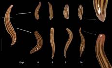
The sectioned animals were kept constantly in the dark. On consecutive days in the first week after the amputation, and subsequently, in longer intervals, each animal was replaced from the container to a moistened glass plate and photographed, taking special care to photograph the surface of the body with the sectioning wound. All photographs were taken with a Canon Eosi5 digital camera with a 100-mm macro lens and a twinlight flash attached.
Animals that recovered the amputated body part, the original body form and the eyes were considered regenerated, even if the original pigmentation was not fully recovered. After the regeneration of the lost parts, the animals continued to be observed to verify the pigment gain of the regenerated portions.
In preliminary experiments, specimens of Imbira marcusi Carbayo et al., 2013 died shortly after amputation. Since the species is abundant in nature, we were able to collect ten additional specimens, which were kept in the same laboratory conditions as the amputated individuals to test whether these conditions could affect the survival rate over time.
RESULTS
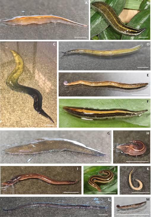
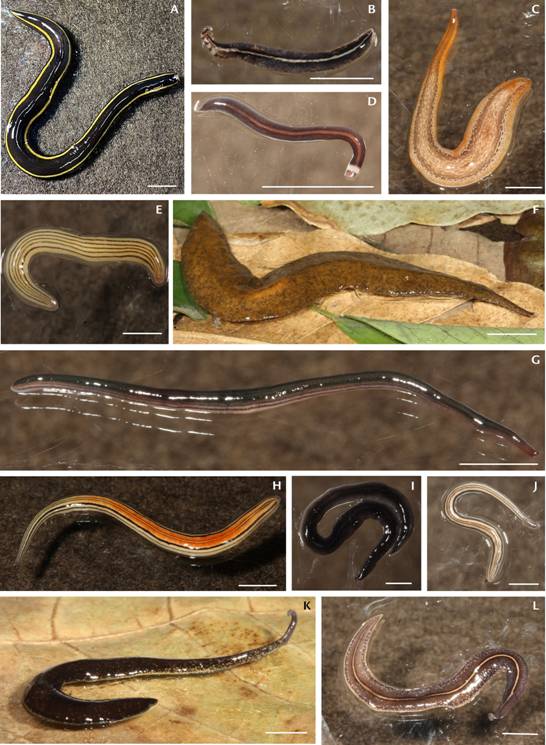
A total of 104 specimens of 25 species of land planarians of the genera Cephaloflexa, Choeradoplana, Cratera, Geoplana, Imbira, Xerapoa, Luteostriata, Notogynaphallia, Obama, Pasipha (Geoplaninae), and the exotic Dolichoplana and Endeavouria (Rhynchodeminae) were collected (Figs 2-3). Regeneration tests were tentatively carried out with all collected specimens, but early death in the laboratory of some animals prevented generalized tests.
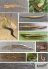
Intact living specimens of the land planarian species studied in this work. (A) Obama burmeisteri; (B) Cratera sp.; (C) Obama braunsi; (D) Cephaloflexa sp.; (E) Choeradoplana banga; (F) Cratera hina; (G) Obama carinata; (H) Pasipha pinima; (I) Obama anthropophila; (J) Issoca rezendei; (K) Xerapoa pseudorhynchodemus L Imbira sp.; (M) Xerapoa sp. Scale bars: ~10.0 mm.
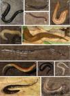
Intact living specimens of the land planarian species studied in this work. (A) Imbira marcusi; (B) Geoplana quagga; (C) Geoplaninae 1; (D) Pasipha sp. 1; (E) Luteostriata ernesti; (F) Obama divae; (G) Endeavouria septemlineata; (H) Notogynaphallia sp.; (I) Pasipha sp. 2; (J) Dolichoplana striata; (K) Cephaloflexa bergi; (L) Pasipha pasipha. Scale bars: ~10.0 mm.
Survival
One specimen of Xerapoa pseudorhynchodemus (Ries ter, 1938) (n = 2, Fig. 2K) did not survive the three days in the laboratory before amputation. Xerapoa sp. (n = 1; Fig. 2M) died immediately after amputation. Obama divae (Marcus, 1951) (n = 1, Fig. 3F) died 11 days after amputation, while it was regenerating. Cratera hina (Marcus, 1951) (n = 1, Fig. 2F), Cratera sp. (n = 1, Fig. 2B), Geoplaninae 1 (n = 1, Fig. 3C), and Imbira sp. (n = 1, Fig. 2L) died within 1-3 days, and it was not possible to see the blastema formation. Most I. marcusi (n = 20; Fig. 3A) specimens died within 3-7 days. Of the individuals in the control group of I. marcusi (n = 10), 3 died whithin 2 days, another 3 whithin 3 days, 2 whithin 7 days, 1 whithin 11 days and the last one died whithin 34 days. The specimens of I. marcusi and Imbira sp. that were sectioned only closed the wound and died within 15 days without developing even a blastema, whereas all other species that regenerated had already developed it within this period of time. The individual that survived longer (15 days) closed the wound with the sides of the body, being the only one to do so.
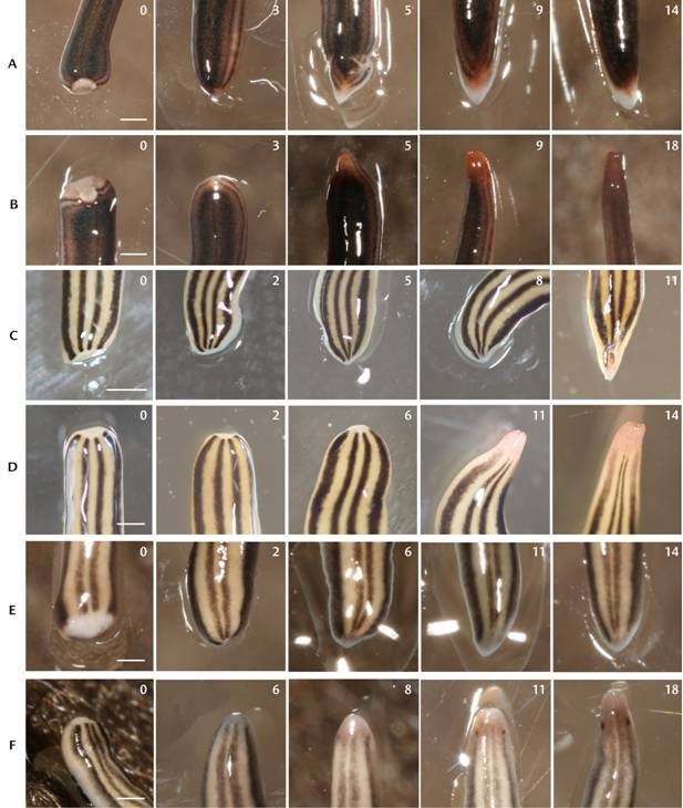
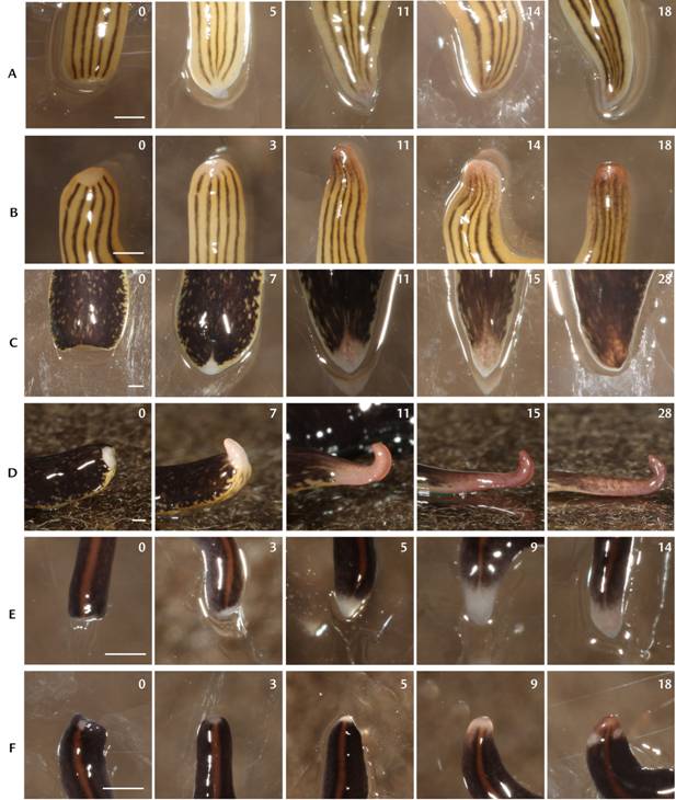
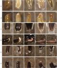
In general, anterior parts had a higher survival rate and survived longer than the posterior ones. The species presented different morphological development of the amputated body parts, regarding the blastema shapes, re-pigmentation and time required for the regeneration process (Figs 4-7).
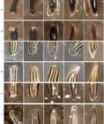
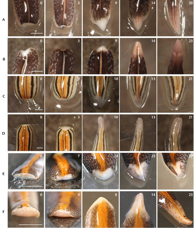
Photographs of the regeneration process of the species that regenerated body parts amputated and at least partially re-pigmented. The numbers on each figure indicate the number of days after the amputation: (A) anterior part (AP) of Endeavoria septemlineata; (B) posterior part (PP) of Endeavoria septemlineata; (C) AP of Issoca rezendei; (D) PP of Issoca rezendei; (E) AP of Dolichoplana striata; (F) PP of Dolichoplana striata. Scale bars: ~1.0 mm.
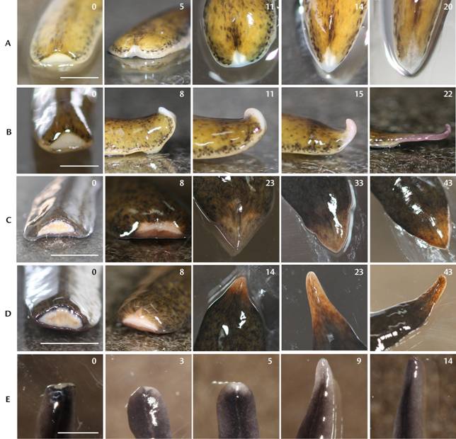
Photographs of the regeneration process of the species that regenerated body parts amputated and at least partially re-pigmented. The numbers on each figure indicate the number of days after the amputation: (A) anterior part (AP) of Luteostriata ernesti; (B) posterior part (PP) of Luteostriata ernesti; (C) AP of Cephaloflexa bergi; (D) PP of Cephaloflexa bergi; (E) AP of Pasipha sp. 1; (F) PP of Pasipha sp. 1. Scale bars: ~1.0 mm.
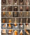
Photographs of the regeneration process of the species that regenerated body parts amputated and at least partially re-pigmented. The numbers on each figure indicate the number of days after the amputation: (A) anterior part (AP) of Pasipha pasipha; (B) posterior part (PP) of Pasipha pasipha; (C) AP of Notogynaphallia sp.; (D) PP of Notogynaphallia sp.; (E) AP of Obama burmeisteri; (F) PP of Obama burmeisteri. Scale bars: ~10.0 mm (E, F), ~5.0 mm (A, B); ~1.0 mm (C, D).
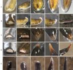
Photographs of the regeneration process of the species that regenerated body parts amputated and at least partially re-pigmented. The numbers on each figure indicate the number of days after the amputation: (A) anterior part (AP) of Cephaloflexa sp.; (B) posterior part (PP) of Cephaloflexa sp.; (C) AP of Obama carinata; (D) PP of Obama carinata; (E) PP of Pasipha sp. 2. AP of Pasipha sp. 2 is not illustrated since it did not survive. Scale bars: ~10.0 mm (C, D), ~5.0 mm (E), ~1.0 mm (A, B).
All other specimens regenerated head and/or tail, with marked specific differences in the time required for regeneration (Table 1).
Average time spent (in days) in regeneration and re-pigmentation of anterior and posterior body regions in the species of land planarians (Geoplanidae), with the number of specimens that survived the regeneration process.
Regeneration speed
Anterior body pieces regenerated the tail in 5-27 days and the posterior ones regenerated the head in 6-22 days (Table 1). Issoca rezendei (Schirch, 1929) (n = 9, Fig. 2J) regenerated fastest among geoplanins: an average of 5.25 days for regenerating the tail, and 11 days for the heads (Table 1 and Fig. 1), while the exotic Endeavouria septemlineata (Hyman, 1939) (n = 1, Fig. 3G) regenerated fastest: 5 days for the anterior part (Table 1). Obama braunsi (Graff, 1899) (n = 1, Fig. 2C) regenerated at the slowest pace: 27 days for the regeneration of the tail (Table 1).
One day after sectioning, the resulting posterior part of the two longest specimens of Dolichoplana striata Moseley, 1877 (n = 8; Fig. 3J) spontaneously divided themselves into five and two parts, respectively. Each new body piece generated the head and tail to a new individual. In species of Obama it was possible to see the formation of a new pharynx, without the formation of its copulatory apparatus during the observation time. Obama carinata (Riester, 1938), O. divae and O. braunsi developed a pigmented blastema in the first days of its formation, projected from the central surface of the wound (Fig. 8).
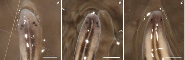
Two specimens of D. striata exhibited three and four eyes, respectively, when collected, instead of a pair of eyes. These animals were also amputated and the posterior parts normally regenerated only two eyes (Fig. 9).
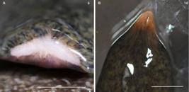
Pigmentation in the head blastema in Obama carinata. The numbers indicate days after sectioning: (A) frontal view of the blastema; (B) dorsal view of the blastema. Scale bars: ~5.0 mm.
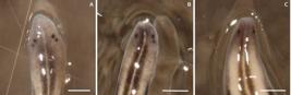
Photographs of the anterior tip of Dolichoplana striata: (A) specimen with four eyes; (B) specimen with three eyes; (C) same specimen with three eyes, regenerated 2. Scale bars: ~1.0 mm (A, B, C).
Blastema formation
Right after the amputation, the wound region in all species contracted to a concave shape (Figs 4-7) for both anterior and posterior parts, minimizing the wound area, and developing the blastema from it.
The process of formation and morphology of the caudal blastema was the same in all species. The wound closed and a non-pigmented tissue emerged from the site, which over time firstly gave origin to the intestine in species that presented a translucid blastema, and then the re-pigmentation with the original colors of an intact animal, or slightly paler.
Blastema shape
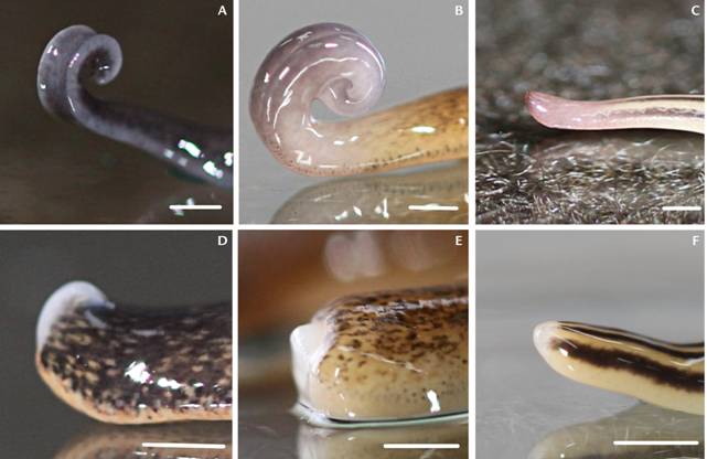
Three different shapes of head blastemas can be recognized in the species (Fig. 10). The blastema of Cephalo flexa sp. and Cephaloflexa bergi (Graff, 1899) was formed as a small tumescence emerging from the dorsal surface of the wound (Fig. 10D). In Choeradoplana banga Carbayo & Froehlich, 2012 the blastema developed as a small tumescence emerging from the central region of the wound (Fig. 10E). In the remaining species, the forming blastema projected from the entire surface of the wound as a frontal tumescence (Fig. 10F).
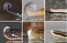
Head regeneration blastemas of different species evaluated in the study, lateral view: (A-C) heads in intact; (D-F) regenerating animals. The blastema is the lighter mass at the tip of each animal’s body: (A, D) Cephaloflexa bergi; (B, E) Choeradoplana banga; (C, F) Issoca rezendei. Scale bars: ~ 1.0 mm.
Re-pigmentation
The regenerated body parts of I. rezendei, Obama anthropophila Amaral, Carbayo & Leal-Zanchet, 2015, Cepha loflexa sp., C. bergi, Notogynaphallia sp., Pasipha pasipha (Marcus, 1951), Pasipha sp. 1, Pasipha sp. 2 and Luteostriata ernesti (Leal-Zanchet & Froehlich, 2006) recovered the pigment in the regenerated region. The same is true for the rhynchodemin species, i.e, D. striata and E. septemlineata. Obama burmeisteri (Schultze & Müller, 1857), C. banga, O. carinata and O. braunsi died before complete re-pigmentation of both the head and tail had been attained.
Cephaloflexa sp. and C. bergi did not fully recover the pigment in the regenerated heads and tails, as they showed the typical light-colored blastema.
Unlike all other species studied, O. carinata, O. divae and O. braunsi developed a pigmented blastema from a very early stage in both anterior and posterior parts, even before regeneration (Figs 6E, 6F, 7C, 7D, 8). All other species developed unpigmented blastemas. Obama burmeisteri also showed a different pigmentation process of the tail from the other species, gaining color from the body sides towards the median zone, which remained colorless even within 30 days following amputation (Fig. 6E).
The tail blastema of Cephaloflexa sp. and C. bergi were translucent in the first days of formation and showed white lateral masses, probably the paired posterior intestinal branches (Fig. 7A).
DISCUSSION
Regeneration capacity of geoplanins
This is the first extensive work in testing the regene ration ability of land planarians of Geoplaninae. Besides the rhynchodemins D. striata and E. septemlineata, 17 out of the 23 geoplanin species regenerated head and/or tail. Therefore, the regeneration ability seems to be extensively spread across the Neotropical subfamily Geoplaninae, in agreement with the few experiments conducted with P. pulla and L. abundans (Goetsch 1933, Boll et al. 2023). It is also possible that the high rate of species of Geoplaninae able to regenerate suffers from a sampling bias since most animals were collected in human-transformed habitats (Table 1, Parque Ecológico do Tietê), and the species dwelling there might be those with pre-adaptations, including regeneration abilities, to those environs.
From our experience in the field, geoplanins reproduce primarily sexually, as individuals of virtually all species observed in field campaigns are sexually mature and sometimes found copulating. This is in agreement with Froehlich (1955) and Boll et al. (2023). In contrast to species that reproduce asexually through regeneration, in the Geoplaninae investigated so far, regeneration might be maintained as an adaptive solution to regain body parts lost in the wild.
Regeneration speed
The regeneration times of our experiments ranged between 5-27 days for the head and 6-22 for the tail, and are similar to those observed for T. tasmaniana (25 days) (Darwin 1844), B. kewense (2-3 weeks to 30 days) (Fletcher 1887, Connella and Stern 1969), B. nobile, B. multilineatum (2-5 weeks) (Shirasawa and Makino 1987), and R. bilineatus (16-18 days) (Lehnert 1891).
Body width (possibly related to body mass) seems to be related to the speed of regeneration. Subcylindrical animals, such as I. rezendei, and the exotic D. striata and E. septem lineata, regenerated the fastest, while flattened animals such as O. braunsi and O. carinata took the longest time to regenerate. There are no studies of this kind with flattened land planarians, which to compare, but faster regeneration rates are possibly related to the relatively smaller wound surface in cylindrical animals than in flattened animals.
Survival
Although the early death of about a third of the anterior parts and almost half of the posterior parts (Table 1) suggests a decline in physiological function under laboratory conditions, it also suggests that the regenerative capacity of the species tested may be higher under natural conditions. The early death of all specimens of I. marcusi seems to be related to the sensitivity of this species to laboratory conditions because all individuals of the experimental group died within 15 days before showing any signal of blastema. Furthermore, the ten specimens of the control group of this species also died in the lab within 34 days. The same situation might be applied to Imbira sp., Xerapoa sp., C. hina, Cratera sp., and Geoplaninae 1, though the sample size is very low. The difficulty of providing adequate culture conditions for some land planarians is well known (Froehlich 1955, Christensen and Mather 2001, Carbayo et al. 2002).
The higher survival rate of the anterior parts is consistent with observations on neotropical P. pulla and L. abundans (Goetsch 1933, Boll et al. 2023). However, individuals of D. multilineatum (Bipaliinae) sectioned into 7-16 pieces showed that all body parts were fully regenerated (Shirasawa and Makino 1987). Boll et al. (2023) wondered whether aspects, such as age or place of origin might influence the survival rate of L. abundans.
Blastema shape
The concave wound in both anterior and posterior body pieces that formed immediately after amputation seems to be caused by muscle contraction as reported for L. abundans (Hauser 1971). As the head regenerated, the blastema took a shape, which depends on the taxa. The variation in the shape of the head-forming blastema (a small tumescence emerging from the dorsal surface of the wound, a small tumescence emerging from the central region of the wound surface or a frontal tumescence) seems to be related with the variety of head shapes: dorsally curled-up and slightly concave ventrally (Fig. 10A), dorsally curled-up with a longitudinal ventral groove (Fig. 10B), and regular (Fig. 10C). The blastema normally does not vary in freshwater planarians and it would only do so if the cut were made at different angles (Reddien and Sánchez-Alvarado 2004). Similarly, freshwater planarians do not present significantly different head shapes as in land ones, which may be the reason why the shape of the head blastema does not vary among freshwater species.
Re-pigmentation
The re-pigmentation process presented some variation in terms of sequence of stages and pigment recovery of the pre-amputation state. All blastemas were unpigmented, except in O. carinata, O. braunsi, and O. divae. In these species the pigment appears in the initial stages of the blastema formation. In freshwater planarians, the blastemas are formed with migration of the undifferentiated cells, growing with the accumulation of these cells in the wound area (Saló and Baguñà 1984), which stay unpigmented until the initial differentiation of the stem cells (Reddien and Sánchez-Alvarado 2004). Pigment is produced by a specialized cell type in these planarians (Lindsay-Mosher and Pearson 2019), so it will appear once the cells differentiate. If this also applies to the three species of Obama, then specialization of the neoblasts in their blastemas would start at earlier regeneration stages than for the other species.
On the other hand, only I. rezendei, C. bergi and Cephaloflexa sp. regenerated a tail with a pigment pattern different from that of intact animals. Possibly, the re-pigmentation process in these species would take longer than the duration of our experiments.
Perspectives
The regenerative abilities of geoplanins shown here open up opportunities for further comparative studies from an evolutionary and ecological perspective. The freshwater planarians Schmidtea mediterranea (Benazzi et al., 1975) and Dugesia japonica Ichikawa & Kawakatsu, 1964 (Dugesiidae) are model species due to their robust and rapid whole-body regeneration (Saló and Agata 2012). In turn, the geoplanid I. rezendei here studied combines some desirable features: it is easily found in urban yards and gardens in southeastern Brazil (Froehlich 1955), its diet is known (Cseh et al. 2017), it thrives well under laboratory conditions, and it also is able to regenerate in a few days. Given these characteristics, I. rezendei may also be a good model organism for further regeneration studies.
Although we tested a limited number of individuals and species, the differences shown in terms of regeneration speed, blastema shape, and repigmentation process, combined with the diverse reproductive and feeding habits, present some challenging predictions to be tested. These predictions include the weight of pleiotropy, phylogenetic inertia, or direct selection in maintaining regeneration (Bely and Nymberg 2010). To be directly selected, animals are predicted to be frequently injured in the field and subsequently regenerate lost parts (Bely and Nymberg 2010, Lindsay 2010, Lin et al. 2017). Geoplanins frequently exhibit injuries in the field (pers. obs.), some of which are apparently caused by their hard-bodied prey (Silva et al. 2018). Also land planarians that prey on invertebrates with hard body structures, such as arthropods, would regenerate lost parts more quickly than species such as O. burmeisteri and O. carinata, which prey on soft-bodied animals like snails and earthworms. Indeed, the regeneration process takes longer in these two Obama species than in I. rezendei and C. bergi, which prey on arthropods.
ACKNOWLEDGEMENTS
We thank SisBio for licensing the fiel work (#11473-4), the reviewers for the valuable comments and suggestions to the manuscript and Miquel Vila-Farré and Emili Saló for helpful discussions on experimental design. VM thanks the University of São Paulo for an undergraduate fellowship (PUB, USP). FC has financial support from São Paulo Research Foundation (FAPESP #2022/11972-2).
LITERATURE CITED
- Bandier J (1936) Histologische Untersuchungen über die Regeneration von Landplanarien. Archiv für Entwicklungsmechanik der Organismen Berlin 135: 316-348.
- Bell FJ (1886) Note on Bipalium kewense, and their generic characters of land-planarians. Proceedings of the Zoological Society of London: 166-168.
-
Bely AE, Nyberg KG (2010) Evolution of animal regeneration: re-emergence of a field. Trends in Ecology and Evolution 25(3): 161-170. https://doi.org/10.1016/j.tree.2009.08.005
» https://doi.org/10.1016/j.tree.2009.08.005 -
Boll PK, Rossi I, Amaral SV, Leal-Zanchet AM (2023) Regeneration in a Neotropical land planarian (Platyhelminthes, Tricladida). Neotropical Biology and Conservation 18(3): 163-176. https://doi.org/10.3897/neotropical.18.e103357
» https://doi.org/10.3897/neotropical.18.e103357 -
Brockes J, Kumar A, Velloso C (2001) Regeneration as an evolutionary variable. Journal of Anatomy 199(1-2): 3-11. https://doi.org/10.1046/j.1469-7580.2001.19910003.x
» https://doi.org/10.1046/j.1469-7580.2001.19910003.x -
Carbayo F, Leal-Zanchet AM, Vieira EM (2002) Terrestrial flatworm (Platyhelminthes: Tricladida: Terricola) diversity vs. man-induced disturbance in a subtropical rainforest from Southern Brazil. Biodiversity and Conservation 11: 1091-1104. https://doi.org/10.1023/A:1015865005604
» https://doi.org/10.1023/A:1015865005604 -
Christensen OM, Mather JG (2001) Long-term study of growth in the New Zealand flatworm Arthurdendyus triangulatus under laboratory conditions. Pedobiologia 45: 535-549. https://doi.org/10.1078/0031-4056-00105
» https://doi.org/10.1078/0031-4056-00105 -
Connella JV, Stern DH (1969) Land Planarians: Sexuality and Occurrence. Transactions of the American Microscopical Society 88(2): 309-311. https://doi.org/10.2307/3224508
» https://doi.org/10.2307/3224508 -
Cseh A, Carbayo F, Froehlich EM (2017) Observations on food preference of Neotropical land planarians (Platyhelminthes), with emphasis on Obama anthropophila, and their phylogenetic diversification. Zoologia 34: e12622. https://doi.org/10.3897/zoologia.34.e12622
» https://doi.org/10.3897/zoologia.34.e12622 - Darwin C (1844) Brief descriptions of several terrestrial planariae, and of some remarkable marine species, with an account of their habits. Annals and Magazine of Natural History, Annales de Sciences Naturelles 14: 241-251.
-
Fletcher JJ (1887) Remarks on an introduced species of land planarian apparently Bipalium kewense, Moseley. Proceedings of the Linnean Society of New South Wales 2: 244-249. https://doi.org/10.5962/bhl.part.29178
» https://doi.org/10.5962/bhl.part.29178 -
Froehlich CG (1955) On the biology of land planarians. Boletim da Faculdade de Filosofia, Ciências e Letras, Universidade de São Paulo, Zoologia 20: 263-271. https://doi.org/10.11606/issn.2526-3382.bffclzoologia.1955.120214
» https://doi.org/10.11606/issn.2526-3382.bffclzoologia.1955.120214 -
Gazave E, Röttinger E (2019) 7th Euro Evo Devo meeting: Report on the “Evolution of regeneration in Metazoa” symposium. Journal of Experimental Zoology, B: Molecular and Developmental Evolution 336: 89-93. https://doi.org/10.1002/jez.b.22897
» https://doi.org/10.1002/jez.b.22897 - Goetsch W (1933) Verbreitung und Biologie der Landplanarien Chiles. Zoologische Jahrbücher, Abteilung für Systematik 64: 245-288.
- Graff L von (1912-1917) Tricladida. In: Bronn HG (Ed.) Klassen und Ordnungen des Tier-Reichs, Band. IV Vermes, Abtheilung IC: Turbellaria, II Abtheilung: Tricladida. CF Winter, Leipzig, 2601-3369 pp.
-
Hauser J (1971) Non-cellular regeneration processes in the integument of the flatworm Geoplana abundans Oncology 25: 258-268. https://doi.org/10.1159/000224576
» https://doi.org/10.1159/000224576 - Lehnert GH (1891) Beobachten an Landplanarien. Archiv für Naturgeschichte 56: 306-350.
-
Lin JW, Chen YR, Wang YH, Hung KC, Lin SM (2017) Tail regeneration after autotomy revives survival: a case from a long-term monitored lizard population under avian predation. Proceedings of the Royal Society B: Biological Sciences 284: 20162538. https://doi.org/10.1098/rspb.2016.2538
» https://doi.org/10.1098/rspb.2016.2538 -
Lindsay SM (2010) Frequency of injury and the ecology of regeneration in marine benthic invertebrates. Integrative and Comparative Biology 50(4): 479-493. https://doi.org/10.1093/icb/icq099
» https://doi.org/10.1093/icb/icq099 -
Lindsay-Mosher, Pearson BJ (2019) The true colours of the flatworm: Mechanisms of pigment biosynthesis and pigment cell lineage development in planarians. Seminars in Cell & Developmental Biology 87: 37-44. https://doi.org/10.1016/j.semcdb.2018.05.010
» https://doi.org/10.1016/j.semcdb.2018.05.010 - Morgan TH (1900a) Regeneration in planarians. Archiv für Entwicklungsmechanik der Organismen 10: 58-119.
- Morgan TH (1900b) Regeneration in Bipalium Archiv für Entwicklungsmechanik der Organismen 9: 563-586.
-
Ogren RE (1957) Developmental observations, the egg capsule and sexual maturity of the land planarian Rhynchodemus sylvaticus Transactions of the American Microscopical Society 76(1): 45-52. https://doi.org/10.1007/BF00046245
» https://doi.org/10.1007/BF00046245 - Ogren RE, Sheldon JK (1991) Ecological observations on the land planarian Bipalium pennsylvanicum Ogren, with references to phenology, reproduction, growth rate and food niche. Journal of the Pennsylvania Academy of Science 65(1): 3-9.
-
Reddien P, Sánchez-Alvarado A (2004) Fundamentals of planarian regeneration. Annual Review of Cell and Developmental Biology 20: 725-757. https://doi.org/10.1146/annurev.cellbio.20.010403.095114
» https://doi.org/10.1146/annurev.cellbio.20.010403.095114 -
Rink JC (2013) Stem cell systems and regeneration in planaria. Development Genes and Evolution 223: 67-84. https://doi.org/10.1007/s00427-012-0426-4
» https://doi.org/10.1007/s00427-012-0426-4 -
Saló E, Agata K (2012) Planarian regeneration: a classic topic claiming new attention. The International Journal of Developmental Biology 56: 3-4. https://doi.org/10.1387/ijdb.123495es
» https://doi.org/10.1387/ijdb.123495es - Saló E, Baguñà J (1984) Regeneration and pattern formation in planarians. I. The pattern of mitosis in anterior and posterior regeneration in Dugesia (G) tigrina, and a new proposal for blastema formation. Journal of Embryology and Experimental Morphology 83: 63-80.
-
Sánchez-Alvarado A (2000) Regeneration in the Metazoans: Why does it happen? BioEssays 22: 578-590. https://doi.org/10.1002/(SICI)1521-1878(200006)22:6<578::AID-BIES11>3.0.CO;2-%23
» https://doi.org/10.1002/(SICI)1521-1878(200006)22:6<578::AID-BIES11>3.0.CO;2-%23 -
Sánchez-Alvarado A (2003) Regeneration in the metazoa. In: Hall BK, Olson WM (Eds) Keywords and concepts in evolutionary developmental biology. Harvard University Press, Cambridge, 318-325. https://doi.org/10.2307/j.ctv228vqrq.47
» https://doi.org/10.2307/j.ctv228vqrq.47 - Shirasawa Y, Makino N (1978) Study on regeneration on Bipalium kewense Moseley. Zoological Magazine 87: 506.
- Shirasawa Y, Makino N (1979) Comparative studies of several land planarians (Bipalium fuscatum, B. sp. Type I, B. sp. Type II, and B. sp. Type III) on the starvation and regeneration. Zoological Magazine 5: 25-38.
- Shirasawa Y, Makino N (1981) Light and electron microscopic studies on the normal and regenerating photoreceptor of a land planarian (Bipalium fuscatum). Bulletin of Tokyo Medical College 7: 35-50.
- Shirasawa Y, Makino N (1983) On the regeneration of the pharynx in the land planarian of a new species, Bipalium sp. Type III. Bulletin of Tokyo Medical College 9: 123-141.
- Shirasawa Y, Makino N (1984) On the regeneration of the pharynx in the land planarian of a new species (Bipalium nobile Kawakatsu et Makino). Bulletin of Tokyo Medical College 10: 111-125.
- Shirasawa Y, Makino N (1985) Cytological study on the intestinal tissue in the regenerating land planarian, Bipalium nobile Bulletin of Tokyo Medical College, 12(Suppl.) : 99-110.
- Shirasawa Y, Makino N (1987) Studies on the morphological changes and the body weight of the regenerating piece in the land planarian, Bipalium (Turbellaria, Tricladida, Terricola). Bulletin of Tokyo Medical College 13: 91-102.
- Shirasawa Y, Makino N (1988) Observations on the regeneration of the sagittal pieces in the land planarian, Bipalium nobile Bulletin of Tokyo Medical College 14: 43-60.
-
Shirasawa Y, Makino N (1991) Pharyngeal regeneration in the land planarian Bipalium kewense VI International Symposium on the Biology of the Turbellaria, Hirosaki, Japan, 7-12 August 1990, 57. https://doi.org/10.1007/978-94-011-2775-2_8
» https://doi.org/10.1007/978-94-011-2775-2_8 -
Silva MS, Willemart R, Carbayo F (2018) Sticky flatworms (Platyhelminthes) kill armored harvestmen (Arachnida, Opiliones) but are not immune to the prey’s weapons. Journal of Zoology 306: 88. https://doi.org/10.1111/jzo.12570
» https://doi.org/10.1111/jzo.12570 -
Sluys R, Kawakatsu M, Riutort M, Baguñà J (2009) A new higher classification of planarian flatworms (Platyhelminthes, Tricladida). Journal of Natural History 43: 1763-1777. https://doi.org/10.1080/00222930902741669
» https://doi.org/10.1080/00222930902741669 -
Vila-Farré M, Rozanski A, Ivanković M, Cleland J, Brand JN, Thalen F, et al. (2023) Evolutionary dynamics of whole-body regeneration across planarian flatworms. Nature Ecology & Evolution 7: 2108-2124. https://doi.org/10.1038/s41559-023-02221-7
» https://doi.org/10.1038/s41559-023-02221-7 -
Wagner DE, Wang IE, Reddien PW (2011) Clonogenic neoblasts are pluripotent adult stem cells that underlie planarian regeneration. Science 332(6031): 811-816. https://doi.org/10.1126/science.1203983
» https://doi.org/10.1126/science.1203983
ADDITIONAL NOTES
- ZooBank register
-
How to cite this article
Milanese V, Carbayo F (2024) Do Geoplaninae, a neotropi cal subfamily of land planarians (Platyhelminthes: Tricladida: Geoplanidae), regenerate well? Zoologia 41: e23094. https://doi.org/10.1590/S1984-4689.v41.e23094
-
Published by
Sociedade Brasileira de Zoologia at Scientific Electronic Library Online - https://www.scielo.br/zool