Abstract
The middle course of the Tocantins river is located in the eastern portion of the “Legal Amazon” region of Brazil and the Dantas river is one of its tributaries. Among the components of the aquatic biota, eukaryote microparasites establish direct relationships with several species of fish and have zoonotic potential that is still little known. Myxozoans stand out among these parasites: they cause myxosporidiosis, a disease that gives rise to high mortality rates worldwide. The genus Myxobolus accounts for the largest number of species that have been described. Thirty specimens of Astyanax aff. bimaculatus that had been caught in the Dantas river were examined. The prevalence of cysts with spores morphologically compatible with myxozoans of the genus Myxobolus in the arcuate and gill filaments of these specimens was 20%.
Keywords:
Parasites; Myxozoa; Bivalvulida; Characiformes
Resumo
O curso médio do rio Tocantins está localizado na porção leste da região da “Amazônia Legal” do Brasil, e o rio Dantas é um dos seus afluentes. Dentre os componentes da biota aquática, os microparasitos eucarióticos estabelecem relações diretas com várias espécies de peixes e possuem potencial zoonótico ainda pouco conhecido. Os mixozoários destacam-se entre esses parasitos causando mixosporidiose, doença que dá origem a altas taxas de mortalidade em todo o mundo. O gênero Myxobolus é responsável pelo maior número de espécies descritas de mixozoários. Trinta espécimes de Astyanax aff. bimaculatus capturados no rio Dantas foram examinados. A prevalência de cistos com esporos morfologicamente compatíveis com mixozoários do gênero Myxobolus nos filamentos arqueados e branquiais desses espécimes foi de 20%.
Palavras-chave:
parasitos; Myxozoa; Bivalvulida; Characiformes
Introduction
Fish of the genus Astyanax (Baird & Girard, 1854) have wide distribution in the neotropical region, with occurrence of 146 valid species, of which 91% occur in South America (ABDALLAH et al., 2004; LUCENA & SOARES, 2016). They are fish of low commercial value and, for this reason, are usually used to feed the riverine population and the lower-income communities of urban centers. In addition, it is one of the main components of the food base of carnivorous fish in rivers in tropical regions.
The term Astyanax bimaculatus, which refers to species of characiform fish in the Suriname region, is also used to denote the “bimaculatus group” composed of approximately 22 species of generalist and migratory fish. These are well suited to both lotic and lentic environments and are widely distributed in Brazilian freshwater basins (GARUTTI & BRITSKI, 2000; LUCENA et al., 2013).
Studies on occurrences of microparasites in the aquatic fauna of the tropical region have frequently been conducted (BÉKÉSI et al., 2002; AZEVEDO et al., 2002; LUQUE, 2004; AZEVEDO et al., 2010; MILANIN et al., 2010; AZEVEDO et al, 2011a; MACIEL et al, 2011; CARRIERO et al, 2013). These studies gain greater importance, given that parasites can be used as bioindicators to determine stocks (LUQUE, 2004). In addition, evaluation of the ecology of parasitism, which includes studies on population dynamics, enables assessment of the zoonotic potential of the parasites, which may indicate that parasitism could be a limiting factor in relation to rearing a certain species of fish (KENT et al., 2001; HARTIGAN et al., 2016; VIDEIRA et al., 2016).
Material and Methods
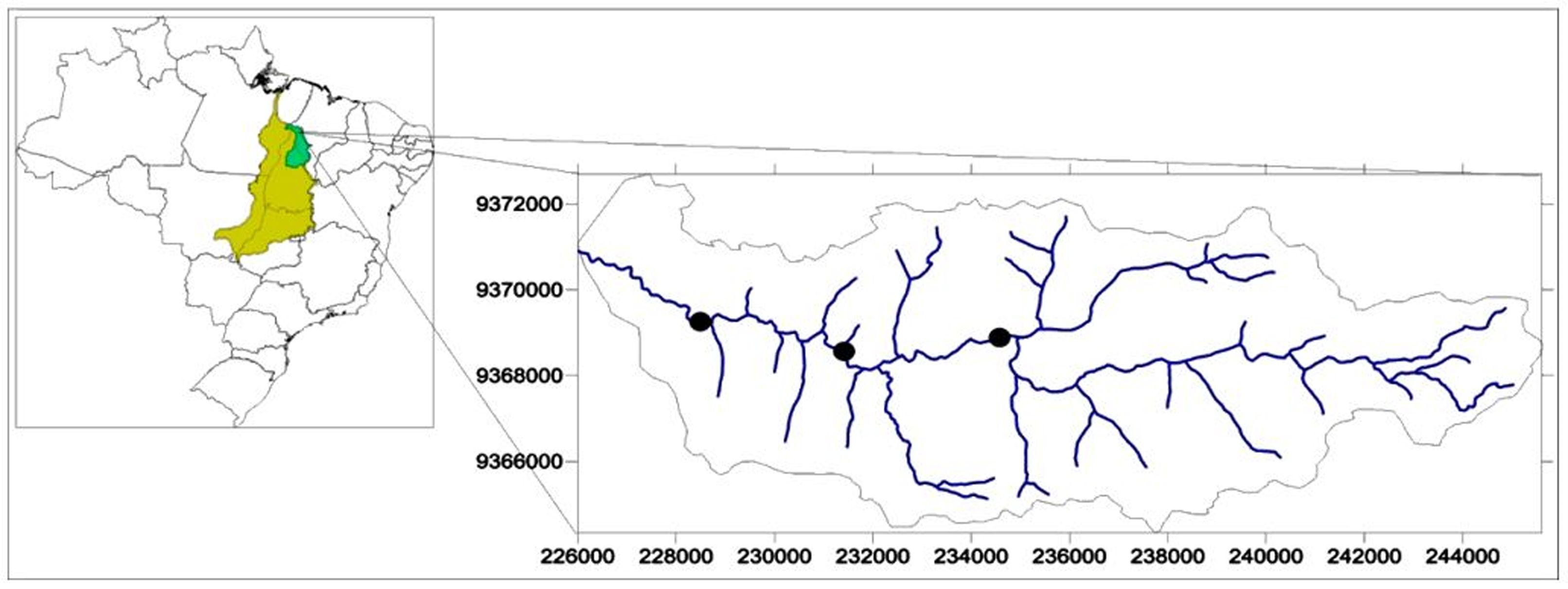
Thirty specimens of Astyanax aff. bimaculatus, collected in the period from February to July of 2018 in three sampling points in the river Dantas (23M 227839.79 E 9369879.17 S), were examined. This river is a tributary of the Tocantins river in the municipality of Governador Edson Lobão, which is located in the mesoregion of Imperatriz in the state of Maranhão, Brazil (Figure 1). The specimens were transported alive in plastic bags with water from the habitat, under conditions of artificial aeration to the Ecology and Limnology Laboratory at UEMASUL, in Imperatriz. In this laboratory, they were kept in an aquarium at temperatures of between 26 and 28 °C.
Location of collection points in the Dantas river basin, middle Tocantins river basin, eastern Brazilian Amazon region.
To investigate the presence of parasites and cysts, the specimens were anesthetized with tricaine methanesulfonate (MS222; Sigma) at a concentration of 50 mg L-1. They were then dissected under a stereoscopic microscope. Fragments of the organs were removed for observation under an optical microscope and were analyzed with the objective of determining the presence of parasites. The procedures adopted in the present study were approved by the Animal Ethics and Experimentation Committee of the Federal Rural University of Amazonia (CEUA/UFRA no. 013/2014). Photomicrographs of the microparasite spores that were observed were captured through a Zeiss Axiocam ICc 1 camera that was coupled to a Zeiss Axioskop 40 microscope, in order to measure the spores (n=50) with the aid of the AxioZen Blue software.
Fragments of the infected organs were fixed in Davidson solution for 24 h and were then processed using the method of embedment in paraffin. Through this, blocks and histological sections of thickness 6 μm thickness were prepared. These sections were stained with hematoxylin-eosin (HE) and Ziehl-Neelsen (ZN). Fixed fragments were later dissociated for preparation of slides to obtain spore images by means of the differential interference contrast (DIC) technique, at the Carlos Azevedo Research Laboratory of UFRA, in Belém.
Results and Discussion
Investigation of eukaryote microparasites in the specimens of Astyanax aff. bimaculatus that were sampled revealed that the prevalence of cyst infection in the gill arches and lamellae caused by myxosporeans was 20% (6/30) (Figure 2A). Observation under a microscope after rupture of the cysts (Figure 2B) revealed the presence of spores with a morphological pattern consistent with myxozoa of the genus Myxobolus, as described by Lom & Dyková (2006), consisting of a spore body of ellipsoid shape, with two polar caps, two valves positioned in parallel and a suture line, and with a binucleate sporoplasma cell (Figure 2C).
Myxobolus sp. on the gills of Astyanax aff. bimaculatus. A - Cysts in the gill´s arches, white arrow, and small cysts in gill filaments, black arrows; B - Fresh photomicrography an optical microscope of small cyst (*) dissociated from gills filament e; C - Sporocysts with morphological pattern of the genus Myxobolus; D - Differential interference contrast (DIC) microscopy of Myxobolus sp. with extruded polar filament., seen using. Scale: 10 μm.
The morphometric analysis showed that the spores of Myxobolus sp. present in cysts on the gills of Astyanax aff. bimaculatus in the Dantas river basin had mean total length of 10.9 (0.4) μm and width of 7.7 (0.6) μm. The polar capsules were morphologically similar, with length of 4.3 (0.2) μm and width of 2.0 (0.2) μm, and the number of turns of the filaments within the polar capsules ranged from 6 to 7. The results from the measurements and morphometric comparisons with Myxobolus species are presented in Table 1.
Comparison of measurements of spores of the parasite Myxobolus spp. from the gills of Characiform fish in Brazil. LS - spore length; WS - spore width; PCL - polar capsules length, PCW - polar capsules width, PF - Polar filament turns. All measurements are in micrometers.
When compared to the morphological aspects of the sporocysts with other Myxobolus spp described parasitizing gills of Characiform fish in Brazil, the present myxozoa presents greater similarity in relation to the shape of the sporocysts and number of turns of the polar filaments with M. oliverai (MILANIN et al., 2010) and in relation to the form of the polar capsules to M. brycom (AZEVEDO et al. 2011b), both described parasitizing Brycon hilarii in flooded fields of the Brazilian Pantanal.
The hematoxylin-eosin (HE) histological technique enabled visualization of the interaction between the spores of Myxobolus sp. and the gill tissue (Figure 3A and B), while the staining by Ziehl-Neelsen made it possible to detect the polar capsules and sporoplasm (Figure 3C). Development of the parasite led to compression of adjacent connective and epithelial tissues. However, no granulocytic cells were observed at the site of infection. The differential interference contrast microscopy technique (Figure 2D) allowed better observation of spore specificity and arrangement, and also highlighted the polar capsules and filaments.
Histological section of the gills of Astyanax aff. bimaculatus, stained with hematoxylin-eosin (A, B) and Sporocysts of Myxosbolus sp. stained by Ziehl-Neelsen (C). Asterisk - cyst, arrow head - cyst wall, black arrow - polar capsule, white sporoplasma arrow. Scale: 50 μm.
Despite the moderate prevalence of parasitism, the hosts did not present clinical signs of disease. This corroborated observations that were made by Tavares-Dias et al. (2006) and Thatcher (2006), who described asymptomatic Myxobolus infections in the gills of other fish species in the Amazon region.
Infections of the gill system caused by myxosporeans have been described worldwide. Such infections result in direct or indirect harm to the health of their hosts (AZEVEDO et al., 2014; VIDEIRA et al., 2016; DE ARAUJO et al, 2018). Thus, investigations like the present study are of fundamental importance for gaining knowledge regarding myxosporidiosis in fish in the Tocantins river basin.
Conclusions
From the findings described in this study, it was possible to characterize infection of the gills of Astyanax aff. bimaculatus, caused by Myxobolus sp. This was the first report of occurrence of this genus of parasite in the middle course of the Tocantins river basin.
The morphometric patterns observed in the spores of Myxobolus sp. of the present study, regarding total length and width, polar capsule size and number of turns of the polar filaments, were divergent in relation to all other studies reporting infection by Myxobolus spp. in the gills of Characiformes in Brazil. Because of these records of Myxobolus sp. in Astyanax aff. bimaculatus and the characteristics presented by this parasite, continuation of this work addressing molecular and ultrastructural characteristics is of fundamental importance for determination and classification of this parasite species in the Brazilian “Legal Amazon” region in the state of Maranhão.
Acknowledgements
This work was financially supported by Coordination of Improvement of Higher Level Personnel - CAPES (Special Visiting Researcher Program 88887.125002 / 2-14-00); National Council for Scientific and Technological Development – CNPQ (Universal Announcement 441645-2014-3; Productivity in Research, No. 301497 / 2016-8); The Amazonia Foundation for Studies and Research (EDITAL, 006/2014, ICAAF 162/2014). The Foundation for Support of Research in the State of Maranhão (FAPEMA) and the State University of the Tocantina Region of Maranhão (UEMASUL).
References
- Abdallah VD, Azevedo RK, Luque JL. Metazoários parasitos dos lambaris Astyanax bimaculatus (Linnaeus, 1758), A. parahybae Eigenmann, 1908 e Oligosarcus hepsetus (Cuvier, 1829) (Osteichthyes: Characidae), do rio Guandu, estado do Rio de Janeiro, Brasil. Rev Bras Parasitol Vet 2004; 13(2): 57-63.
-
Adriano EA, Arana S, Carriero MM, Naldoni J, Ceccarelli PS, Maia AA. Light, electron microscopy and histopathology of Myxobolus salminus n. sp., a parasite of Salminus brasiliensis from the Brazilian Pantanal. Vet Parasitol 2009; 165(1-2): 25-29. http://dx.doi.org/10.1016/j.vetpar.2009.07.001 PMid:19640650.
» http://dx.doi.org/10.1016/j.vetpar.2009.07.001 -
Araújo RS, Corrêa F, De Sousa FB, Ramos ABMA, Neto JLS, Matos ER. Ocorrência de Myxobolus sp. (Myxozoa) em Thoracocharax stellatus (Kner, 1858) (Characiformes) em um igarapé da floresta amazônica, Pará, Brasil. Braz J Aquat Sci Technol 2018; 21(1): 16-20. https://doi.org/10.14210/bjast.v21n1.11102
» https://doi.org/10.14210/bjast.v21n1.11102 -
Azevedo C, Casal G, Matos P, Alves A, Matos E. Henneguya torpedo sp. nov. (Myxozoa), a parasite from the nervous system of the Amazonian teleost Brachyhypopomus pinnicaudatus (Hypopomidae). Dis Aquat Organ 2011a; 93(3): 235-242. http://dx.doi.org/10.3354/dao02292 PMid:21516976.
» http://dx.doi.org/10.3354/dao02292 -
Azevedo C, Casal G, Marques D, Silva E, Matos E. Ultrastructure of Myxobolus brycon n. sp. (Phylum Myxozoa), parasite of the piraputanga fish Brycon hilarii (Teleostei) from Pantanal (Brazil). J Eukaryot Microbiol 2011b; 58(2): 88-93. http://dx.doi.org/10.1111/j.1550-7408.2010.00525.x PMid:21309886.
» http://dx.doi.org/10.1111/j.1550-7408.2010.00525.x -
Azevedo C, Corral L, Matos E. Myxobolus desaequalis n. sp. (Myxozoa, Myxosporea), parasite of the Amazonian freshwater fish, Apteronotus albifrons (Teleostei, Apteronotidae). J Eukaryot Microbiol 2002; 49(6): 485-488. http://dx.doi.org/10.1111/j.1550-7408.2002.tb00233.x PMid:12503685.
» http://dx.doi.org/10.1111/j.1550-7408.2002.tb00233.x -
Azevedo C, Casal G, Mendonça I, Carvalho E, Matos P, Matos E. Light and electron microscopy of Myxobolus sciades n. sp. (Myxozoa), a parasite of the gills of the Brazilian fish Sciades herzbergii (Block, 1794) (Teleostei: Ariidae). Mem Inst Oswaldo Cruz 2010; 105(2): 203-207. http://dx.doi.org/10.1590/S0074-02762010000200016 PMid:20428682.
» http://dx.doi.org/10.1590/S0074-02762010000200016 -
Azevedo RK, Vieira DH, Vieira GH, Silva RJ, Matos E, Abdallah VD. Phylogeny, ultrastructure and histopathology of Myxobolus lomi sp. nov., a parasite of Prochilodus lineatus (Valenciennes, 1836) (Characiformes: Prochilodontidae) from the Peixes River, São Paulo State, Brazil. Parasitol Int 2014; 63(2): 303-307. http://dx.doi.org/10.1016/j.parint.2013.11.008 PMid:24291290.
» http://dx.doi.org/10.1016/j.parint.2013.11.008 -
Békési L, Székely C, Molnár K. Atuais conhecimentos sobre Myxosporea (Myxozoa), parasitas de peixes. Um estágio alternativo dos parasitas no Brasil. Braz J Vet Res Anim Sci 2002; 39(5): 271-276. http://dx.doi.org/10.1590/S1413-95962002000500010
» http://dx.doi.org/10.1590/S1413-95962002000500010 -
Carriero MM, Adriano EA, Silva MRM, Ceccarelli PS, Maia AAM. Molecular phylogeny of the Myxobolus and Henneguya genera with several new south American species. PLoS One 2013; 8(9): e73713. http://dx.doi.org/10.1371/journal.pone.0073713 PMid:24040037.
» http://dx.doi.org/10.1371/journal.pone.0073713 - Garutti V, Britski H. Descrição de uma nova espécie de Astyanax (Teleostei: Characidae) da bacia do alto rio Paraná e considerações sobre as demais espécies do gênero. Comum Mus Ciên Tec PUCRS 2000; 13: 65-88.
-
Hartigan A, Wilkinson M, Gower DJ, Streicher JW, Holzer AS, Okamura B. Myxozoan infections of caecilians demonstrate broad host specificity and indicate a link with human activity. Int J Parasitol 2016; 4(5-6): 375-381. http://dx.doi.org/10.1016/j.ijpara.2016.02.001 PMid:26945641.
» http://dx.doi.org/10.1016/j.ijpara.2016.02.001 -
Kent ML, Andree KB, Bartholomew JL, El-Matbouli M, Desser SS, Devlin RH, et al. Recent advances in our knowledge of the Myxozoa. J Eukaryot Microbiol 2001; 48(4): 395-413. http://dx.doi.org/10.1111/j.1550-7408.2001.tb00173.x PMid:11456316.
» http://dx.doi.org/10.1111/j.1550-7408.2001.tb00173.x -
Lom J, Dyková I. Myxozoan genera: definition and notes on taxonomy, life-cycle terminology and oathogenic species. Folia Parasitol (Praha) 2006; 53(1): 1-36. http://dx.doi.org/10.14411/fp.2006.001 PMid:16696428.
» http://dx.doi.org/10.14411/fp.2006.001 -
Lucena CAS, Castro JB, Bertaco VA. Three new species of Astyanax from drainages of southern Brazil (Characiformes: Characidae). Neotrop Ichthyol 2013; 11(3): 537-552. http://dx.doi.org/10.1590/S1679-62252013000300007
» http://dx.doi.org/10.1590/S1679-62252013000300007 -
Lucena CAS, Soares HG. Review of species of the Astyanax bimaculatus “caudal peduncle spot” subgroup sensu Garutti & Langeani (Characiformes, Characidae) from the rio La Plata and rio São Francisco drainages and coastal systems of southern Brazil and Uruguay. Zootaxa 2016; 4072(1): 101-125. http://dx.doi.org/10.11646/zootaxa.4072.1.5 PMid:27395912.
» http://dx.doi.org/10.11646/zootaxa.4072.1.5 - Luque JL. Biologia, epidemiologia e controle de parasitos de peixes. Rev Bras Parasitol Vet 2004; 13(Suppl Supl.1): 161-165.
-
Maciel PO, Affonso EG, Boijink CL, Tavares-Dias M, Inoue LAKA. Myxobolus sp. (Myxozoa) in the circulating blood of Colossoma macropomum (Osteichthyes, Characidae). Rev Bras Parasitol Vet 2011; 20(1): 82-84. http://dx.doi.org/10.1590/S1984-29612011000100018 PMid:21439240.
» http://dx.doi.org/10.1590/S1984-29612011000100018 -
Milanin T, Eiras JC, Arana S, Maia AAM, Alves AL, Silva MRM, et al. Phylogeny, ultrastructure, histopathology and prevalence of Myxobolus oliveirai sp. nov., a parasite of Brycon hilarii (Characidae) in the Pantanal wetland, Brazil. Mem Inst Oswaldo Cruz 2010; 105(6): 762-769. http://dx.doi.org/10.1590/S0074-02762010000600006 PMid:20944990.
» http://dx.doi.org/10.1590/S0074-02762010000600006 -
Naldoni J, Zatti AS, Capodifoglio KRH, Milanin T, Maia AAM, Silva MRM, Adriano EA. Host-parasite and phylogenetic relationships of Myxobolus filamentum sp. n. (Myxozoa: Myxosporea), a parasite of Brycon orthotaenia (Characiformes: Bryconidae) in Brazil. Folia Parasitol 2015; 62:14. https://doi.org/10.14411/fp.2015.014
» https://doi.org/10.14411/fp.2015.014 - Tavares-Dias M, Lemos JRG, Andrade SMS, Pereira SLA. Ocorrência de ectoparasitos em Colossoma macropomum Cuvier, 1818 (Characidae) cultivados em estação de piscicultura na Amazônia Central. CIVA 2006; p.726-731
- Thatcher VE. Amazon fish parasites Sofia-Moscow: Pensoft Publishers. 2006. 508 p.
-
Videira M, Velasco M, Malcher CS, Santos P, Matos P, Matos E. An outbreak of myxozoan parasites in farmed freshwater fish Colossoma macropomum (Cuvier, 1818) (Characidae, Serrasalminae) in the Amazon region, Brazil. Aquacult Rep 2016; 3: 31-34. http://dx.doi.org/10.1016/j.aqrep.2015.11.004
» http://dx.doi.org/10.1016/j.aqrep.2015.11.004

 Myxobolus sp. (Myxozoa; Myxosporea) causing asymptomatic parasitic gill disease in Astyanax aff. bimaculatus (Characiformes; Characidae) in the Tocantins river basin, amazon region, Brazil


