Abstract
We present a case of disseminated cutaneous histoplasmosis in a male patient, rural worker, HIV positive for 20 years, with a history of irregular use of antiretroviral therapy, T cell counts below 50 cells/mm3 and with good response to treatment with Itraconazole. We highlight importance of skin lesions in clarifying early diagnosis, since this co-infection often leads patients to death.
Keywords: HIV; HIV infections; Histoplasmosis
INTRODUCTION
Histoplasmosis is a systemic mycosis caused by a dimorphic fungus, Histoplasma capsulatum var capsulatum, which is endemic in Latin America and other tropical countries.1It is a saprophytic fungus found in soil contaminated by birds feces. Primary infection is acquired through inhalation of conidia present in nature (caves with bats, chicken coops, etc.). It is a self-limited disease with clinical signs absent in healthy individuals.2In individuals exposed to large numbers of spores, late cavitary pulmonary histoplasmosis, granulomatous mediastinitis or mediastinal fibrosis may occur.3
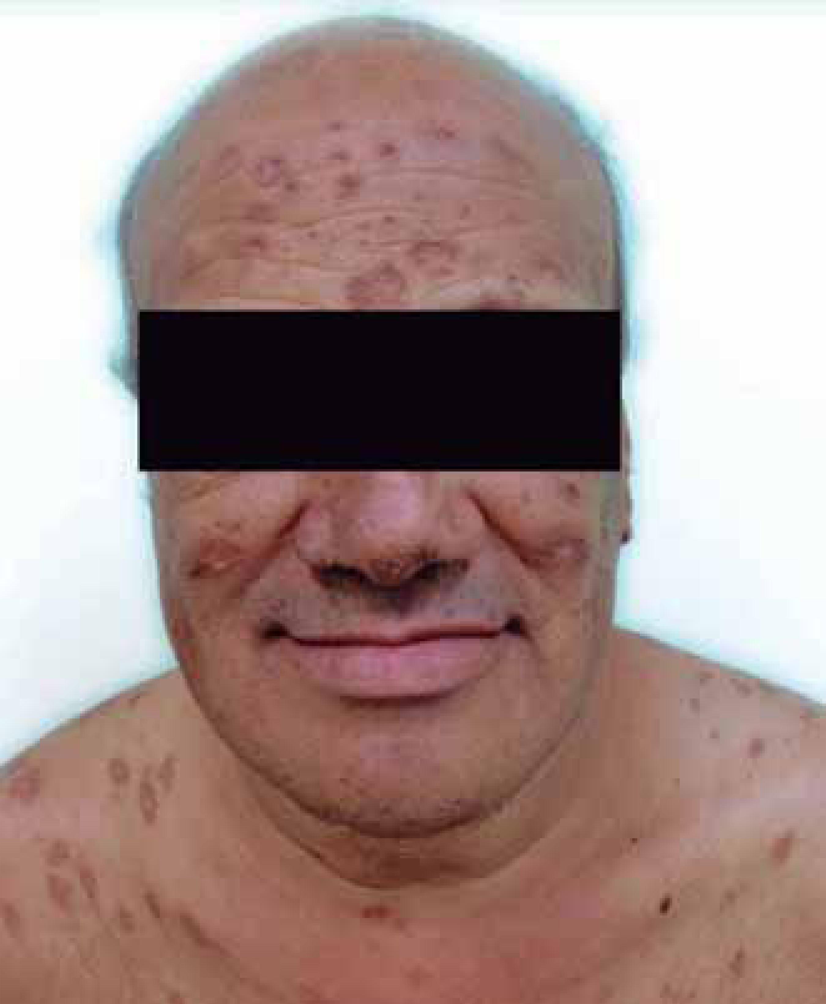
Clinical presentations are: acute and chronic pulmonary histoplasmosis, disseminated and primary cutaneous2. Infection is limited and restricted to the lungs in 99% of cases; the rest progresses to disseminated or chronic form. Advanced disease may occur as a progression of acute infection or late reactivation of focus with viable fungi.4In the disseminated form, the main findings are weight loss, fever, hepatosplenomegaly, generalized lymphadenopathy, and involvement of bone marrow, CNS, skin and mucous membranes. Regarding skin lesions, they are generally nonspecific, manifesting as macules, papules, pustules, verrucous plaques and ulcers.2Ulcers may also be found on oral and labial mucosa.5
In immunocompromised subjects, particularly in AIDS patients when their CD4 T-lymphocyte count is less than 50 cells/mm3, histoplasmosis may be presented in the form of a severe and widespread infection.5Skin lesions occur in 4-11% of patients and result from secondary invasion of skin in widespread form of infection, and it may be the first sign of the disease in immunocompromised individuals.6
The gold standard for diagnosis is the anatomopathological examination and culture for fungi of the involved skin tissues.2Fungus may also be evidenced in the sputum, blood, bone marrow and urine sediment.2Histopathological examination can detect the fungus through PAS and Grocott stains. The material for culture can be obtained by biopsy, aspiration, bronchial lavage and blood or marrow punction.2Detection of polysaccharide antigens in fluids such as urine or serum are also helpful, but there may be false-positive results, especially in patients with paracoccidioidomycosis.3
Complement fixation and immunodiffusion tests are usually negative in patients with HIV.3PCR on samples of blood and tissue is highly sensitive and specific. Histoplasmin skin test is indicated for non-endemic areas.2
The objective of this study is to report the case of a subject with large skin and mucosa lesions, which allowed the diagnosis and early treatment of the disease. The patient was infected with HIV for 20 years and had severe immunosuppression, with disseminated fungal disease and satisfactory clinical outcome despite the severity of co-infection.
CASE REPORT
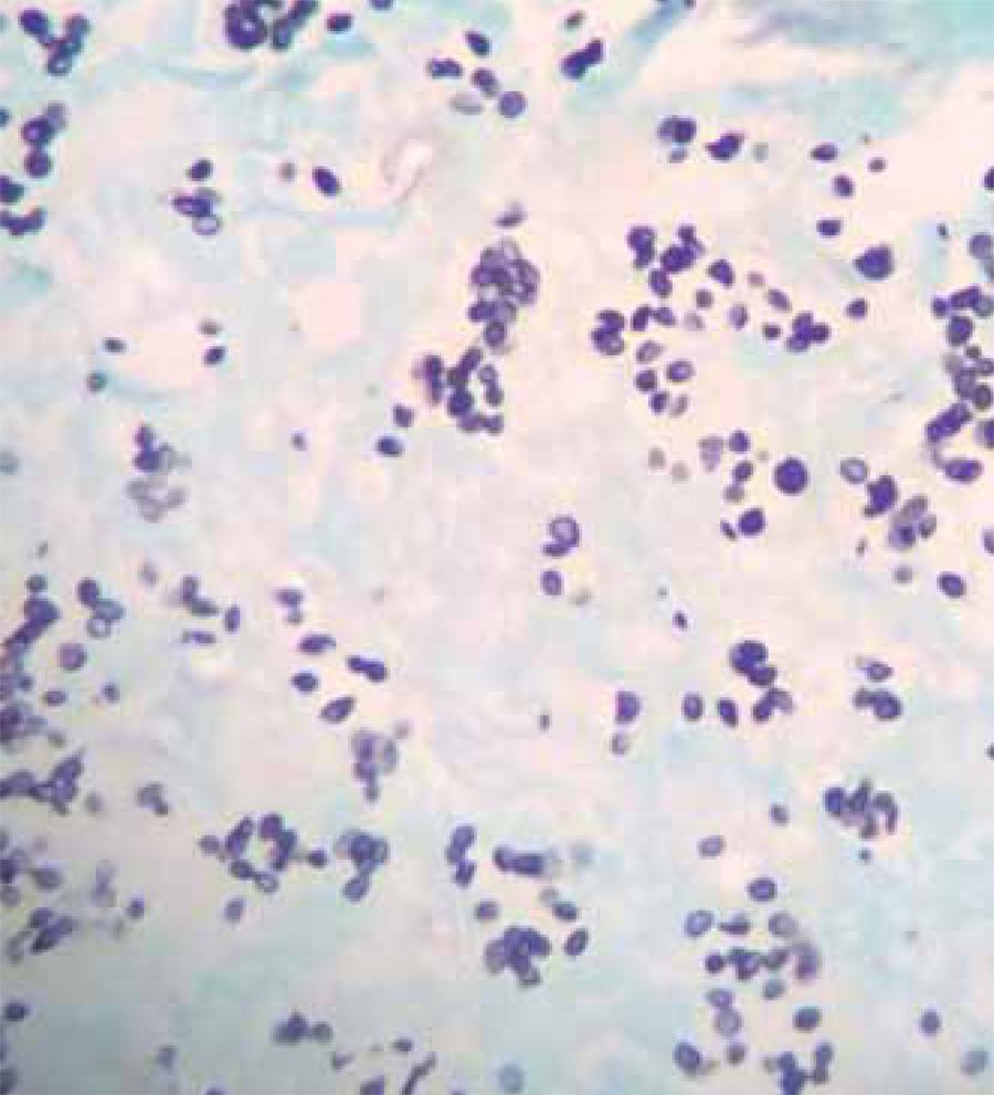
Man, 52 years old, rural worker, infected with HIV, with severe immunosuppression due to the irregular treatment with antiretroviral drugs. He has presented for 2 months poor general condition and verrucous lesions throughout integument, predominantly in the face and trunk (Figure 1). Examination showed pancytopenia and renal dysfunction (creatinine clearance 27 ml/min). Chest tomography revealed diffuse accentuation of the lung interstitium. Pathological examination (HE) showed epidermis with central ulceration, necrosis and numerous macrophages with clear cytoplasm and oval structures (Figure 2). Grocott stain was positive for Histoplasma (Figure 3). Direct mycological examination revealed fungal structures compatible with Histoplasma. Culture was performed from skin biopsy sample in Sabouraud-dextrose agar, at room temperature. On macroscopic examination white cotton-wool spots colonies were observed and, on microscopic examination, presence of hyphae, with rounded microconidia and macroconidia, was noted (Figure 4and5). Myeloculture and blood culture for fungus also showed the presence of Histoplasma capsulatum var capsulatum. Serology for specific antibodies was negative for the fungus. During hospitalization, the patient developed febrile neutropenia and therapy with Cefepime, Granulokine and reintroduction of antiretrovirals was started. CD4 was 7 cel/m3and viral load was 598,342 copies. A chemoprophylaxis with trimethoprim-sulfamethoxazole and azithromycin was performed. Therapy adopted was Itraconazole - 200 mg 3 times daily for 3 days in a row followed by 400 mg/day. It was of utmost importance the reintroduction of antiretrovirals, which led to an CD4 increase.
Pili torti. Polarized light microscopy, 10x magnification: Hair twisted about its longitudinal axis
100X HE: Skin sample containing histiocytic infi ltration in the dermis, which show punctate structures in the cytoplasm
Groccot 400X: In the silver staining it’s possible to note oval and uniform intracellular and grouped structures compatible with Histoplasma
The evolution of the case was satisfactory, with progressive improvement soon after the institution of specific antifungal treatment. After monitoring for 11 months, the patient had a weight gain of 20 kilos, clinical resolution of all lesions and improvement in general condition (Figure 6).
DISCUSSION
The most important risk factor for reactivation of infection and progression to disease is HIV-induced immunosuppression.3
Co-infection of histoplasmosis and AIDS, particularly when CD4 T-lymphocytes count is less than 50 cells/mm3, leads to a severe and disseminated infection.5In our case, the importance of skin lesions as a sign of systemic disease was evident, and it also may assist in the early diagnosis of histoplasmosis. This allowed the immediate start of specific treatment, thus reducing the risk of disease progression, which has a high mortality rate in immunocompromised patients.3
Skin lesions are present in most Brazilian cases (38-85%) and they are often more extensive compared with those reported in the USA, thus justifying the tropism for skin of strains diagnosed in South America.3Skin lesions predominate when H. duboisii is the etiologic agent.7Histoplasmosis mortality in immunosuppressed patients is greater than 33%, while in immunocompetent individuals is approximately 17%.3
In the literature, the drug of choice for disseminated histoplasmosis is Amphotericin B, 0.5-0.7 mg/kg/day for 10 weeks, followed by Itraconazole, 200 mg 3 times a day and maintenance dose of 400 mg/day for 12 weeks.8-9
For our patient, the treatment of choice was Itraconazole, due to the associated comorbidities. The patient presented excellent therapeutic response, and the drug was kept at a dose of 400 mg/day until reversal of immunosuppression state. The patient remains well during outpatient follow up, already for 11 months. In conclusion, it should be emphasized that histoplasmosis, particularly in HIV-infected patients, needs early diagnostic clarification, seeking immediate specific therapy, which will surely bring better progress.
-
Financial Support: None.
-
How to cite this article: voss Gonzalez T, Mattos e Dinato SL, Sementilli A, Romiti N, Beltrame PM, Veiga APR. Atypical presentation of histoplasmosis in na immunocompromised patient.. An Bras Dermatol. 2015;90 (3 Suppl 1): S32-5.
-
*
Study performed at Centro Universitário Lusíada (UNILUS) and Hospital Guilherme Álvaro– Santos (SP), Brazil.
References
- 1Vasudevan B, Ashish B, Amitabh S, A P M. Primary cutaneous histoplasmosis in a HIV-positive individual. J Glob Infect Dis. 2010;2:112-5.
- 2Kauls L, Blauvelt A, Skin disease in acute and chronic immunosuppression. In: Wolff K, Goldsmith LA, Katz SI, Gilchrest BA, Paller AS, Leffell DJ, editors. Fitzpatrick's Dermatology in General Medicine. 7th ed. New York: McGraw Hill; 2008. p. 275 - 6.
- 3Cunha VS, Zampese MS, Aquino VR, Cestari TF, Goldani LZ. Mucocutaneous manifestations of disseminated histoplasmosis in patients with acquired immunodeficiency syndrome: particular aspects in a Latin-American population. Clin Exp Dermatol. 2007;32:250-5.
- 4Kauffman CA. Histoplasmosis: a clinical and laboratory update. Clin Microbiol Rev. 2007;20:115-32.
- 5Orsi AT, Nogueira L, Chrusciak-Talhari A, Santos M, Ferreira LC, Talhari S, et al. Histoplasmosis and AIDS co-infection. An Bras Dermatol. 2011;86:1025-6.
- 6Saeki NM, Schubach AO, Salgueiro MM, Silva CF, et al. Histoplasmose cutânea primária: relato de caso em paciente imunocompetente e revisão de literatura. Rev Soc Bras Med Trop. 2008;41:680-2.
- 7Burns T, Breathnach S, Cox N, Griffiths C. Rook's Textbook of Dermatology 8th ed. Oxford: Wiley-Blackwell Publishing Ltd; 2010. p 36.82-84
- 8Wheat LJ, Freifeld AG, Kleiman MB, Baddley JW, McKinsey DS, Loyd JE, et al. Clinical practice guidelines for management of patients with histoplasmosis:2007 update by the infectious Disease of America. Clin Infect Dis. 2007;45:807-25.
- 9Bhagwat PV, Hanumanthayya K, Tophakhane RS, Rathod RM. Two unusual cases of histoplasmosis in human immunodeficiency virus-infected individuals. Indian J Dermatol Venereol Leprol. 2009;75:173-6.
Publication Dates
-
Publication in this collection
June 2015
History
-
Received
18 June 2014 -
Accepted
11 May 2014

 Atypical presentation of histoplasmosis in an immunocompromised patient


