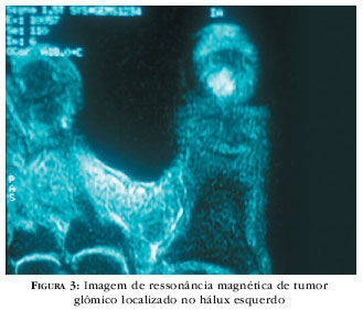
BACKGROUND: The subungual glomus tumor is a benign neoplasm of glomus cells, most frequently observed as a unique lesion on distal phalanx of fingers, and represents from 1 to 4.5% of hand neoplasms. OBJECTIVE: To evaluate the epidemiological and clinical aspects and diagnostic exams, such as histopathology and imaging methods. METHOD: Twenty cases of glomus tumor seen at the Dermatology Outpatient´s Clinics of Hospital das Clínicas and Hospital do Servidor Público Municipal of São Paulo, from 1991 to 2003, were studied. RESULTS: The epidemiological findings of this study did not significantly differ from the bibliographic search carried out. The preference for fingers and greater prevalence in women were confirmed in the patients observed. The histological data were similar to those in the literature. CONCLUSIONS: The imaging methods were not used in a systematic manner in diagnosis of glomus tumor, but they are very helpful to confirm and circumscribe the tumor, especially high definition magnetic resonance imaging. Although rare, relapses occurred in 15% of cases; thus there is no need for a prolonged surgical follow-up.
Diagnostic imaging; Glomus tumor; Glomus tumor; Glomus tumor; Nails


